
Dear Freesurfer experts,
I am a medical student and am currently doing a fellowship at the JPK Stroke Research Center - MGH. My current research project involves measuring the area of a cortical hypointense brain MRI signal (cortical superficial siderosis) , a marker that lays along the subarachnoid space/pial surface, only visible in blood sensitive MRI (SWI in my case).
In summary: We are trying to do a parallel to fMRI studies (fMRI x T1w // SWI x T1w), analyzing SWI signal intensity on a registered inflated brain surface; Projecting SWI surface intensity would then allow manual area demarcation of hipointensities in a flat surface.
Here is our step-by-step plan (analogous to fMRIxT1w registration/BOLD signal intensity projection to surface):
1. Perform freesurfer recon-all of the subject in question to generate subject-surface models. 2. BET and register the image of interest (SWI) into the freesurfer-subject space (register both SWI and MEMPRAGE sequences) [bbregister] 3. Having SWI in subject space and a surface model (generated in 1); * Split registered SWI into LH and RH * Tkregister2 to get LTA to mri_vol2surf format * Mri_vol2surf each hemisphere to the recon output 4. Display the SWI hemi surface as an overlay 5. Inflate T1 surface and observe its SWI overlay; * Manual demarcation of characteristic SWI hypointense signals 6. Analyze measured signal We did try this approach, but yet with these steps we could only obtain an inflated cortical surface representation that has no correspondence to SWI`s intensity.
So, here comes the question: If BOLD signal intensity can be projected onto surface area; is there a way to project SWI intensity onto the cortical surface? Or Can we preserve the info of SWI surface voxel intensity while doing the surface inflation? If not Is it possible to use the positional information of t1w voxel (used in inflation process) as an index to retrieve its correspondent SWI intensity value?
Sorry for the long question. In advance I thank you already for being so attentive.
Best, Pedro
Pedro Augusto Assis Lopes Student Researcher J. Philip Kistler Stroke Research Center Massachusetts General Hospital 175 Cambridge Street, Suite 300 Boston, MA 02114 Phone: (617) 643-3940

Hi Pedro, what you are trying to do is certainly possible. It looks like you are doing the right thing as far as I can tell, but I got a little lost after step 4, particularly the "Analyze measured signal" step. One other thing that may be an issue is what projection fraction you used in the mri_vol2surf step.
On 12/30/19 2:21 PM, Assis Lopes, Pedro Augusto wrote:
Dear Freesurfer experts,
I am a medical student and am currently doing a fellowship at the JPK Stroke Research Center - MGH. My current research project involves measuring the area of a cortical hypointense brain MRI signal (cortical superficial siderosis) , a marker that lays along the subarachnoid space/pial surface, only visible in blood sensitive MRI (SWI in my case).
In summary:
We are trying to do a parallel to fMRI studies (fMRI x T1w // SWI x T1w), analyzing SWI signal intensity on a registered inflated brain surface;
Projecting SWI surface intensity would then allow manual area demarcation of hipointensities in a flat surface.
Here is our step-by-step plan (analogous to fMRIxT1w registration/BOLD signal intensity projection to surface):
- Perform freesurfer recon-all of the subject in question to generate subject-surface models.
- BET and register the image of interest (SWI) into the freesurfer-subject space (register both SWI and MEMPRAGE sequences) [bbregister]
- Having SWI in subject space and a surface model (generated in 1);
- Split registered SWI into LH and RH
- Tkregister2 to get LTA to mri_vol2surf format
- Mri_vol2surf each hemisphere to the recon output
- Display the SWI hemi surface as an overlay
- Inflate T1 surface and observe its SWI overlay;
- Manual demarcation of characteristic SWI hypointense signals
- Analyze measured signal
_We did try this approach_, but yet with these steps we could only obtain an inflated cortical surface representation that has _no correspondence to SWI`s intensity_.
So, here comes the question:
*If BOLD signal intensity can be projected onto surface area; is there a way to project SWI intensity onto the cortical surface?*
Or
Can we preserve the info of SWI surface voxel intensity while doing the surface inflation?
If not
Is it possible to use the positional information of t1w voxel (used in inflation process) as an index to retrieve its correspondent SWI intensity value?
Sorry for the long question. In advance I thank you already for being so attentive.
Best,
Pedro
Pedro Augusto Assis Lopes
Student Researcher J. Philip Kistler Stroke Research Center Massachusetts General Hospital 175 Cambridge Street, Suite 300 Boston, MA 02114
Phone: (617) 643-3940
Freesurfer mailing list Freesurfer@nmr.mgh.harvard.edu https://mail.nmr.mgh.harvard.edu/mailman/listinfo/freesurfer
Dear Freesurfer experts,
Thank you for the quick response Dr. Greve! It`s an attempt to measure siderosis area, a thin (pathologically), small and blooming cortical signal seen through SWI (due to B0 inhomogeneity). We did: Recon-all
SWI BET and registration to T1 #BET bet $SUBJECTS_DIR/$subject/SWI_Surface/original/$subject_SWI.nii.gz $SUBJECTS_DIR/$subject/SWI_Surface/bet/$subject_SWI-bet.nii.gz -R
#bbregister (SWI to T1 skull stripped output from recon all) bbregister --s $subject --mov $SUBJECTS_DIR/$subject/SWI_Surface/bet/$subject_SWI-bet.nii.gz --reg $SUBJECTS_DIR/$subject/SWI_Surface/bbreg/$subject_SWI_bbreg.dat --o $SUBJECTS_DIR/$subject/SWI_Surface/bbreg/$subject_SWI_bbreg.nii.gz --t1
#Divided hemispheres #LH and RH mri_binarize --i $SUBJECTS_DIR/$subject/SWI_Surface/hemi/$subject_filled.nii.gz --match 255 --o $SUBJECTS_DIR/$subject/SWI_Surface/hemi/$subject_filled_LH.nii.gz mri_mask $SUBJECTS_DIR/$subject/SWI_Surface/bbreg/$subject_SWI_bbreg.nii.gz $SUBJECTS_DIR/$subject/SWI_Surface/hemi/$subject_filled_LH.nii.gz $SUBJECTS_DIR/$subject/SWI_Surface/hemi/$subject_SWI-LH.nii.gz
#Transform the volume into a surface file with mri_vol2surf #Left Hemi mri_vol2surf --mov $SUBJECTS_DIR/$subject/SWI_Surface/hemi/$subject_SWI-LH.nii.gz --srcreg $SUBJECTS_DIR/$subject/SWI_Surface/bbreg/$subject_SWI_bbreg.lta --hemi lh --o $SUBJECTS_DIR/$subject/SWI_Surface/vol2surf/$subject_SWI-LH_surface.nii.gz --regheader $subject #and right mri_vol2surf --mov $SUBJECTS_DIR/$subject/SWI_Surface/hemi/$subject_SWI-RH.nii.gz --srcreg $SUBJECTS_DIR/$subject/SWI_Surface/bbreg/$subject_SWI_bbreg.lta --hemi rh --o $SUBJECTS_DIR/$subject/SWI_Surface/vol2surf/$subject_SWI-RH_surface.nii.gz --regheader $subject
#Freeview and project SWI as an overlay onto the inflated surface freeview -f $SUBJECTS_DIR/$subject/surf/lh.inflated:annot=aparc.annot:annot_outline=1:overlay=$SUBJECTS_DIR/$subject/SWI_Surface/vol2surf/$subject_SWI-LH_surface.nii.gz -f $SUBJECTS_DIR/$subject/surf/rh.inflated:annot=aparc.annot:annot_outline=1:overlay=$SUBJECTS_DIR/$subject/SWI_Surface/vol2surf/$subject_SWI-RH_surface.nii.gz
We did not specify any projection fraction in our mri_vol2surf. Sorry for not being clearer in the step of analyze measured signal. We aim to make a brain mask of such hypointense signal and obtain its proportional area to total brain pial surface.
Thank you so much for the attention Dr. Greve, I look forward in receiving your response!
-----Original Message----- From: freesurfer-bounces@nmr.mgh.harvard.edu freesurfer-bounces@nmr.mgh.harvard.edu On Behalf Of Greve, Douglas N.,Ph.D. Sent: Monday, January 06, 2020 12:33 PM To: freesurfer@nmr.mgh.harvard.edu Subject: Re: [Freesurfer] Cortical inflation and maintenance of surface voxel intensity
Hi Pedro, what you are trying to do is certainly possible. It looks like you are doing the right thing as far as I can tell, but I got a little lost after step 4, particularly the "Analyze measured signal" step. One other thing that may be an issue is what projection fraction you used in the mri_vol2surf step.
On 12/30/19 2:21 PM, Assis Lopes, Pedro Augusto wrote:
Dear Freesurfer experts,
I am a medical student and am currently doing a fellowship at the JPK Stroke Research Center - MGH. My current research project involves measuring the area of a cortical hypointense brain MRI signal (cortical superficial siderosis) , a marker that lays along the subarachnoid space/pial surface, only visible in blood sensitive MRI (SWI in my case).
In summary:
We are trying to do a parallel to fMRI studies (fMRI x T1w // SWI x T1w), analyzing SWI signal intensity on a registered inflated brain surface;
Projecting SWI surface intensity would then allow manual area demarcation of hipointensities in a flat surface.
Here is our step-by-step plan (analogous to fMRIxT1w registration/BOLD signal intensity projection to surface):
- Perform freesurfer recon-all of the subject in question to generate subject-surface models.
- BET and register the image of interest (SWI) into the freesurfer-subject space (register both SWI and MEMPRAGE sequences) [bbregister]
- Having SWI in subject space and a surface model (generated in 1);
- Split registered SWI into LH and RH
- Tkregister2 to get LTA to mri_vol2surf format
- Mri_vol2surf each hemisphere to the recon output 4. Display
the SWI hemi surface as an overlay 5. Inflate T1 surface and observe its SWI overlay; 1. Manual demarcation of characteristic SWI hypointense signals 6. Analyze measured signal
_We did try this approach_, but yet with these steps we could only obtain an inflated cortical surface representation that has _no correspondence to SWI`s intensity_.
So, here comes the question:
*If BOLD signal intensity can be projected onto surface area; is there a way to project SWI intensity onto the cortical surface?*
Or
Can we preserve the info of SWI surface voxel intensity while doing the surface inflation?
If not
Is it possible to use the positional information of t1w voxel (used in inflation process) as an index to retrieve its correspondent SWI intensity value?
Sorry for the long question. In advance I thank you already for being so attentive.
Best,
Pedro
Pedro Augusto Assis Lopes
Student Researcher J. Philip Kistler Stroke Research Center Massachusetts General Hospital 175 Cambridge Street, Suite 300 Boston, MA 02114
Phone: (617) 643-3940
Freesurfer mailing list Freesurfer@nmr.mgh.harvard.edu https://mail.nmr.mgh.harvard.edu/mailman/listinfo/freesurfer
_______________________________________________ Freesurfer mailing list Freesurfer@nmr.mgh.harvard.edu https://mail.nmr.mgh.harvard.edu/mailman/listinfo/freesurfer
I think the problem is that you apply the registration twice instead of once. the first time is on the volume output of bbregister, but then you do it again when you run mri_vol2surf. I would just use SWI-bet.nii.gz (input to bbregister) as input to mri_vol2surf. Also, it is unnecessary to do bet or mri_binarize or mri_mask for this process (though you may need it for other things). Also, I would use --projfrac 0.5 when running mri_vol2surf
On 1/6/2020 10:03 PM, Assis Lopes, Pedro Augusto wrote:
Dear Freesurfer experts,
Thank you for the quick response Dr. Greve! It`s an attempt to measure siderosis area, a thin (pathologically), small and blooming cortical signal seen through SWI (due to B0 inhomogeneity). We did: Recon-all
SWI BET and registration to T1 #BET bet $SUBJECTS_DIR/$subject/SWI_Surface/original/$subject_SWI.nii.gz $SUBJECTS_DIR/$subject/SWI_Surface/bet/$subject_SWI-bet.nii.gz -R
#bbregister (SWI to T1 skull stripped output from recon all) bbregister --s $subject --mov $SUBJECTS_DIR/$subject/SWI_Surface/bet/$subject_SWI-bet.nii.gz --reg $SUBJECTS_DIR/$subject/SWI_Surface/bbreg/$subject_SWI_bbreg.dat --o $SUBJECTS_DIR/$subject/SWI_Surface/bbreg/$subject_SWI_bbreg.nii.gz --t1
#Divided hemispheres #LH and RH mri_binarize --i $SUBJECTS_DIR/$subject/SWI_Surface/hemi/$subject_filled.nii.gz --match 255 --o $SUBJECTS_DIR/$subject/SWI_Surface/hemi/$subject_filled_LH.nii.gz mri_mask $SUBJECTS_DIR/$subject/SWI_Surface/bbreg/$subject_SWI_bbreg.nii.gz $SUBJECTS_DIR/$subject/SWI_Surface/hemi/$subject_filled_LH.nii.gz $SUBJECTS_DIR/$subject/SWI_Surface/hemi/$subject_SWI-LH.nii.gz
#Transform the volume into a surface file with mri_vol2surf #Left Hemi mri_vol2surf --mov $SUBJECTS_DIR/$subject/SWI_Surface/hemi/$subject_SWI-LH.nii.gz --srcreg $SUBJECTS_DIR/$subject/SWI_Surface/bbreg/$subject_SWI_bbreg.lta --hemi lh --o $SUBJECTS_DIR/$subject/SWI_Surface/vol2surf/$subject_SWI-LH_surface.nii.gz --regheader $subject #and right mri_vol2surf --mov $SUBJECTS_DIR/$subject/SWI_Surface/hemi/$subject_SWI-RH.nii.gz --srcreg $SUBJECTS_DIR/$subject/SWI_Surface/bbreg/$subject_SWI_bbreg.lta --hemi rh --o $SUBJECTS_DIR/$subject/SWI_Surface/vol2surf/$subject_SWI-RH_surface.nii.gz --regheader $subject
#Freeview and project SWI as an overlay onto the inflated surface freeview -f $SUBJECTS_DIR/$subject/surf/lh.inflated:annot=aparc.annot:annot_outline=1:overlay=$SUBJECTS_DIR/$subject/SWI_Surface/vol2surf/$subject_SWI-LH_surface.nii.gz -f $SUBJECTS_DIR/$subject/surf/rh.inflated:annot=aparc.annot:annot_outline=1:overlay=$SUBJECTS_DIR/$subject/SWI_Surface/vol2surf/$subject_SWI-RH_surface.nii.gz
We did not specify any projection fraction in our mri_vol2surf. Sorry for not being clearer in the step of analyze measured signal. We aim to make a brain mask of such hypointense signal and obtain its proportional area to total brain pial surface.
Thank you so much for the attention Dr. Greve, I look forward in receiving your response!
-----Original Message----- From: freesurfer-bounces@nmr.mgh.harvard.edu freesurfer-bounces@nmr.mgh.harvard.edu On Behalf Of Greve, Douglas N.,Ph.D. Sent: Monday, January 06, 2020 12:33 PM To: freesurfer@nmr.mgh.harvard.edu Subject: Re: [Freesurfer] Cortical inflation and maintenance of surface voxel intensity
Hi Pedro, what you are trying to do is certainly possible. It looks like you are doing the right thing as far as I can tell, but I got a little lost after step 4, particularly the "Analyze measured signal" step. One other thing that may be an issue is what projection fraction you used in the mri_vol2surf step.
On 12/30/19 2:21 PM, Assis Lopes, Pedro Augusto wrote:
Dear Freesurfer experts,
I am a medical student and am currently doing a fellowship at the JPK Stroke Research Center - MGH. My current research project involves measuring the area of a cortical hypointense brain MRI signal (cortical superficial siderosis) , a marker that lays along the subarachnoid space/pial surface, only visible in blood sensitive MRI (SWI in my case).
In summary:
We are trying to do a parallel to fMRI studies (fMRI x T1w // SWI x T1w), analyzing SWI signal intensity on a registered inflated brain surface;
Projecting SWI surface intensity would then allow manual area demarcation of hipointensities in a flat surface.
Here is our step-by-step plan (analogous to fMRIxT1w registration/BOLD signal intensity projection to surface):
- Perform freesurfer recon-all of the subject in question to generate subject-surface models.
- BET and register the image of interest (SWI) into the freesurfer-subject space (register both SWI and MEMPRAGE sequences) [bbregister]
- Having SWI in subject space and a surface model (generated in 1);
- Split registered SWI into LH and RH
- Tkregister2 to get LTA to mri_vol2surf format
- Mri_vol2surf each hemisphere to the recon output 4. Display
the SWI hemi surface as an overlay 5. Inflate T1 surface and observe its SWI overlay; 1. Manual demarcation of characteristic SWI hypointense signals 6. Analyze measured signal
_We did try this approach_, but yet with these steps we could only obtain an inflated cortical surface representation that has _no correspondence to SWI`s intensity_.
So, here comes the question:
*If BOLD signal intensity can be projected onto surface area; is there a way to project SWI intensity onto the cortical surface?*
Or
Can we preserve the info of SWI surface voxel intensity while doing the surface inflation?
If not
Is it possible to use the positional information of t1w voxel (used in inflation process) as an index to retrieve its correspondent SWI intensity value?
Sorry for the long question. In advance I thank you already for being so attentive.
Best,
Pedro
Pedro Augusto Assis Lopes
Student Researcher J. Philip Kistler Stroke Research Center Massachusetts General Hospital 175 Cambridge Street, Suite 300 Boston, MA 02114
Phone: (617) 643-3940
Freesurfer mailing list Freesurfer@nmr.mgh.harvard.edu https://mail.nmr.mgh.harvard.edu/mailman/listinfo/freesurfer
Freesurfer mailing list Freesurfer@nmr.mgh.harvard.edu https://mail.nmr.mgh.harvard.edu/mailman/listinfo/freesurfer
Freesurfer mailing list Freesurfer@nmr.mgh.harvard.edu https://mail.nmr.mgh.harvard.edu/mailman/listinfo/freesurfer
Hello Freesurfer experts!
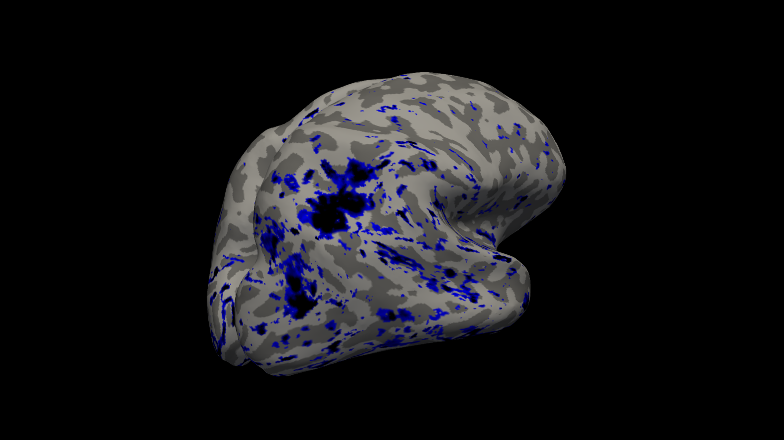
Thank you so much for the explanation Dr. Greve! Attached is the output of our overlaid surfaces:
1 - Grossly speaking, sorry for any jargon mistaken, this image show the intensity value given a projection, from the WM surf. towards pial surf., of 80% (mri_vol2surf --projfrac 0.8) of a src volume (a registered SWI/T1). (We tried surfproj of 1 but too noisy, and if 0.5, loses much info, that is why we chose the 0.8). - However, my question is: is the intensity value projected onto the surface an average of this vector of intensities (the projfrac from WM surf to PS) OR just the intensity value of a single voxel (voxel at the "tip" of vector) ?
2 - I plan to first make a mask that covers the range of intensities in black and and blue, then manually select only the black and the surrounding blue. - Is there a way to make this mask out of my vol2surf surfaces? - And later, how would one manually select a given ROI of this surface? (3Dslicer/ matlab or freesurfer can do it?)
Thank you so much for the attention! Best, Pedro
-----Original Message----- From: freesurfer-bounces@nmr.mgh.harvard.edu freesurfer-bounces@nmr.mgh.harvard.edu On Behalf Of Greve, Douglas N.,Ph.D. Sent: Friday, January 10, 2020 11:12 AM To: freesurfer@nmr.mgh.harvard.edu Subject: Re: [Freesurfer] Cortical inflation and maintenance of surface voxel intensity
I think the problem is that you apply the registration twice instead of once. the first time is on the volume output of bbregister, but then you do it again when you run mri_vol2surf. I would just use SWI-bet.nii.gz (input to bbregister) as input to mri_vol2surf. Also, it is unnecessary to do bet or mri_binarize or mri_mask for this process (though you may need it for other things). Also, I would use --projfrac 0.5 when running mri_vol2surf
On 1/6/2020 10:03 PM, Assis Lopes, Pedro Augusto wrote:
Dear Freesurfer experts,
Thank you for the quick response Dr. Greve! It`s an attempt to measure siderosis area, a thin (pathologically), small and blooming cortical signal seen through SWI (due to B0 inhomogeneity). We did: Recon-all
SWI BET and registration to T1 #BET bet $SUBJECTS_DIR/$subject/SWI_Surface/original/$subject_SWI.nii.gz $SUBJECTS_DIR/$subject/SWI_Surface/bet/$subject_SWI-bet.nii.gz -R
#bbregister (SWI to T1 skull stripped output from recon all) bbregister --s $subject --mov $SUBJECTS_DIR/$subject/SWI_Surface/bet/$subject_SWI-bet.nii.gz --reg $SUBJECTS_DIR/$subject/SWI_Surface/bbreg/$subject_SWI_bbreg.dat --o $SUBJECTS_DIR/$subject/SWI_Surface/bbreg/$subject_SWI_bbreg.nii.gz --t1
#Divided hemispheres #LH and RH mri_binarize --i $SUBJECTS_DIR/$subject/SWI_Surface/hemi/$subject_filled.nii.gz --match 255 --o $SUBJECTS_DIR/$subject/SWI_Surface/hemi/$subject_filled_LH.nii.gz mri_mask $SUBJECTS_DIR/$subject/SWI_Surface/bbreg/$subject_SWI_bbreg.nii.gz $SUBJECTS_DIR/$subject/SWI_Surface/hemi/$subject_filled_LH.nii.gz $SUBJECTS_DIR/$subject/SWI_Surface/hemi/$subject_SWI-LH.nii.gz
#Transform the volume into a surface file with mri_vol2surf #Left Hemi mri_vol2surf --mov $SUBJECTS_DIR/$subject/SWI_Surface/hemi/$subject_SWI-LH.nii.gz --srcreg $SUBJECTS_DIR/$subject/SWI_Surface/bbreg/$subject_SWI_bbreg.lta --hemi lh --o $SUBJECTS_DIR/$subject/SWI_Surface/vol2surf/$subject_SWI-LH_surface.n ii.gz --regheader $subject #and right mri_vol2surf --mov $SUBJECTS_DIR/$subject/SWI_Surface/hemi/$subject_SWI-RH.nii.gz --srcreg $SUBJECTS_DIR/$subject/SWI_Surface/bbreg/$subject_SWI_bbreg.lta --hemi rh --o $SUBJECTS_DIR/$subject/SWI_Surface/vol2surf/$subject_SWI-RH_surface.n ii.gz --regheader $subject
#Freeview and project SWI as an overlay onto the inflated surface freeview -f $SUBJECTS_DIR/$subject/surf/lh.inflated:annot=aparc.annot:annot_outlin e=1:overlay=$SUBJECTS_DIR/$subject/SWI_Surface/vol2surf/$subject_SWI- LH_surface.nii.gz -f $SUBJECTS_DIR/$subject/surf/rh.inflated:annot=aparc.annot:annot_outlin e=1:overlay=$SUBJECTS_DIR/$subject/SWI_Surface/vol2surf/$subject_SWI- RH_surface.nii.gz
We did not specify any projection fraction in our mri_vol2surf. Sorry for not being clearer in the step of analyze measured signal. We aim to make a brain mask of such hypointense signal and obtain its proportional area to total brain pial surface.
Thank you so much for the attention Dr. Greve, I look forward in receiving your response!
-----Original Message----- From: freesurfer-bounces@nmr.mgh.harvard.edu freesurfer-bounces@nmr.mgh.harvard.edu On Behalf Of Greve, Douglas N.,Ph.D. Sent: Monday, January 06, 2020 12:33 PM To: freesurfer@nmr.mgh.harvard.edu Subject: Re: [Freesurfer] Cortical inflation and maintenance of surface voxel intensity
Hi Pedro, what you are trying to do is certainly possible. It looks like you are doing the right thing as far as I can tell, but I got a little lost after step 4, particularly the "Analyze measured signal" step. One other thing that may be an issue is what projection fraction you used in the mri_vol2surf step.
On 12/30/19 2:21 PM, Assis Lopes, Pedro Augusto wrote:
Dear Freesurfer experts,
I am a medical student and am currently doing a fellowship at the JPK Stroke Research Center - MGH. My current research project involves measuring the area of a cortical hypointense brain MRI signal (cortical superficial siderosis) , a marker that lays along the subarachnoid space/pial surface, only visible in blood sensitive MRI (SWI in my case).
In summary:
We are trying to do a parallel to fMRI studies (fMRI x T1w // SWI x T1w), analyzing SWI signal intensity on a registered inflated brain surface;
Projecting SWI surface intensity would then allow manual area demarcation of hipointensities in a flat surface.
Here is our step-by-step plan (analogous to fMRIxT1w registration/BOLD signal intensity projection to surface):
- Perform freesurfer recon-all of the subject in question to generate subject-surface models.
- BET and register the image of interest (SWI) into the freesurfer-subject space (register both SWI and MEMPRAGE sequences) [bbregister]
- Having SWI in subject space and a surface model (generated in 1);
- Split registered SWI into LH and RH
- Tkregister2 to get LTA to mri_vol2surf format
- Mri_vol2surf each hemisphere to the recon output 4. Display
the SWI hemi surface as an overlay 5. Inflate T1 surface and observe its SWI overlay; 1. Manual demarcation of characteristic SWI hypointense signals 6. Analyze measured signal
_We did try this approach_, but yet with these steps we could only obtain an inflated cortical surface representation that has _no correspondence to SWI`s intensity_.
So, here comes the question:
*If BOLD signal intensity can be projected onto surface area; is there a way to project SWI intensity onto the cortical surface?*
Or
Can we preserve the info of SWI surface voxel intensity while doing the surface inflation?
If not
Is it possible to use the positional information of t1w voxel (used in inflation process) as an index to retrieve its correspondent SWI intensity value?
Sorry for the long question. In advance I thank you already for being so attentive.
Best,
Pedro
Pedro Augusto Assis Lopes
Student Researcher J. Philip Kistler Stroke Research Center Massachusetts General Hospital 175 Cambridge Street, Suite 300 Boston, MA 02114
Phone: (617) 643-3940
Freesurfer mailing list Freesurfer@nmr.mgh.harvard.edu https://mail.nmr.mgh.harvard.edu/mailman/listinfo/freesurfer
Freesurfer mailing list Freesurfer@nmr.mgh.harvard.edu https://mail.nmr.mgh.harvard.edu/mailman/listinfo/freesurfer
Freesurfer mailing list Freesurfer@nmr.mgh.harvard.edu https://mail.nmr.mgh.harvard.edu/mailman/listinfo/freesurfer
_______________________________________________ Freesurfer mailing list Freesurfer@nmr.mgh.harvard.edu https://mail.nmr.mgh.harvard.edu/mailman/listinfo/freesurfer
I've answered one of your questions below.
Ruopeng, there is a question for you too
On 1/21/20 11:02 AM, Assis Lopes, Pedro Augusto wrote:
Hello Freesurfer experts!
Thank you so much for the explanation Dr. Greve! Attached is the output of our overlaid surfaces:
1 - Grossly speaking, sorry for any jargon mistaken, this image show the intensity value given a projection, from the WM surf. towards pial surf., of 80% (mri_vol2surf --projfrac 0.8) of a src volume (a registered SWI/T1). (We tried surfproj of 1 but too noisy, and if 0.5, loses much info, that is why we chose the 0.8). - However, my question is: is the intensity value projected onto the surface an average of this vector of intensities (the projfrac from WM surf to PS) OR just the intensity value of a single voxel (voxel at the "tip" of vector) ?
Just a single voxel
2 - I plan to first make a mask that covers the range of intensities in black and and blue, then manually select only the black and the surrounding blue. - Is there a way to make this mask out of my vol2surf surfaces?
If you know the intensity threshold, you can use mri_binarize
- And later, how would one manually select a given ROI of this surface? (3Dslicer/ matlab or freesurfer can do it?)
Not sure, Ruopeng? I think he just wants to create a label from a thresholded overlay.
Thank you so much for the attention! Best, Pedro
-----Original Message----- From: freesurfer-bounces@nmr.mgh.harvard.edu freesurfer-bounces@nmr.mgh.harvard.edu On Behalf Of Greve, Douglas N.,Ph.D. Sent: Friday, January 10, 2020 11:12 AM To: freesurfer@nmr.mgh.harvard.edu Subject: Re: [Freesurfer] Cortical inflation and maintenance of surface voxel intensity
I think the problem is that you apply the registration twice instead of once. the first time is on the volume output of bbregister, but then you do it again when you run mri_vol2surf. I would just use SWI-bet.nii.gz (input to bbregister) as input to mri_vol2surf. Also, it is unnecessary to do bet or mri_binarize or mri_mask for this process (though you may need it for other things). Also, I would use --projfrac 0.5 when running mri_vol2surf
On 1/6/2020 10:03 PM, Assis Lopes, Pedro Augusto wrote:
Dear Freesurfer experts,
Thank you for the quick response Dr. Greve! It`s an attempt to measure siderosis area, a thin (pathologically), small and blooming cortical signal seen through SWI (due to B0 inhomogeneity). We did: Recon-all
SWI BET and registration to T1 #BET bet $SUBJECTS_DIR/$subject/SWI_Surface/original/$subject_SWI.nii.gz $SUBJECTS_DIR/$subject/SWI_Surface/bet/$subject_SWI-bet.nii.gz -R
#bbregister (SWI to T1 skull stripped output from recon all) bbregister --s $subject --mov $SUBJECTS_DIR/$subject/SWI_Surface/bet/$subject_SWI-bet.nii.gz --reg $SUBJECTS_DIR/$subject/SWI_Surface/bbreg/$subject_SWI_bbreg.dat --o $SUBJECTS_DIR/$subject/SWI_Surface/bbreg/$subject_SWI_bbreg.nii.gz --t1
#Divided hemispheres #LH and RH mri_binarize --i $SUBJECTS_DIR/$subject/SWI_Surface/hemi/$subject_filled.nii.gz --match 255 --o $SUBJECTS_DIR/$subject/SWI_Surface/hemi/$subject_filled_LH.nii.gz mri_mask $SUBJECTS_DIR/$subject/SWI_Surface/bbreg/$subject_SWI_bbreg.nii.gz $SUBJECTS_DIR/$subject/SWI_Surface/hemi/$subject_filled_LH.nii.gz $SUBJECTS_DIR/$subject/SWI_Surface/hemi/$subject_SWI-LH.nii.gz
#Transform the volume into a surface file with mri_vol2surf #Left Hemi mri_vol2surf --mov $SUBJECTS_DIR/$subject/SWI_Surface/hemi/$subject_SWI-LH.nii.gz --srcreg $SUBJECTS_DIR/$subject/SWI_Surface/bbreg/$subject_SWI_bbreg.lta --hemi lh --o $SUBJECTS_DIR/$subject/SWI_Surface/vol2surf/$subject_SWI-LH_surface.n ii.gz --regheader $subject #and right mri_vol2surf --mov $SUBJECTS_DIR/$subject/SWI_Surface/hemi/$subject_SWI-RH.nii.gz --srcreg $SUBJECTS_DIR/$subject/SWI_Surface/bbreg/$subject_SWI_bbreg.lta --hemi rh --o $SUBJECTS_DIR/$subject/SWI_Surface/vol2surf/$subject_SWI-RH_surface.n ii.gz --regheader $subject
#Freeview and project SWI as an overlay onto the inflated surface freeview -f $SUBJECTS_DIR/$subject/surf/lh.inflated:annot=aparc.annot:annot_outlin e=1:overlay=$SUBJECTS_DIR/$subject/SWI_Surface/vol2surf/$subject_SWI- LH_surface.nii.gz -f $SUBJECTS_DIR/$subject/surf/rh.inflated:annot=aparc.annot:annot_outlin e=1:overlay=$SUBJECTS_DIR/$subject/SWI_Surface/vol2surf/$subject_SWI- RH_surface.nii.gz
We did not specify any projection fraction in our mri_vol2surf. Sorry for not being clearer in the step of analyze measured signal. We aim to make a brain mask of such hypointense signal and obtain its proportional area to total brain pial surface.
Thank you so much for the attention Dr. Greve, I look forward in receiving your response!
-----Original Message----- From: freesurfer-bounces@nmr.mgh.harvard.edu freesurfer-bounces@nmr.mgh.harvard.edu On Behalf Of Greve, Douglas N.,Ph.D. Sent: Monday, January 06, 2020 12:33 PM To: freesurfer@nmr.mgh.harvard.edu Subject: Re: [Freesurfer] Cortical inflation and maintenance of surface voxel intensity
Hi Pedro, what you are trying to do is certainly possible. It looks like you are doing the right thing as far as I can tell, but I got a little lost after step 4, particularly the "Analyze measured signal" step. One other thing that may be an issue is what projection fraction you used in the mri_vol2surf step.
On 12/30/19 2:21 PM, Assis Lopes, Pedro Augusto wrote:
Dear Freesurfer experts,
I am a medical student and am currently doing a fellowship at the JPK Stroke Research Center - MGH. My current research project involves measuring the area of a cortical hypointense brain MRI signal (cortical superficial siderosis) , a marker that lays along the subarachnoid space/pial surface, only visible in blood sensitive MRI (SWI in my case).
In summary:
We are trying to do a parallel to fMRI studies (fMRI x T1w // SWI x T1w), analyzing SWI signal intensity on a registered inflated brain surface;
Projecting SWI surface intensity would then allow manual area demarcation of hipointensities in a flat surface.
Here is our step-by-step plan (analogous to fMRIxT1w registration/BOLD signal intensity projection to surface):
- Perform freesurfer recon-all of the subject in question to generate subject-surface models.
- BET and register the image of interest (SWI) into the freesurfer-subject space (register both SWI and MEMPRAGE sequences) [bbregister]
- Having SWI in subject space and a surface model (generated in 1);
- Split registered SWI into LH and RH
- Tkregister2 to get LTA to mri_vol2surf format
- Mri_vol2surf each hemisphere to the recon output 4. Display
the SWI hemi surface as an overlay 5. Inflate T1 surface and observe its SWI overlay; 1. Manual demarcation of characteristic SWI hypointense signals 6. Analyze measured signal
_We did try this approach_, but yet with these steps we could only obtain an inflated cortical surface representation that has _no correspondence to SWI`s intensity_.
So, here comes the question:
*If BOLD signal intensity can be projected onto surface area; is there a way to project SWI intensity onto the cortical surface?*
Or
Can we preserve the info of SWI surface voxel intensity while doing the surface inflation?
If not
Is it possible to use the positional information of t1w voxel (used in inflation process) as an index to retrieve its correspondent SWI intensity value?
Sorry for the long question. In advance I thank you already for being so attentive.
Best,
Pedro
Pedro Augusto Assis Lopes
Student Researcher J. Philip Kistler Stroke Research Center Massachusetts General Hospital 175 Cambridge Street, Suite 300 Boston, MA 02114
Phone: (617) 643-3940
Freesurfer mailing list Freesurfer@nmr.mgh.harvard.edu https://mail.nmr.mgh.harvard.edu/mailman/listinfo/freesurfer
Freesurfer mailing list Freesurfer@nmr.mgh.harvard.edu https://mail.nmr.mgh.harvard.edu/mailman/listinfo/freesurfer
Freesurfer mailing list Freesurfer@nmr.mgh.harvard.edu https://mail.nmr.mgh.harvard.edu/mailman/listinfo/freesurfer
Freesurfer mailing list Freesurfer@nmr.mgh.harvard.edu https://mail.nmr.mgh.harvard.edu/mailman/listinfo/freesurfer
Freesurfer mailing list Freesurfer@nmr.mgh.harvard.edu https://mail.nmr.mgh.harvard.edu/mailman/listinfo/freesurfer
freesurfer@nmr.mgh.harvard.edu
-
 Assis Lopes, Pedro Augusto
 -
 Greve, Douglas N.,Ph.D.