Reposting a Question RE: Hippocampal/Amygdala subfield segmentation - Medial Nucleus Errors

External Email - Use Caution
Hello Freesurfer Experts,
I wanted to re-post a portion of a question that a previous team member posted on June 20th, 2019 (See: Reposting a Question RE: Hippocampal/Amygdala subfield segmentation - Anterior Amygdala Area / Medial Nucleus Errors)
We have visually checked 133 subjects that have been processed using Freesurfer's latest hippocampal/amygdala subfield segmentation algorithm (Development version 20180518 https://surfer.nmr.mgh.harvard.edu/fswiki/HippocampalSubfieldsAndNucleiOfAmygdala) and came across a few cases that had questionable segmentation. The T1s had were processed using FS 5.3, and we used the command "segmentHA_T1.sh" from the latest Dev version.
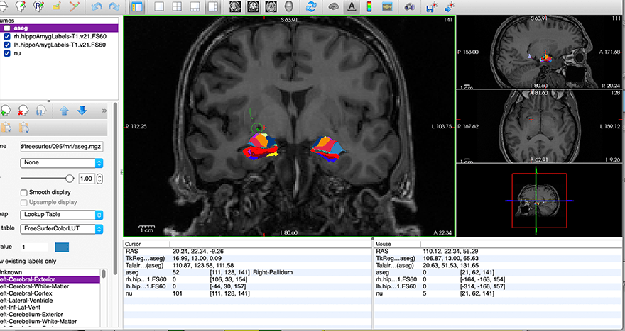
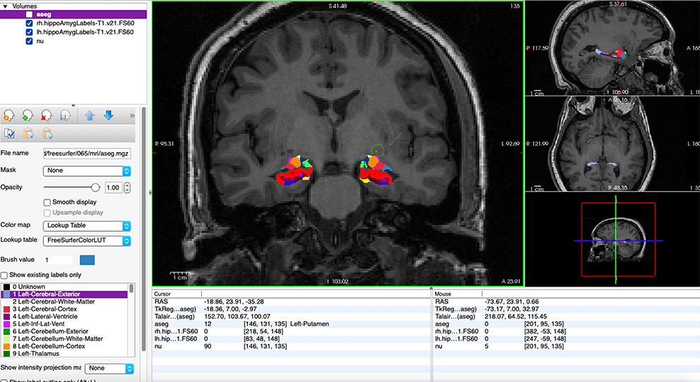
-We have come across about 5 subjects' medical nuclei masks that are located in the Putamen region (more superior/detached from the rest of the amygdala) on one side of the brain (in some cases just the right side, in other cases just the left side). In all of the other subjects, we don't see that same detached looking segmentation into the putamen on one side as these few show. I will attach screenshots to show this.
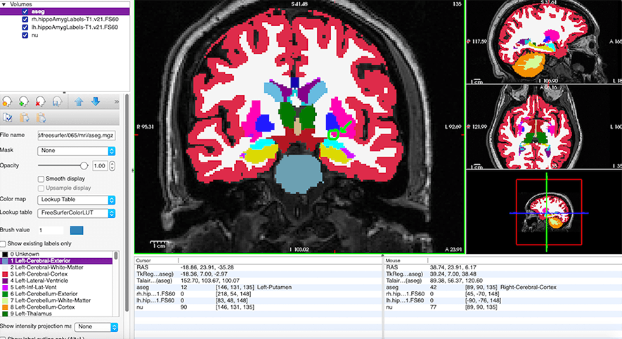
-I have checked the aseg for these subjects as suggested in the previous question, and the hippocampus, amygdala, and putamen all look fine there. I have also checked the output volumes for the medial nucleus of these subjects and the numbers all look fine.
-So, my main question is: If the placement of the medial nucleus is wrong in the segmentation (detached/in the putamen as shown) but the aseg looks correct, can we still use those values reliably?
Thank you!
Angel Hammond
Research Specialist
Center for Healthy Minds
University of Wisconsin-Madison
freesurfer@nmr.mgh.harvard.edu
-
 Angel Hammond