
Hi Freesurfer team I am wondering if there would be a way to improve surfaces on the medial wall, not for the measurement of cortical thickness, but also for laminar analysis in the cortex. We would need them to be accurate even for the medial wall. What would you suggest? Thanks Celine
Hi Freesurfer team I am wondering if there would be a way to improve surfaces on the medial wall, not for the measurement of cortical thickness, but also for laminar analysis in the cortex. We would need them to be accurate even for the medial wall. What would you suggest? Thanks Celine
-- Celine Louapre, MD Research Fellow at Massachusetts General Hospital Department of Radiology, MGH
Building 149, Room 2301 13th Street Charlestown, MA 02129 _______________________________________________ Freesurfer mailing list Freesurfer@nmr.mgh.harvard.edu https://mail.nmr.mgh.harvard.edu/mailman/listinfo/freesurfer
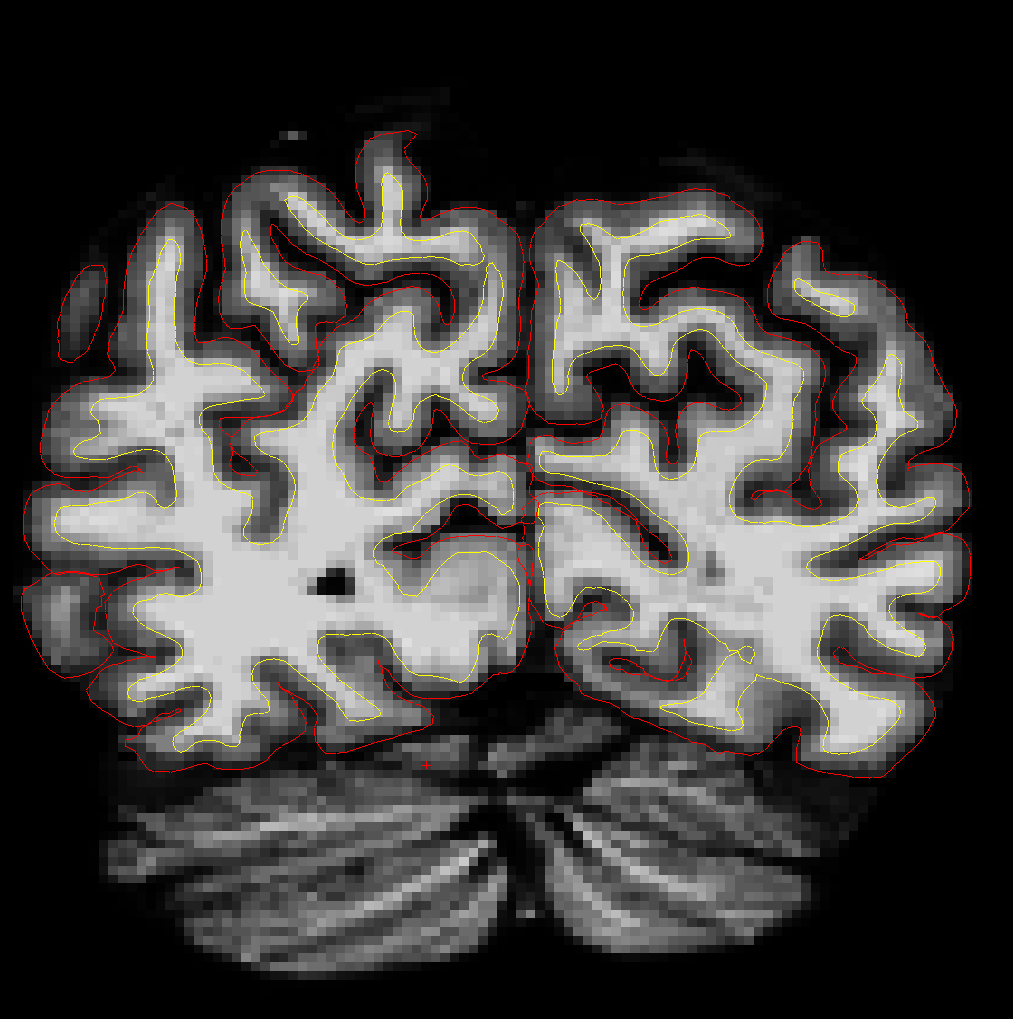
Sorry here is a snapshot for an example.
Hi Freesurfer team I am wondering if there would be a way to improve surfaces on the medial wall, not for the measurement of cortical thickness, but also for laminar analysis in the cortex. We would need them to be accurate even for the medial wall. What would you suggest? Thanks Celine
-- Celine Louapre, MD Research Fellow at Massachusetts General Hospital Department of Radiology, MGH
Building 149, Room 2301 13th Street Charlestown, MA 02129 _______________________________________________ Freesurfer mailing list Freesurfer@nmr.mgh.harvard.edu https://mail.nmr.mgh.harvard.edu/mailman/listinfo/freesurfer

Hi Celine
is the aseg correctly labeled in these regions? Is the filled.mgz? That's a tough case as it's not clear from looking at it which hemi those voxels belong to (probably some of each)
Bruce
On Tue, 18 Jun 2013, celine@nmr.mgh.harvard.edu wrote:
Sorry here is a snapshot for an example.
Hi Freesurfer team I am wondering if there would be a way to improve surfaces on the medial wall, not for the measurement of cortical thickness, but also for laminar analysis in the cortex. We would need them to be accurate even for the medial wall. What would you suggest? Thanks Celine
-- Celine Louapre, MD Research Fellow at Massachusetts General Hospital Department of Radiology, MGH
Building 149, Room 2301 13th Street Charlestown, MA 02129 _______________________________________________ Freesurfer mailing list Freesurfer@nmr.mgh.harvard.edu https://mail.nmr.mgh.harvard.edu/mailman/listinfo/freesurfer
Hi Bruce The aseg is correctly labeled as regards right and left GM cortex, but definitely some voxels are belonging to both hemisphere. The filled.mgz is fine too. Should we delete from aseg the voxels that are at the border between the lh and rh? Thanks Celine
Hi Celine
is the aseg correctly labeled in these regions? Is the filled.mgz? That's a tough case as it's not clear from looking at it which hemi those voxels belong to (probably some of each)
Bruce
On Tue, 18 Jun 2013, celine@nmr.mgh.harvard.edu wrote:
Sorry here is a snapshot for an example.
Hi Freesurfer team I am wondering if there would be a way to improve surfaces on the medial wall, not for the measurement of cortical thickness, but also for laminar analysis in the cortex. We would need them to be accurate even for the medial wall. What would you suggest? Thanks Celine
-- Celine Louapre, MD Research Fellow at Massachusetts General Hospital Department of Radiology, MGH
Building 149, Room 2301 13th Street Charlestown, MA 02129 _______________________________________________ Freesurfer mailing list Freesurfer@nmr.mgh.harvard.edu https://mail.nmr.mgh.harvard.edu/mailman/listinfo/freesurfer
Hi Celine the only existing way to correct this would be to create two volumes in addition to brain.finalsurfs.mgz (say, lh.brain.finalsurfs.mgz and rh.brain.finalsurfs.mgz) and edit each of them to remove voxels in the contralateral hemisphere that are causing problems, then rerun mris_make_surfaces for each hemi, using the appropriate volume. At some point I guess we should put an intersection check into it for the contra hemi
cheers Bruce
On Tue, 18 Jun 2013, celine@nmr.mgh.harvard.edu wrote:
Hi Bruce The aseg is correctly labeled as regards right and left GM cortex, but definitely some voxels are belonging to both hemisphere. The filled.mgz is fine too. Should we delete from aseg the voxels that are at the border between the lh and rh? Thanks Celine
Hi Celine
is the aseg correctly labeled in these regions? Is the filled.mgz? That's a tough case as it's not clear from looking at it which hemi those voxels belong to (probably some of each)
Bruce
On Tue, 18 Jun 2013, celine@nmr.mgh.harvard.edu wrote:
Sorry here is a snapshot for an example.
Hi Freesurfer team I am wondering if there would be a way to improve surfaces on the medial wall, not for the measurement of cortical thickness, but also for laminar analysis in the cortex. We would need them to be accurate even for the medial wall. What would you suggest? Thanks Celine
-- Celine Louapre, MD Research Fellow at Massachusetts General Hospital Department of Radiology, MGH
Building 149, Room 2301 13th Street Charlestown, MA 02129 _______________________________________________ Freesurfer mailing list Freesurfer@nmr.mgh.harvard.edu https://mail.nmr.mgh.harvard.edu/mailman/listinfo/freesurfer
freesurfer@nmr.mgh.harvard.edu
-
 Bruce Fischl
 -
 celine@nmr.mgh.harvard.edu