
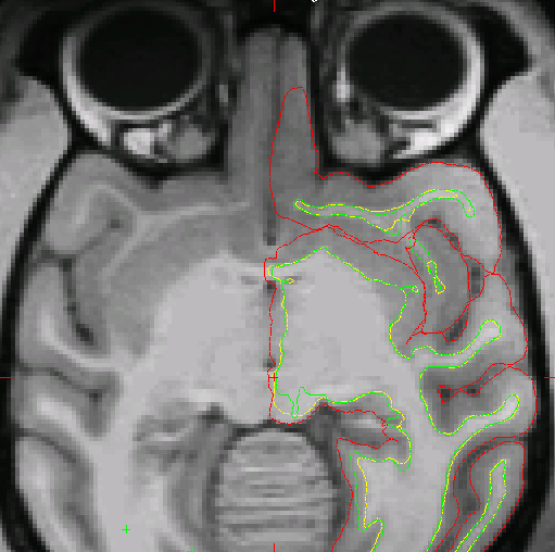
Hi! I am failing to get a proper pial surface in orbitofrontal cortex. See attached screenshot. This is NHP data, processed with version 4.5. I am not sure how to proceed here, as there is no clear with matter. Any advice would be appreciated. Thanks! Caspar

Hi Caspar
it's hard to tell from a single slice. There does seem to be some darkening there, probably due to B0. Is there wm on the next slice inferior/superior? If you upload the whole subject dir I'll take a look.
cheers Bruce
p.s. NHP data?
On Thu, 16 May 2013, Caspar M. Schwiedrzik wrote:
Hi! I am failing to get a proper pial surface in orbitofrontal cortex. See attached screenshot. This is NHP data, processed with version 4.5. I am not sure how to proceed here, as there is no clear with matter. Any advice would be appreciated. Thanks! Caspar

Is the brain extraction removing that part of the brain? Do you know where the OFC ends and olfactory bulb begins?
Peace,
Matt.
On 5/16/13 11:43 AM, "Caspar M. Schwiedrzik" cschwiedrz@mail.rockefeller.edu wrote:
Hi! I am failing to get a proper pial surface in orbitofrontal cortex. See attached screenshot. This is NHP data, processed with version 4.5. I am not sure how to proceed here, as there is no clear with matter. Any advice would be appreciated. Thanks! Caspar _______________________________________________ Freesurfer mailing list Freesurfer@nmr.mgh.harvard.edu https://mail.nmr.mgh.harvard.edu/mailman/listinfo/freesurfer
The information in this e-mail is intended only for the person to whom it is addressed. If you believe this e-mail was sent to you in error and the e-mail contains patient information, please contact the Partners Compliance HelpLine at http://www.partners.org/complianceline . If the e-mail was sent to you in error but does not contain patient information, please contact the sender and properly dispose of the e-mail.
Hi Bruce and Matt, yes, it is primate data. I just dropped an archive on your FTP server in transfer/incoming. Thanks! Caspar
2013/5/16 Matt Glasser matt@ma-tea.com:
Is the brain extraction removing that part of the brain? Do you know where the OFC ends and olfactory bulb begins?
Peace,
Matt.
On 5/16/13 11:43 AM, "Caspar M. Schwiedrzik" cschwiedrz@mail.rockefeller.edu wrote:
Hi! I am failing to get a proper pial surface in orbitofrontal cortex. See attached screenshot. This is NHP data, processed with version 4.5. I am not sure how to proceed here, as there is no clear with matter. Any advice would be appreciated. Thanks! Caspar _______________________________________________ Freesurfer mailing list Freesurfer@nmr.mgh.harvard.edu https://mail.nmr.mgh.harvard.edu/mailman/listinfo/freesurfer
The information in this e-mail is intended only for the person to whom it is addressed. If you believe this e-mail was sent to you in error and the e-mail contains patient information, please contact the Partners Compliance HelpLine at http://www.partners.org/complianceline . If the e-mail was sent to you in error but does not contain patient information, please contact the sender and properly dispose of the e-mail.
Hi Caspar
if it's primate I don't think I'm going to be able to help - you'll need someone more familiar with primate anatomy
sorry Bruce On Thu, 16 May 2013, Caspar M. Schwiedrzik wrote:
Hi Bruce and Matt, yes, it is primate data. I just dropped an archive on your FTP server in transfer/incoming. Thanks! Caspar
2013/5/16 Matt Glasser matt@ma-tea.com:
Is the brain extraction removing that part of the brain? Do you know where the OFC ends and olfactory bulb begins?
Peace,
Matt.
On 5/16/13 11:43 AM, "Caspar M. Schwiedrzik" cschwiedrz@mail.rockefeller.edu wrote:
Hi! I am failing to get a proper pial surface in orbitofrontal cortex. See attached screenshot. This is NHP data, processed with version 4.5. I am not sure how to proceed here, as there is no clear with matter. Any advice would be appreciated. Thanks! Caspar _______________________________________________ Freesurfer mailing list Freesurfer@nmr.mgh.harvard.edu https://mail.nmr.mgh.harvard.edu/mailman/listinfo/freesurfer
The information in this e-mail is intended only for the person to whom it is addressed. If you believe this e-mail was sent to you in error and the e-mail contains patient information, please contact the Partners Compliance HelpLine at http://www.partners.org/complianceline . If the e-mail was sent to you in error but does not contain patient information, please contact the sender and properly dispose of the e-mail.
Hi Bruce, ok. But in theory, what would you recommend to get around the darkening issue? Unfortunately, I do not have a field map available for this data set. Or should I not expect to get a pial surface in this area since the white matter is not discernable? There are five slices without clear white matter (original voxel size 0.5x0.5x0.5 mm). Thanks, Caspar
2013/5/16 Bruce Fischl fischl@nmr.mgh.harvard.edu:
Hi Caspar
if it's primate I don't think I'm going to be able to help - you'll need someone more familiar with primate anatomy
sorry Bruce On Thu, 16 May 2013, Caspar M. Schwiedrzik wrote:
Hi Bruce and Matt, yes, it is primate data. I just dropped an archive on your FTP server in transfer/incoming. Thanks! Caspar
2013/5/16 Matt Glasser matt@ma-tea.com:
Is the brain extraction removing that part of the brain? Do you know where the OFC ends and olfactory bulb begins?
Peace,
Matt.
On 5/16/13 11:43 AM, "Caspar M. Schwiedrzik" cschwiedrz@mail.rockefeller.edu wrote:
Hi! I am failing to get a proper pial surface in orbitofrontal cortex. See attached screenshot. This is NHP data, processed with version 4.5. I am not sure how to proceed here, as there is no clear with matter. Any advice would be appreciated. Thanks! Caspar _______________________________________________ Freesurfer mailing list Freesurfer@nmr.mgh.harvard.edu https://mail.nmr.mgh.harvard.edu/mailman/listinfo/freesurfer
The information in this e-mail is intended only for the person to whom it is addressed. If you believe this e-mail was sent to you in error and the e-mail contains patient information, please contact the Partners Compliance HelpLine at http://www.partners.org/complianceline . If the e-mail was sent to you in error but does not contain patient information, please contact the sender and properly dispose of the e-mail.
Freesurfer mailing list Freesurfer@nmr.mgh.harvard.edu https://mail.nmr.mgh.harvard.edu/mailman/listinfo/freesurfer
Hi Caspar
is the closest white matter captured by the white surface? What is the intensity of voxels there? If < 110 you could try putting control points in them and seeing if that helps (that is, in the closest voxels that are entirely white matter).
Bruce
On Thu, 16 May 2013, Caspar M. Schwiedrzik wrote:
Hi Bruce, ok. But in theory, what would you recommend to get around the darkening issue? Unfortunately, I do not have a field map available for this data set. Or should I not expect to get a pial surface in this area since the white matter is not discernable? There are five slices without clear white matter (original voxel size 0.5x0.5x0.5 mm). Thanks, Caspar
2013/5/16 Bruce Fischl fischl@nmr.mgh.harvard.edu:
Hi Caspar
if it's primate I don't think I'm going to be able to help - you'll need someone more familiar with primate anatomy
sorry Bruce On Thu, 16 May 2013, Caspar M. Schwiedrzik wrote:
Hi Bruce and Matt, yes, it is primate data. I just dropped an archive on your FTP server in transfer/incoming. Thanks! Caspar
2013/5/16 Matt Glasser matt@ma-tea.com:
Is the brain extraction removing that part of the brain? Do you know where the OFC ends and olfactory bulb begins?
Peace,
Matt.
On 5/16/13 11:43 AM, "Caspar M. Schwiedrzik" cschwiedrz@mail.rockefeller.edu wrote:
Hi! I am failing to get a proper pial surface in orbitofrontal cortex. See attached screenshot. This is NHP data, processed with version 4.5. I am not sure how to proceed here, as there is no clear with matter. Any advice would be appreciated. Thanks! Caspar _______________________________________________ Freesurfer mailing list Freesurfer@nmr.mgh.harvard.edu https://mail.nmr.mgh.harvard.edu/mailman/listinfo/freesurfer
The information in this e-mail is intended only for the person to whom it is addressed. If you believe this e-mail was sent to you in error and the e-mail contains patient information, please contact the Partners Compliance HelpLine at http://www.partners.org/complianceline . If the e-mail was sent to you in error but does not contain patient information, please contact the sender and properly dispose of the e-mail.
Freesurfer mailing list Freesurfer@nmr.mgh.harvard.edu https://mail.nmr.mgh.harvard.edu/mailman/listinfo/freesurfer
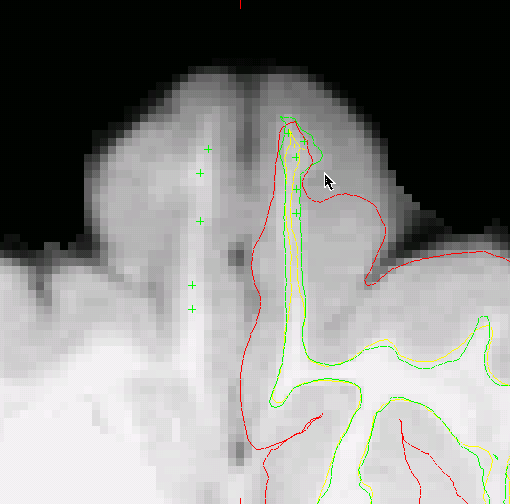
Hi Bruce, I tried adding control points in the white matter in that region, however, it does not seem to fix the issue. Also, the brainmask.mgz does not seem to exclude this part of the brain, so that is not the problem either. I am attaching another screenshot (horizontal 119) to illustrate the problem. White matter pixel values range from the high 80ies to >100 in this area. Included grey matter pixel values are in the low 80ies. Excluded grey matter pixel values range from the low 60ies to low 70ies.
Would it make sense to re-run mri_segment with a different threshold? Thanks, Caspar
2013/5/16 Bruce Fischl fischl@nmr.mgh.harvard.edu:
Hi Caspar
is the closest white matter captured by the white surface? What is the intensity of voxels there? If < 110 you could try putting control points in them and seeing if that helps (that is, in the closest voxels that are entirely white matter).
Bruce
On Thu, 16 May 2013, Caspar M. Schwiedrzik wrote:
Hi Bruce, ok. But in theory, what would you recommend to get around the darkening issue? Unfortunately, I do not have a field map available for this data set. Or should I not expect to get a pial surface in this area since the white matter is not discernable? There are five slices without clear white matter (original voxel size 0.5x0.5x0.5 mm). Thanks, Caspar
2013/5/16 Bruce Fischl fischl@nmr.mgh.harvard.edu:
Hi Caspar
if it's primate I don't think I'm going to be able to help - you'll need someone more familiar with primate anatomy
sorry Bruce On Thu, 16 May 2013, Caspar M. Schwiedrzik wrote:
Hi Bruce and Matt, yes, it is primate data. I just dropped an archive on your FTP server in transfer/incoming. Thanks! Caspar
2013/5/16 Matt Glasser matt@ma-tea.com:
Is the brain extraction removing that part of the brain? Do you know where the OFC ends and olfactory bulb begins?
Peace,
Matt.
On 5/16/13 11:43 AM, "Caspar M. Schwiedrzik" cschwiedrz@mail.rockefeller.edu wrote:
Hi! I am failing to get a proper pial surface in orbitofrontal cortex. See attached screenshot. This is NHP data, processed with version 4.5. I am not sure how to proceed here, as there is no clear with matter. Any advice would be appreciated. Thanks! Caspar _______________________________________________ Freesurfer mailing list Freesurfer@nmr.mgh.harvard.edu https://mail.nmr.mgh.harvard.edu/mailman/listinfo/freesurfer
The information in this e-mail is intended only for the person to whom it is addressed. If you believe this e-mail was sent to you in error and the e-mail contains patient information, please contact the Partners Compliance HelpLine at http://www.partners.org/complianceline . If the e-mail was sent to you in error but does not contain patient information, please contact the sender and properly dispose of the e-mail.
Freesurfer mailing list Freesurfer@nmr.mgh.harvard.edu https://mail.nmr.mgh.harvard.edu/mailman/listinfo/freesurfer
Hi Caspar
yes, that might help. There are expert opts for this. Sorry, I have almost no experience analyzing monkey brains at .5mm, so I'm really not sure what to advise you. Perhaps one of the other people on list who have done a bunch can comment? Bruce On Fri, 17 May 2013, Caspar M. Schwiedrzik wrote:
Hi Bruce, I tried adding control points in the white matter in that region, however, it does not seem to fix the issue. Also, the brainmask.mgz does not seem to exclude this part of the brain, so that is not the problem either. I am attaching another screenshot (horizontal 119) to illustrate the problem. White matter pixel values range from the high 80ies to >100 in this area. Included grey matter pixel values are in the low 80ies. Excluded grey matter pixel values range from the low 60ies to low 70ies.
Would it make sense to re-run mri_segment with a different threshold? Thanks, Caspar
2013/5/16 Bruce Fischl fischl@nmr.mgh.harvard.edu:
Hi Caspar
is the closest white matter captured by the white surface? What is the intensity of voxels there? If < 110 you could try putting control points in them and seeing if that helps (that is, in the closest voxels that are entirely white matter).
Bruce
On Thu, 16 May 2013, Caspar M. Schwiedrzik wrote:
Hi Bruce, ok. But in theory, what would you recommend to get around the darkening issue? Unfortunately, I do not have a field map available for this data set. Or should I not expect to get a pial surface in this area since the white matter is not discernable? There are five slices without clear white matter (original voxel size 0.5x0.5x0.5 mm). Thanks, Caspar
2013/5/16 Bruce Fischl fischl@nmr.mgh.harvard.edu:
Hi Caspar
if it's primate I don't think I'm going to be able to help - you'll need someone more familiar with primate anatomy
sorry Bruce On Thu, 16 May 2013, Caspar M. Schwiedrzik wrote:
Hi Bruce and Matt, yes, it is primate data. I just dropped an archive on your FTP server in transfer/incoming. Thanks! Caspar
2013/5/16 Matt Glasser matt@ma-tea.com:
Is the brain extraction removing that part of the brain? Do you know where the OFC ends and olfactory bulb begins?
Peace,
Matt.
On 5/16/13 11:43 AM, "Caspar M. Schwiedrzik" cschwiedrz@mail.rockefeller.edu wrote:
> Hi! > I am failing to get a proper pial surface in orbitofrontal cortex. > See attached screenshot. > This is NHP data, processed with version 4.5. > I am not sure how to proceed here, as there is no clear with matter. > Any advice would be appreciated. > Thanks! > Caspar > _______________________________________________ > Freesurfer mailing list > Freesurfer@nmr.mgh.harvard.edu > https://mail.nmr.mgh.harvard.edu/mailman/listinfo/freesurfer > > > The information in this e-mail is intended only for the person to whom > it > is > addressed. If you believe this e-mail was sent to you in error and the > e-mail > contains patient information, please contact the Partners Compliance > HelpLine at > http://www.partners.org/complianceline . If the e-mail was sent to you > in > error > but does not contain patient information, please contact the sender > and > properly > dispose of the e-mail.
Freesurfer mailing list Freesurfer@nmr.mgh.harvard.edu https://mail.nmr.mgh.harvard.edu/mailman/listinfo/freesurfer
Hard to fix if that is the receive field and you don't have another image like a T2w to remove it with. Perhaps mri_normalize can be tuned to improve this with expert options as the white matter that is being correctly segmented appears to be darker as well.
Peace,
Matt.
On 5/17/13 9:30 AM, "Bruce Fischl" fischl@nmr.mgh.harvard.edu wrote:
Hi Caspar
yes, that might help. There are expert opts for this. Sorry, I have almost no experience analyzing monkey brains at .5mm, so I'm really not sure what to advise you. Perhaps one of the other people on list who have done a bunch can comment? Bruce On Fri, 17 May 2013, Caspar M. Schwiedrzik wrote:
Hi Bruce, I tried adding control points in the white matter in that region, however, it does not seem to fix the issue. Also, the brainmask.mgz does not seem to exclude this part of the brain, so that is not the problem either. I am attaching another screenshot (horizontal 119) to illustrate the problem. White matter pixel values range from the high 80ies to >100 in this area. Included grey matter pixel values are in the low 80ies. Excluded grey matter pixel values range from the low 60ies to low 70ies.
Would it make sense to re-run mri_segment with a different threshold? Thanks, Caspar
2013/5/16 Bruce Fischl fischl@nmr.mgh.harvard.edu:
Hi Caspar
is the closest white matter captured by the white surface? What is the intensity of voxels there? If < 110 you could try putting control points in them and seeing if that helps (that is, in the closest voxels that are entirely white matter).
Bruce
On Thu, 16 May 2013, Caspar M. Schwiedrzik wrote:
Hi Bruce, ok. But in theory, what would you recommend to get around the darkening issue? Unfortunately, I do not have a field map available for this data set. Or should I not expect to get a pial surface in this area since the white matter is not discernable? There are five slices without clear white matter (original voxel size 0.5x0.5x0.5 mm). Thanks, Caspar
2013/5/16 Bruce Fischl fischl@nmr.mgh.harvard.edu:
Hi Caspar
if it's primate I don't think I'm going to be able to help - you'll need someone more familiar with primate anatomy
sorry Bruce On Thu, 16 May 2013, Caspar M. Schwiedrzik wrote:
Hi Bruce and Matt, yes, it is primate data. I just dropped an archive on your FTP server in transfer/incoming. Thanks! Caspar
2013/5/16 Matt Glasser matt@ma-tea.com: > > Is the brain extraction removing that part of the brain? Do you >know > where the OFC ends and olfactory bulb begins? > > Peace, > > Matt. > > On 5/16/13 11:43 AM, "Caspar M. Schwiedrzik" > cschwiedrz@mail.rockefeller.edu wrote: > >> Hi! >> I am failing to get a proper pial surface in orbitofrontal cortex. >> See attached screenshot. >> This is NHP data, processed with version 4.5. >> I am not sure how to proceed here, as there is no clear with >>matter. >> Any advice would be appreciated. >> Thanks! >> Caspar >> _______________________________________________ >> Freesurfer mailing list >> Freesurfer@nmr.mgh.harvard.edu >> https://mail.nmr.mgh.harvard.edu/mailman/listinfo/freesurfer >> >> >> The information in this e-mail is intended only for the person to >>whom >> it >> is >> addressed. If you believe this e-mail was sent to you in error >>and the >> e-mail >> contains patient information, please contact the Partners >>Compliance >> HelpLine at >> http://www.partners.org/complianceline . If the e-mail was sent >>to you >> in >> error >> but does not contain patient information, please contact the >>sender >> and >> properly >> dispose of the e-mail. > > >
Freesurfer mailing list Freesurfer@nmr.mgh.harvard.edu https://mail.nmr.mgh.harvard.edu/mailman/listinfo/freesurfer
When I try to specify a different threshold using -p, mri_segment reads in the threshold as the input volume. I tried specifying the input with -i (as explained here: http://www.mail-archive.com/freesurfer@nmr.mgh.harvard.edu/msg18826.html), but now it does not recognize the -i flag. Caspar
2013/5/17 Matt Glasser matt@ma-tea.com:
Hard to fix if that is the receive field and you don't have another image like a T2w to remove it with. Perhaps mri_normalize can be tuned to improve this with expert options as the white matter that is being correctly segmented appears to be darker as well.
Peace,
Matt.
On 5/17/13 9:30 AM, "Bruce Fischl" fischl@nmr.mgh.harvard.edu wrote:
Hi Caspar
yes, that might help. There are expert opts for this. Sorry, I have almost no experience analyzing monkey brains at .5mm, so I'm really not sure what to advise you. Perhaps one of the other people on list who have done a bunch can comment? Bruce On Fri, 17 May 2013, Caspar M. Schwiedrzik wrote:
Hi Bruce, I tried adding control points in the white matter in that region, however, it does not seem to fix the issue. Also, the brainmask.mgz does not seem to exclude this part of the brain, so that is not the problem either. I am attaching another screenshot (horizontal 119) to illustrate the problem. White matter pixel values range from the high 80ies to >100 in this area. Included grey matter pixel values are in the low 80ies. Excluded grey matter pixel values range from the low 60ies to low 70ies.
Would it make sense to re-run mri_segment with a different threshold? Thanks, Caspar
2013/5/16 Bruce Fischl fischl@nmr.mgh.harvard.edu:
Hi Caspar
is the closest white matter captured by the white surface? What is the intensity of voxels there? If < 110 you could try putting control points in them and seeing if that helps (that is, in the closest voxels that are entirely white matter).
Bruce
On Thu, 16 May 2013, Caspar M. Schwiedrzik wrote:
Hi Bruce, ok. But in theory, what would you recommend to get around the darkening issue? Unfortunately, I do not have a field map available for this data set. Or should I not expect to get a pial surface in this area since the white matter is not discernable? There are five slices without clear white matter (original voxel size 0.5x0.5x0.5 mm). Thanks, Caspar
2013/5/16 Bruce Fischl fischl@nmr.mgh.harvard.edu:
Hi Caspar
if it's primate I don't think I'm going to be able to help - you'll need someone more familiar with primate anatomy
sorry Bruce On Thu, 16 May 2013, Caspar M. Schwiedrzik wrote:
> Hi Bruce and Matt, > yes, it is primate data. I just dropped an archive on your FTP >server > in transfer/incoming. > Thanks! > Caspar > > 2013/5/16 Matt Glasser matt@ma-tea.com: >> >> Is the brain extraction removing that part of the brain? Do you >>know >> where the OFC ends and olfactory bulb begins? >> >> Peace, >> >> Matt. >> >> On 5/16/13 11:43 AM, "Caspar M. Schwiedrzik" >> cschwiedrz@mail.rockefeller.edu wrote: >> >>> Hi! >>> I am failing to get a proper pial surface in orbitofrontal cortex. >>> See attached screenshot. >>> This is NHP data, processed with version 4.5. >>> I am not sure how to proceed here, as there is no clear with >>>matter. >>> Any advice would be appreciated. >>> Thanks! >>> Caspar >>> _______________________________________________ >>> Freesurfer mailing list >>> Freesurfer@nmr.mgh.harvard.edu >>> https://mail.nmr.mgh.harvard.edu/mailman/listinfo/freesurfer >>> >>> >>> The information in this e-mail is intended only for the person to >>>whom >>> it >>> is >>> addressed. If you believe this e-mail was sent to you in error >>>and the >>> e-mail >>> contains patient information, please contact the Partners >>>Compliance >>> HelpLine at >>> http://www.partners.org/complianceline . If the e-mail was sent >>>to you >>> in >>> error >>> but does not contain patient information, please contact the >>>sender >>> and >>> properly >>> dispose of the e-mail. >> >> >> > > > _______________________________________________ Freesurfer mailing list Freesurfer@nmr.mgh.harvard.edu https://mail.nmr.mgh.harvard.edu/mailman/listinfo/freesurfer
what is your command line? You are probably better off setting gray_hi, gray_low, wm_hi, wm_low, etc... Bruce On Fri, 17 May 2013, Caspar M. Schwiedrzik wrote:
When I try to specify a different threshold using -p, mri_segment reads in the threshold as the input volume. I tried specifying the input with -i (as explained here: http://www.mail-archive.com/freesurfer@nmr.mgh.harvard.edu/msg18826.html), but now it does not recognize the -i flag. Caspar
2013/5/17 Matt Glasser matt@ma-tea.com:
Hard to fix if that is the receive field and you don't have another image like a T2w to remove it with. Perhaps mri_normalize can be tuned to improve this with expert options as the white matter that is being correctly segmented appears to be darker as well.
Peace,
Matt.
On 5/17/13 9:30 AM, "Bruce Fischl" fischl@nmr.mgh.harvard.edu wrote:
Hi Caspar
yes, that might help. There are expert opts for this. Sorry, I have almost no experience analyzing monkey brains at .5mm, so I'm really not sure what to advise you. Perhaps one of the other people on list who have done a bunch can comment? Bruce On Fri, 17 May 2013, Caspar M. Schwiedrzik wrote:
Hi Bruce, I tried adding control points in the white matter in that region, however, it does not seem to fix the issue. Also, the brainmask.mgz does not seem to exclude this part of the brain, so that is not the problem either. I am attaching another screenshot (horizontal 119) to illustrate the problem. White matter pixel values range from the high 80ies to >100 in this area. Included grey matter pixel values are in the low 80ies. Excluded grey matter pixel values range from the low 60ies to low 70ies.
Would it make sense to re-run mri_segment with a different threshold? Thanks, Caspar
2013/5/16 Bruce Fischl fischl@nmr.mgh.harvard.edu:
Hi Caspar
is the closest white matter captured by the white surface? What is the intensity of voxels there? If < 110 you could try putting control points in them and seeing if that helps (that is, in the closest voxels that are entirely white matter).
Bruce
On Thu, 16 May 2013, Caspar M. Schwiedrzik wrote:
Hi Bruce, ok. But in theory, what would you recommend to get around the darkening issue? Unfortunately, I do not have a field map available for this data set. Or should I not expect to get a pial surface in this area since the white matter is not discernable? There are five slices without clear white matter (original voxel size 0.5x0.5x0.5 mm). Thanks, Caspar
2013/5/16 Bruce Fischl fischl@nmr.mgh.harvard.edu: > > Hi Caspar > > if it's primate I don't think I'm going to be able to help - you'll > need > someone more familiar with primate anatomy > > sorry > Bruce > On Thu, 16 May 2013, Caspar M. > Schwiedrzik wrote: > >> Hi Bruce and Matt, >> yes, it is primate data. I just dropped an archive on your FTP >> server >> in transfer/incoming. >> Thanks! >> Caspar >> >> 2013/5/16 Matt Glasser matt@ma-tea.com: >>> >>> Is the brain extraction removing that part of the brain? Do you >>> know >>> where the OFC ends and olfactory bulb begins? >>> >>> Peace, >>> >>> Matt. >>> >>> On 5/16/13 11:43 AM, "Caspar M. Schwiedrzik" >>> cschwiedrz@mail.rockefeller.edu wrote: >>> >>>> Hi! >>>> I am failing to get a proper pial surface in orbitofrontal cortex. >>>> See attached screenshot. >>>> This is NHP data, processed with version 4.5. >>>> I am not sure how to proceed here, as there is no clear with >>>> matter. >>>> Any advice would be appreciated. >>>> Thanks! >>>> Caspar >>>> _______________________________________________ >>>> Freesurfer mailing list >>>> Freesurfer@nmr.mgh.harvard.edu >>>> https://mail.nmr.mgh.harvard.edu/mailman/listinfo/freesurfer >>>> >>>> >>>> The information in this e-mail is intended only for the person to >>>> whom >>>> it >>>> is >>>> addressed. If you believe this e-mail was sent to you in error >>>> and the >>>> e-mail >>>> contains patient information, please contact the Partners >>>> Compliance >>>> HelpLine at >>>> http://www.partners.org/complianceline . If the e-mail was sent >>>> to you >>>> in >>>> error >>>> but does not contain patient information, please contact the >>>> sender >>>> and >>>> properly >>>> dispose of the e-mail. >>> >>> >>> >> >> >> > _______________________________________________ > Freesurfer mailing list > Freesurfer@nmr.mgh.harvard.edu > https://mail.nmr.mgh.harvard.edu/mailman/listinfo/freesurfer
mri_segment \ -v \ -fillv \ -fillbg \ -wlo 104 \ -ghi 118 \ -whi 140 \ -n 4 \ -keep \ brain.mgz wm.mgz
The pial surface in the rest of the brain is ok, it is only the orbitofrontal/piriform cortex that is problematic. I now wanted to add -p 0.7.
Caspar
2013/5/17 Bruce Fischl fischl@nmr.mgh.harvard.edu:
what is your command line? You are probably better off setting gray_hi, gray_low, wm_hi, wm_low, etc...
Bruce On Fri, 17 May 2013, Caspar M. Schwiedrzik wrote:
When I try to specify a different threshold using -p, mri_segment reads in the threshold as the input volume. I tried specifying the input with -i (as explained here: http://www.mail-archive.com/freesurfer@nmr.mgh.harvard.edu/msg18826.html), but now it does not recognize the -i flag. Caspar
2013/5/17 Matt Glasser matt@ma-tea.com:
Hard to fix if that is the receive field and you don't have another image like a T2w to remove it with. Perhaps mri_normalize can be tuned to improve this with expert options as the white matter that is being correctly segmented appears to be darker as well.
Peace,
Matt.
On 5/17/13 9:30 AM, "Bruce Fischl" fischl@nmr.mgh.harvard.edu wrote:
Hi Caspar
yes, that might help. There are expert opts for this. Sorry, I have almost no experience analyzing monkey brains at .5mm, so I'm really not sure what to advise you. Perhaps one of the other people on list who have done a bunch can comment? Bruce On Fri, 17 May 2013, Caspar M. Schwiedrzik wrote:
Hi Bruce, I tried adding control points in the white matter in that region, however, it does not seem to fix the issue. Also, the brainmask.mgz does not seem to exclude this part of the brain, so that is not the problem either. I am attaching another screenshot (horizontal 119) to illustrate the problem. White matter pixel values range from the high 80ies to >100 in this area. Included grey matter pixel values are in the low 80ies. Excluded grey matter pixel values range from the low 60ies to low 70ies.
Would it make sense to re-run mri_segment with a different threshold? Thanks, Caspar
2013/5/16 Bruce Fischl fischl@nmr.mgh.harvard.edu:
Hi Caspar
is the closest white matter captured by the white surface? What is the intensity of voxels there? If < 110 you could try putting control points in them and seeing if that helps (that is, in the closest voxels that are entirely white matter).
Bruce
On Thu, 16 May 2013, Caspar M. Schwiedrzik wrote:
> Hi Bruce, > ok. > But in theory, what would you recommend to get around the darkening > issue? Unfortunately, I do not have a field map available for this > data set. > Or should I not expect to get a pial surface in this area since the > white matter is not discernable? > There are five slices without clear white matter (original voxel size > 0.5x0.5x0.5 mm). > Thanks, Caspar > > 2013/5/16 Bruce Fischl fischl@nmr.mgh.harvard.edu: >> >> >> Hi Caspar >> >> if it's primate I don't think I'm going to be able to help - you'll >> need >> someone more familiar with primate anatomy >> >> sorry >> Bruce >> On Thu, 16 May 2013, Caspar M. >> Schwiedrzik wrote: >> >>> Hi Bruce and Matt, >>> yes, it is primate data. I just dropped an archive on your FTP >>> server >>> in transfer/incoming. >>> Thanks! >>> Caspar >>> >>> 2013/5/16 Matt Glasser matt@ma-tea.com: >>>> >>>> >>>> Is the brain extraction removing that part of the brain? Do you >>>> know >>>> where the OFC ends and olfactory bulb begins? >>>> >>>> Peace, >>>> >>>> Matt. >>>> >>>> On 5/16/13 11:43 AM, "Caspar M. Schwiedrzik" >>>> cschwiedrz@mail.rockefeller.edu wrote: >>>> >>>>> Hi! >>>>> I am failing to get a proper pial surface in orbitofrontal >>>>> cortex. >>>>> See attached screenshot. >>>>> This is NHP data, processed with version 4.5. >>>>> I am not sure how to proceed here, as there is no clear with >>>>> matter. >>>>> Any advice would be appreciated. >>>>> Thanks! >>>>> Caspar >>>>> _______________________________________________ >>>>> Freesurfer mailing list >>>>> Freesurfer@nmr.mgh.harvard.edu >>>>> https://mail.nmr.mgh.harvard.edu/mailman/listinfo/freesurfer >>>>> >>>>> >>>>> The information in this e-mail is intended only for the person to >>>>> whom >>>>> it >>>>> is >>>>> addressed. If you believe this e-mail was sent to you in error >>>>> and the >>>>> e-mail >>>>> contains patient information, please contact the Partners >>>>> Compliance >>>>> HelpLine at >>>>> http://www.partners.org/complianceline . If the e-mail was sent >>>>> to you >>>>> in >>>>> error >>>>> but does not contain patient information, please contact the >>>>> sender >>>>> and >>>>> properly >>>>> dispose of the e-mail. >>>> >>>> >>>> >>>> >>> >>> >>> >> _______________________________________________ >> Freesurfer mailing list >> Freesurfer@nmr.mgh.harvard.edu >> https://mail.nmr.mgh.harvard.edu/mailman/listinfo/freesurfer > > > > >
and what happens? Can you send the full screen output? On Fri, 17 May 2013, Caspar M. Schwiedrzik wrote:
mri_segment \ -v \ -fillv \ -fillbg \ -wlo 104 \ -ghi 118 \ -whi 140 \ -n 4 \ -keep \ brain.mgz wm.mgz
The pial surface in the rest of the brain is ok, it is only the orbitofrontal/piriform cortex that is problematic. I now wanted to add -p 0.7.
Caspar
2013/5/17 Bruce Fischl fischl@nmr.mgh.harvard.edu:
what is your command line? You are probably better off setting gray_hi, gray_low, wm_hi, wm_low, etc...
Bruce On Fri, 17 May 2013, Caspar M. Schwiedrzik wrote:
When I try to specify a different threshold using -p, mri_segment reads in the threshold as the input volume. I tried specifying the input with -i (as explained here: http://www.mail-archive.com/freesurfer@nmr.mgh.harvard.edu/msg18826.html), but now it does not recognize the -i flag. Caspar
2013/5/17 Matt Glasser matt@ma-tea.com:
Hard to fix if that is the receive field and you don't have another image like a T2w to remove it with. Perhaps mri_normalize can be tuned to improve this with expert options as the white matter that is being correctly segmented appears to be darker as well.
Peace,
Matt.
On 5/17/13 9:30 AM, "Bruce Fischl" fischl@nmr.mgh.harvard.edu wrote:
Hi Caspar
yes, that might help. There are expert opts for this. Sorry, I have almost no experience analyzing monkey brains at .5mm, so I'm really not sure what to advise you. Perhaps one of the other people on list who have done a bunch can comment? Bruce On Fri, 17 May 2013, Caspar M. Schwiedrzik wrote:
Hi Bruce, I tried adding control points in the white matter in that region, however, it does not seem to fix the issue. Also, the brainmask.mgz does not seem to exclude this part of the brain, so that is not the problem either. I am attaching another screenshot (horizontal 119) to illustrate the problem. White matter pixel values range from the high 80ies to >100 in this area. Included grey matter pixel values are in the low 80ies. Excluded grey matter pixel values range from the low 60ies to low 70ies.
Would it make sense to re-run mri_segment with a different threshold? Thanks, Caspar
2013/5/16 Bruce Fischl fischl@nmr.mgh.harvard.edu: > > Hi Caspar > > is the closest white matter captured by the white surface? What is the > intensity of voxels there? If < 110 you could try putting control > points in > them and seeing if that helps (that is, in the closest voxels that are > entirely white matter). > > > Bruce > > On Thu, 16 May 2013, Caspar M. Schwiedrzik wrote: > >> Hi Bruce, >> ok. >> But in theory, what would you recommend to get around the darkening >> issue? Unfortunately, I do not have a field map available for this >> data set. >> Or should I not expect to get a pial surface in this area since the >> white matter is not discernable? >> There are five slices without clear white matter (original voxel size >> 0.5x0.5x0.5 mm). >> Thanks, Caspar >> >> 2013/5/16 Bruce Fischl fischl@nmr.mgh.harvard.edu: >>> >>> >>> Hi Caspar >>> >>> if it's primate I don't think I'm going to be able to help - you'll >>> need >>> someone more familiar with primate anatomy >>> >>> sorry >>> Bruce >>> On Thu, 16 May 2013, Caspar M. >>> Schwiedrzik wrote: >>> >>>> Hi Bruce and Matt, >>>> yes, it is primate data. I just dropped an archive on your FTP >>>> server >>>> in transfer/incoming. >>>> Thanks! >>>> Caspar >>>> >>>> 2013/5/16 Matt Glasser matt@ma-tea.com: >>>>> >>>>> >>>>> Is the brain extraction removing that part of the brain? Do you >>>>> know >>>>> where the OFC ends and olfactory bulb begins? >>>>> >>>>> Peace, >>>>> >>>>> Matt. >>>>> >>>>> On 5/16/13 11:43 AM, "Caspar M. Schwiedrzik" >>>>> cschwiedrz@mail.rockefeller.edu wrote: >>>>> >>>>>> Hi! >>>>>> I am failing to get a proper pial surface in orbitofrontal >>>>>> cortex. >>>>>> See attached screenshot. >>>>>> This is NHP data, processed with version 4.5. >>>>>> I am not sure how to proceed here, as there is no clear with >>>>>> matter. >>>>>> Any advice would be appreciated. >>>>>> Thanks! >>>>>> Caspar >>>>>> _______________________________________________ >>>>>> Freesurfer mailing list >>>>>> Freesurfer@nmr.mgh.harvard.edu >>>>>> https://mail.nmr.mgh.harvard.edu/mailman/listinfo/freesurfer >>>>>> >>>>>> >>>>>> The information in this e-mail is intended only for the person to >>>>>> whom >>>>>> it >>>>>> is >>>>>> addressed. If you believe this e-mail was sent to you in error >>>>>> and the >>>>>> e-mail >>>>>> contains patient information, please contact the Partners >>>>>> Compliance >>>>>> HelpLine at >>>>>> http://www.partners.org/complianceline . If the e-mail was sent >>>>>> to you >>>>>> in >>>>>> error >>>>>> but does not contain patient information, please contact the >>>>>> sender >>>>>> and >>>>>> properly >>>>>> dispose of the e-mail. >>>>> >>>>> >>>>> >>>>> >>>> >>>> >>>> >>> _______________________________________________ >>> Freesurfer mailing list >>> Freesurfer@nmr.mgh.harvard.edu >>> https://mail.nmr.mgh.harvard.edu/mailman/listinfo/freesurfer >> >> >> >> >> >
filling ventricles filling basal ganglia using white lolim = 104.0 using gray hilim = 118.0 using white hilim = 140.0 running border classification 4 times preserving editing changes in output volume... using 70% threshold mri_read(): couldn't determine type of file /.../.7000000000000000000 mri_segment: could not read source volume from .7000000000000000000 ../mri/brain.mgz: Permission denied
if I add a -i to the input volume, it says flag not recognized. to get a floating point number into my tcsh script, I am using `echo "7/10" | bc -l`
according to this previous discussion on the mailing list, there should be a fix available somewhere: http://www.mail-archive.com/freesurfer@nmr.mgh.harvard.edu/msg18824.html
Thanks, Caspar
2013/5/17 Bruce Fischl fischl@nmr.mgh.harvard.edu:
and what happens? Can you send the full screen output?
On Fri, 17 May 2013, Caspar M. Schwiedrzik wrote:
mri_segment \ -v \ -fillv \ -fillbg \ -wlo 104 \ -ghi 118 \ -whi 140 \ -n 4 \ -keep \ brain.mgz wm.mgz
The pial surface in the rest of the brain is ok, it is only the orbitofrontal/piriform cortex that is problematic. I now wanted to add -p 0.7.
Caspar
2013/5/17 Bruce Fischl fischl@nmr.mgh.harvard.edu:
what is your command line? You are probably better off setting gray_hi, gray_low, wm_hi, wm_low, etc...
Bruce On Fri, 17 May 2013, Caspar M. Schwiedrzik wrote:
When I try to specify a different threshold using -p, mri_segment reads in the threshold as the input volume. I tried specifying the input with -i (as explained here:
http://www.mail-archive.com/freesurfer@nmr.mgh.harvard.edu/msg18826.html), but now it does not recognize the -i flag. Caspar
2013/5/17 Matt Glasser matt@ma-tea.com:
Hard to fix if that is the receive field and you don't have another image like a T2w to remove it with. Perhaps mri_normalize can be tuned to improve this with expert options as the white matter that is being correctly segmented appears to be darker as well.
Peace,
Matt.
On 5/17/13 9:30 AM, "Bruce Fischl" fischl@nmr.mgh.harvard.edu wrote:
Hi Caspar
yes, that might help. There are expert opts for this. Sorry, I have almost no experience analyzing monkey brains at .5mm, so I'm really not sure what to advise you. Perhaps one of the other people on list who have done a bunch can comment? Bruce On Fri, 17 May 2013, Caspar M. Schwiedrzik wrote:
> Hi Bruce, > I tried adding control points in the white matter in that region, > however, it does not seem to fix the issue. Also, the brainmask.mgz > does not seem to exclude this part of the brain, so that is not the > problem either. > I am attaching another screenshot (horizontal 119) to illustrate the > problem. > White matter pixel values range from the high 80ies to >100 in this > area. > Included grey matter pixel values are in the low 80ies. > Excluded grey matter pixel values range from the low 60ies to low > 70ies. > > Would it make sense to re-run mri_segment with a different threshold? > Thanks, Caspar > > > > 2013/5/16 Bruce Fischl fischl@nmr.mgh.harvard.edu: >> >> >> Hi Caspar >> >> is the closest white matter captured by the white surface? What is >> the >> intensity of voxels there? If < 110 you could try putting control >> points in >> them and seeing if that helps (that is, in the closest voxels that >> are >> entirely white matter). >> >> >> Bruce >> >> On Thu, 16 May 2013, Caspar M. Schwiedrzik wrote: >> >>> Hi Bruce, >>> ok. >>> But in theory, what would you recommend to get around the darkening >>> issue? Unfortunately, I do not have a field map available for this >>> data set. >>> Or should I not expect to get a pial surface in this area since the >>> white matter is not discernable? >>> There are five slices without clear white matter (original voxel >>> size >>> 0.5x0.5x0.5 mm). >>> Thanks, Caspar >>> >>> 2013/5/16 Bruce Fischl fischl@nmr.mgh.harvard.edu: >>>> >>>> >>>> >>>> Hi Caspar >>>> >>>> if it's primate I don't think I'm going to be able to help - >>>> you'll >>>> need >>>> someone more familiar with primate anatomy >>>> >>>> sorry >>>> Bruce >>>> On Thu, 16 May 2013, Caspar M. >>>> Schwiedrzik wrote: >>>> >>>>> Hi Bruce and Matt, >>>>> yes, it is primate data. I just dropped an archive on your FTP >>>>> server >>>>> in transfer/incoming. >>>>> Thanks! >>>>> Caspar >>>>> >>>>> 2013/5/16 Matt Glasser matt@ma-tea.com: >>>>>> >>>>>> >>>>>> >>>>>> Is the brain extraction removing that part of the brain? Do you >>>>>> know >>>>>> where the OFC ends and olfactory bulb begins? >>>>>> >>>>>> Peace, >>>>>> >>>>>> Matt. >>>>>> >>>>>> On 5/16/13 11:43 AM, "Caspar M. Schwiedrzik" >>>>>> cschwiedrz@mail.rockefeller.edu wrote: >>>>>> >>>>>>> Hi! >>>>>>> I am failing to get a proper pial surface in orbitofrontal >>>>>>> cortex. >>>>>>> See attached screenshot. >>>>>>> This is NHP data, processed with version 4.5. >>>>>>> I am not sure how to proceed here, as there is no clear with >>>>>>> matter. >>>>>>> Any advice would be appreciated. >>>>>>> Thanks! >>>>>>> Caspar >>>>>>> _______________________________________________ >>>>>>> Freesurfer mailing list >>>>>>> Freesurfer@nmr.mgh.harvard.edu >>>>>>> https://mail.nmr.mgh.harvard.edu/mailman/listinfo/freesurfer >>>>>>> >>>>>>> >>>>>>> The information in this e-mail is intended only for the person >>>>>>> to >>>>>>> whom >>>>>>> it >>>>>>> is >>>>>>> addressed. If you believe this e-mail was sent to you in error >>>>>>> and the >>>>>>> e-mail >>>>>>> contains patient information, please contact the Partners >>>>>>> Compliance >>>>>>> HelpLine at >>>>>>> http://www.partners.org/complianceline . If the e-mail was sent >>>>>>> to you >>>>>>> in >>>>>>> error >>>>>>> but does not contain patient information, please contact the >>>>>>> sender >>>>>>> and >>>>>>> properly >>>>>>> dispose of the e-mail. >>>>>> >>>>>> >>>>>> >>>>>> >>>>>> >>>>> >>>>> >>>>> >>>> _______________________________________________ >>>> Freesurfer mailing list >>>> Freesurfer@nmr.mgh.harvard.edu >>>> https://mail.nmr.mgh.harvard.edu/mailman/listinfo/freesurfer >>> >>> >>> >>> >>> >>> >> >
Hi Caspar
can you include the command line and all the output?
Bruce On Fri, 17 May 2013, Caspar M. Schwiedrzik wrote:
filling ventricles filling basal ganglia using white lolim = 104.0 using gray hilim = 118.0 using white hilim = 140.0 running border classification 4 times preserving editing changes in output volume... using 70% threshold mri_read(): couldn't determine type of file /.../.7000000000000000000 mri_segment: could not read source volume from .7000000000000000000 ../mri/brain.mgz: Permission denied
if I add a -i to the input volume, it says flag not recognized. to get a floating point number into my tcsh script, I am using `echo "7/10" | bc -l`
according to this previous discussion on the mailing list, there should be a fix available somewhere: http://www.mail-archive.com/freesurfer@nmr.mgh.harvard.edu/msg18824.html
Thanks, Caspar
2013/5/17 Bruce Fischl fischl@nmr.mgh.harvard.edu:
and what happens? Can you send the full screen output?
On Fri, 17 May 2013, Caspar M. Schwiedrzik wrote:
mri_segment \ -v \ -fillv \ -fillbg \ -wlo 104 \ -ghi 118 \ -whi 140 \ -n 4 \ -keep \ brain.mgz wm.mgz
The pial surface in the rest of the brain is ok, it is only the orbitofrontal/piriform cortex that is problematic. I now wanted to add -p 0.7.
Caspar
2013/5/17 Bruce Fischl fischl@nmr.mgh.harvard.edu:
what is your command line? You are probably better off setting gray_hi, gray_low, wm_hi, wm_low, etc...
Bruce On Fri, 17 May 2013, Caspar M. Schwiedrzik wrote:
When I try to specify a different threshold using -p, mri_segment reads in the threshold as the input volume. I tried specifying the input with -i (as explained here:
http://www.mail-archive.com/freesurfer@nmr.mgh.harvard.edu/msg18826.html), but now it does not recognize the -i flag. Caspar
2013/5/17 Matt Glasser matt@ma-tea.com:
Hard to fix if that is the receive field and you don't have another image like a T2w to remove it with. Perhaps mri_normalize can be tuned to improve this with expert options as the white matter that is being correctly segmented appears to be darker as well.
Peace,
Matt.
On 5/17/13 9:30 AM, "Bruce Fischl" fischl@nmr.mgh.harvard.edu wrote:
> Hi Caspar > > yes, that might help. There are expert opts for this. Sorry, I have > almost no experience analyzing monkey brains at .5mm, so I'm really > not > sure what to advise you. Perhaps one of the other people on list who > have > done a bunch can comment? > Bruce > On Fri, 17 May 2013, Caspar M. Schwiedrzik wrote: > >> Hi Bruce, >> I tried adding control points in the white matter in that region, >> however, it does not seem to fix the issue. Also, the brainmask.mgz >> does not seem to exclude this part of the brain, so that is not the >> problem either. >> I am attaching another screenshot (horizontal 119) to illustrate the >> problem. >> White matter pixel values range from the high 80ies to >100 in this >> area. >> Included grey matter pixel values are in the low 80ies. >> Excluded grey matter pixel values range from the low 60ies to low >> 70ies. >> >> Would it make sense to re-run mri_segment with a different threshold? >> Thanks, Caspar >> >> >> >> 2013/5/16 Bruce Fischl fischl@nmr.mgh.harvard.edu: >>> >>> >>> Hi Caspar >>> >>> is the closest white matter captured by the white surface? What is >>> the >>> intensity of voxels there? If < 110 you could try putting control >>> points in >>> them and seeing if that helps (that is, in the closest voxels that >>> are >>> entirely white matter). >>> >>> >>> Bruce >>> >>> On Thu, 16 May 2013, Caspar M. Schwiedrzik wrote: >>> >>>> Hi Bruce, >>>> ok. >>>> But in theory, what would you recommend to get around the darkening >>>> issue? Unfortunately, I do not have a field map available for this >>>> data set. >>>> Or should I not expect to get a pial surface in this area since the >>>> white matter is not discernable? >>>> There are five slices without clear white matter (original voxel >>>> size >>>> 0.5x0.5x0.5 mm). >>>> Thanks, Caspar >>>> >>>> 2013/5/16 Bruce Fischl fischl@nmr.mgh.harvard.edu: >>>>> >>>>> >>>>> >>>>> Hi Caspar >>>>> >>>>> if it's primate I don't think I'm going to be able to help - >>>>> you'll >>>>> need >>>>> someone more familiar with primate anatomy >>>>> >>>>> sorry >>>>> Bruce >>>>> On Thu, 16 May 2013, Caspar M. >>>>> Schwiedrzik wrote: >>>>> >>>>>> Hi Bruce and Matt, >>>>>> yes, it is primate data. I just dropped an archive on your FTP >>>>>> server >>>>>> in transfer/incoming. >>>>>> Thanks! >>>>>> Caspar >>>>>> >>>>>> 2013/5/16 Matt Glasser matt@ma-tea.com: >>>>>>> >>>>>>> >>>>>>> >>>>>>> Is the brain extraction removing that part of the brain? Do you >>>>>>> know >>>>>>> where the OFC ends and olfactory bulb begins? >>>>>>> >>>>>>> Peace, >>>>>>> >>>>>>> Matt. >>>>>>> >>>>>>> On 5/16/13 11:43 AM, "Caspar M. Schwiedrzik" >>>>>>> cschwiedrz@mail.rockefeller.edu wrote: >>>>>>> >>>>>>>> Hi! >>>>>>>> I am failing to get a proper pial surface in orbitofrontal >>>>>>>> cortex. >>>>>>>> See attached screenshot. >>>>>>>> This is NHP data, processed with version 4.5. >>>>>>>> I am not sure how to proceed here, as there is no clear with >>>>>>>> matter. >>>>>>>> Any advice would be appreciated. >>>>>>>> Thanks! >>>>>>>> Caspar >>>>>>>> _______________________________________________ >>>>>>>> Freesurfer mailing list >>>>>>>> Freesurfer@nmr.mgh.harvard.edu >>>>>>>> https://mail.nmr.mgh.harvard.edu/mailman/listinfo/freesurfer >>>>>>>> >>>>>>>> >>>>>>>> The information in this e-mail is intended only for the person >>>>>>>> to >>>>>>>> whom >>>>>>>> it >>>>>>>> is >>>>>>>> addressed. If you believe this e-mail was sent to you in error >>>>>>>> and the >>>>>>>> e-mail >>>>>>>> contains patient information, please contact the Partners >>>>>>>> Compliance >>>>>>>> HelpLine at >>>>>>>> http://www.partners.org/complianceline . If the e-mail was sent >>>>>>>> to you >>>>>>>> in >>>>>>>> error >>>>>>>> but does not contain patient information, please contact the >>>>>>>> sender >>>>>>>> and >>>>>>>> properly >>>>>>>> dispose of the e-mail. >>>>>>> >>>>>>> >>>>>>> >>>>>>> >>>>>>> >>>>>> >>>>>> >>>>>> >>>>> _______________________________________________ >>>>> Freesurfer mailing list >>>>> Freesurfer@nmr.mgh.harvard.edu >>>>> https://mail.nmr.mgh.harvard.edu/mailman/listinfo/freesurfer >>>> >>>> >>>> >>>> >>>> >>>> >>> >>
set threshold = `echo "7/10" | bc -l` set segment_options = "-v -fillv -fillbg -wlo 104 -ghi 118 -whi 140 -n 4 -p $threshold -keep"
mri_segment ${segment_options} \ -i brain.mgz -seg wm.mgz
filling ventricles filling basal ganglia using white lolim = 104.0 using gray hilim = 118.0 using white hilim = 140.0 running border classification 4 times preserving editing changes in output volume... using 70% threshold mri_read(): couldn't determine type of file mri_segment: could not read source volume from .7000000000000000000 -i: Command not found
2013/5/17 Bruce Fischl fischl@nmr.mgh.harvard.edu:
Hi Caspar
can you include the command line and all the output?
Bruce On Fri, 17 May 2013, Caspar M. Schwiedrzik wrote:
filling ventricles filling basal ganglia using white lolim = 104.0 using gray hilim = 118.0 using white hilim = 140.0 running border classification 4 times preserving editing changes in output volume... using 70% threshold mri_read(): couldn't determine type of file /.../.7000000000000000000 mri_segment: could not read source volume from .7000000000000000000 ../mri/brain.mgz: Permission denied
if I add a -i to the input volume, it says flag not recognized. to get a floating point number into my tcsh script, I am using `echo "7/10" | bc -l`
according to this previous discussion on the mailing list, there should be a fix available somewhere: http://www.mail-archive.com/freesurfer@nmr.mgh.harvard.edu/msg18824.html
Thanks, Caspar
2013/5/17 Bruce Fischl fischl@nmr.mgh.harvard.edu:
and what happens? Can you send the full screen output?
On Fri, 17 May 2013, Caspar M. Schwiedrzik wrote:
mri_segment \ -v \ -fillv \ -fillbg \ -wlo 104 \ -ghi 118 \ -whi 140 \ -n 4 \ -keep \ brain.mgz wm.mgz
The pial surface in the rest of the brain is ok, it is only the orbitofrontal/piriform cortex that is problematic. I now wanted to add -p 0.7.
Caspar
2013/5/17 Bruce Fischl fischl@nmr.mgh.harvard.edu:
what is your command line? You are probably better off setting gray_hi, gray_low, wm_hi, wm_low, etc...
Bruce On Fri, 17 May 2013, Caspar M. Schwiedrzik wrote:
When I try to specify a different threshold using -p, mri_segment reads in the threshold as the input volume. I tried specifying the input with -i (as explained here:
http://www.mail-archive.com/freesurfer@nmr.mgh.harvard.edu/msg18826.html), but now it does not recognize the -i flag. Caspar
2013/5/17 Matt Glasser matt@ma-tea.com: > > > > Hard to fix if that is the receive field and you don't have another > image > like a T2w to remove it with. Perhaps mri_normalize can be tuned to > improve this with expert options as the white matter that is being > correctly segmented appears to be darker as well. > > Peace, > > Matt. > > On 5/17/13 9:30 AM, "Bruce Fischl" fischl@nmr.mgh.harvard.edu > wrote: > >> Hi Caspar >> >> yes, that might help. There are expert opts for this. Sorry, I have >> almost no experience analyzing monkey brains at .5mm, so I'm really >> not >> sure what to advise you. Perhaps one of the other people on list who >> have >> done a bunch can comment? >> Bruce >> On Fri, 17 May 2013, Caspar M. Schwiedrzik wrote: >> >>> Hi Bruce, >>> I tried adding control points in the white matter in that region, >>> however, it does not seem to fix the issue. Also, the brainmask.mgz >>> does not seem to exclude this part of the brain, so that is not the >>> problem either. >>> I am attaching another screenshot (horizontal 119) to illustrate >>> the >>> problem. >>> White matter pixel values range from the high 80ies to >100 in this >>> area. >>> Included grey matter pixel values are in the low 80ies. >>> Excluded grey matter pixel values range from the low 60ies to low >>> 70ies. >>> >>> Would it make sense to re-run mri_segment with a different >>> threshold? >>> Thanks, Caspar >>> >>> >>> >>> 2013/5/16 Bruce Fischl fischl@nmr.mgh.harvard.edu: >>>> >>>> >>>> >>>> Hi Caspar >>>> >>>> is the closest white matter captured by the white surface? What is >>>> the >>>> intensity of voxels there? If < 110 you could try putting control >>>> points in >>>> them and seeing if that helps (that is, in the closest voxels that >>>> are >>>> entirely white matter). >>>> >>>> >>>> Bruce >>>> >>>> On Thu, 16 May 2013, Caspar M. Schwiedrzik wrote: >>>> >>>>> Hi Bruce, >>>>> ok. >>>>> But in theory, what would you recommend to get around the >>>>> darkening >>>>> issue? Unfortunately, I do not have a field map available for >>>>> this >>>>> data set. >>>>> Or should I not expect to get a pial surface in this area since >>>>> the >>>>> white matter is not discernable? >>>>> There are five slices without clear white matter (original voxel >>>>> size >>>>> 0.5x0.5x0.5 mm). >>>>> Thanks, Caspar >>>>> >>>>> 2013/5/16 Bruce Fischl fischl@nmr.mgh.harvard.edu: >>>>>> >>>>>> >>>>>> >>>>>> >>>>>> Hi Caspar >>>>>> >>>>>> if it's primate I don't think I'm going to be able to help - >>>>>> you'll >>>>>> need >>>>>> someone more familiar with primate anatomy >>>>>> >>>>>> sorry >>>>>> Bruce >>>>>> On Thu, 16 May 2013, Caspar M. >>>>>> Schwiedrzik wrote: >>>>>> >>>>>>> Hi Bruce and Matt, >>>>>>> yes, it is primate data. I just dropped an archive on your FTP >>>>>>> server >>>>>>> in transfer/incoming. >>>>>>> Thanks! >>>>>>> Caspar >>>>>>> >>>>>>> 2013/5/16 Matt Glasser matt@ma-tea.com: >>>>>>>> >>>>>>>> >>>>>>>> >>>>>>>> >>>>>>>> Is the brain extraction removing that part of the brain? Do >>>>>>>> you >>>>>>>> know >>>>>>>> where the OFC ends and olfactory bulb begins? >>>>>>>> >>>>>>>> Peace, >>>>>>>> >>>>>>>> Matt. >>>>>>>> >>>>>>>> On 5/16/13 11:43 AM, "Caspar M. Schwiedrzik" >>>>>>>> cschwiedrz@mail.rockefeller.edu wrote: >>>>>>>> >>>>>>>>> Hi! >>>>>>>>> I am failing to get a proper pial surface in orbitofrontal >>>>>>>>> cortex. >>>>>>>>> See attached screenshot. >>>>>>>>> This is NHP data, processed with version 4.5. >>>>>>>>> I am not sure how to proceed here, as there is no clear with >>>>>>>>> matter. >>>>>>>>> Any advice would be appreciated. >>>>>>>>> Thanks! >>>>>>>>> Caspar >>>>>>>>> _______________________________________________ >>>>>>>>> Freesurfer mailing list >>>>>>>>> Freesurfer@nmr.mgh.harvard.edu >>>>>>>>> https://mail.nmr.mgh.harvard.edu/mailman/listinfo/freesurfer >>>>>>>>> >>>>>>>>> >>>>>>>>> The information in this e-mail is intended only for the >>>>>>>>> person >>>>>>>>> to >>>>>>>>> whom >>>>>>>>> it >>>>>>>>> is >>>>>>>>> addressed. If you believe this e-mail was sent to you in >>>>>>>>> error >>>>>>>>> and the >>>>>>>>> e-mail >>>>>>>>> contains patient information, please contact the Partners >>>>>>>>> Compliance >>>>>>>>> HelpLine at >>>>>>>>> http://www.partners.org/complianceline . If the e-mail was >>>>>>>>> sent >>>>>>>>> to you >>>>>>>>> in >>>>>>>>> error >>>>>>>>> but does not contain patient information, please contact the >>>>>>>>> sender >>>>>>>>> and >>>>>>>>> properly >>>>>>>>> dispose of the e-mail. >>>>>>>> >>>>>>>> >>>>>>>> >>>>>>>> >>>>>>>> >>>>>>>> >>>>>>> >>>>>>> >>>>>>> >>>>>> _______________________________________________ >>>>>> Freesurfer mailing list >>>>>> Freesurfer@nmr.mgh.harvard.edu >>>>>> https://mail.nmr.mgh.harvard.edu/mailman/listinfo/freesurfer >>>>> >>>>> >>>>> >>>>> >>>>> >>>>> >>>>> >>>> >>> > >
Your line continuation isn't working for some reason? Type the command line all on one line and see if that fixes the problem.
Matt.
On 5/17/13 1:37 PM, "Caspar M. Schwiedrzik" cschwiedrz@mail.rockefeller.edu wrote:
set threshold = `echo "7/10" | bc -l` set segment_options = "-v -fillv -fillbg -wlo 104 -ghi 118 -whi 140 -n 4 -p $threshold -keep"
mri_segment ${segment_options} \ -i brain.mgz -seg wm.mgz
filling ventricles filling basal ganglia using white lolim = 104.0 using gray hilim = 118.0 using white hilim = 140.0 running border classification 4 times preserving editing changes in output volume... using 70% threshold mri_read(): couldn't determine type of file mri_segment: could not read source volume from .7000000000000000000 -i: Command not found
2013/5/17 Bruce Fischl fischl@nmr.mgh.harvard.edu:
Hi Caspar
can you include the command line and all the output?
Bruce On Fri, 17 May 2013, Caspar M. Schwiedrzik wrote:
filling ventricles filling basal ganglia using white lolim = 104.0 using gray hilim = 118.0 using white hilim = 140.0 running border classification 4 times preserving editing changes in output volume... using 70% threshold mri_read(): couldn't determine type of file /.../.7000000000000000000 mri_segment: could not read source volume from .7000000000000000000 ../mri/brain.mgz: Permission denied
if I add a -i to the input volume, it says flag not recognized. to get a floating point number into my tcsh script, I am using `echo "7/10" | bc -l`
according to this previous discussion on the mailing list, there should be a fix available somewhere:
http://www.mail-archive.com/freesurfer@nmr.mgh.harvard.edu/msg18824.html
Thanks, Caspar
2013/5/17 Bruce Fischl fischl@nmr.mgh.harvard.edu:
and what happens? Can you send the full screen output?
On Fri, 17 May 2013, Caspar M. Schwiedrzik wrote:
mri_segment \ -v \ -fillv \ -fillbg \ -wlo 104 \ -ghi 118 \ -whi 140 \ -n 4 \ -keep \ brain.mgz wm.mgz
The pial surface in the rest of the brain is ok, it is only the orbitofrontal/piriform cortex that is problematic. I now wanted to add -p 0.7.
Caspar
2013/5/17 Bruce Fischl fischl@nmr.mgh.harvard.edu:
what is your command line? You are probably better off setting gray_hi, gray_low, wm_hi, wm_low, etc...
Bruce On Fri, 17 May 2013, Caspar M. Schwiedrzik wrote:
> When I try to specify a different threshold using -p, mri_segment > reads in the threshold as the input volume. > I tried specifying the input with -i (as explained here: > > > >http://www.mail-archive.com/freesurfer@nmr.mgh.harvard.edu/msg18826. >html), > but now it does not recognize the -i flag. > Caspar > > > 2013/5/17 Matt Glasser matt@ma-tea.com: >> >> >> >> Hard to fix if that is the receive field and you don't have >>another >> image >> like a T2w to remove it with. Perhaps mri_normalize can be tuned >>to >> improve this with expert options as the white matter that is being >> correctly segmented appears to be darker as well. >> >> Peace, >> >> Matt. >> >> On 5/17/13 9:30 AM, "Bruce Fischl" fischl@nmr.mgh.harvard.edu >> wrote: >> >>> Hi Caspar >>> >>> yes, that might help. There are expert opts for this. Sorry, I >>>have >>> almost no experience analyzing monkey brains at .5mm, so I'm >>>really >>> not >>> sure what to advise you. Perhaps one of the other people on list >>>who >>> have >>> done a bunch can comment? >>> Bruce >>> On Fri, 17 May 2013, Caspar M. Schwiedrzik wrote: >>> >>>> Hi Bruce, >>>> I tried adding control points in the white matter in that >>>>region, >>>> however, it does not seem to fix the issue. Also, the >>>>brainmask.mgz >>>> does not seem to exclude this part of the brain, so that is not >>>>the >>>> problem either. >>>> I am attaching another screenshot (horizontal 119) to illustrate >>>> the >>>> problem. >>>> White matter pixel values range from the high 80ies to >100 in >>>>this >>>> area. >>>> Included grey matter pixel values are in the low 80ies. >>>> Excluded grey matter pixel values range from the low 60ies to >>>>low >>>> 70ies. >>>> >>>> Would it make sense to re-run mri_segment with a different >>>> threshold? >>>> Thanks, Caspar >>>> >>>> >>>> >>>> 2013/5/16 Bruce Fischl fischl@nmr.mgh.harvard.edu: >>>>> >>>>> >>>>> >>>>> Hi Caspar >>>>> >>>>> is the closest white matter captured by the white surface? >>>>>What is >>>>> the >>>>> intensity of voxels there? If < 110 you could try putting >>>>>control >>>>> points in >>>>> them and seeing if that helps (that is, in the closest voxels >>>>>that >>>>> are >>>>> entirely white matter). >>>>> >>>>> >>>>> Bruce >>>>> >>>>> On Thu, 16 May 2013, Caspar M. Schwiedrzik wrote: >>>>> >>>>>> Hi Bruce, >>>>>> ok. >>>>>> But in theory, what would you recommend to get around the >>>>>> darkening >>>>>> issue? Unfortunately, I do not have a field map available for >>>>>> this >>>>>> data set. >>>>>> Or should I not expect to get a pial surface in this area >>>>>>since >>>>>> the >>>>>> white matter is not discernable? >>>>>> There are five slices without clear white matter (original >>>>>>voxel >>>>>> size >>>>>> 0.5x0.5x0.5 mm). >>>>>> Thanks, Caspar >>>>>> >>>>>> 2013/5/16 Bruce Fischl fischl@nmr.mgh.harvard.edu: >>>>>>> >>>>>>> >>>>>>> >>>>>>> >>>>>>> Hi Caspar >>>>>>> >>>>>>> if it's primate I don't think I'm going to be able to help - >>>>>>> you'll >>>>>>> need >>>>>>> someone more familiar with primate anatomy >>>>>>> >>>>>>> sorry >>>>>>> Bruce >>>>>>> On Thu, 16 May 2013, Caspar M. >>>>>>> Schwiedrzik wrote: >>>>>>> >>>>>>>> Hi Bruce and Matt, >>>>>>>> yes, it is primate data. I just dropped an archive on your >>>>>>>>FTP >>>>>>>> server >>>>>>>> in transfer/incoming. >>>>>>>> Thanks! >>>>>>>> Caspar >>>>>>>> >>>>>>>> 2013/5/16 Matt Glasser matt@ma-tea.com: >>>>>>>>> >>>>>>>>> >>>>>>>>> >>>>>>>>> >>>>>>>>> Is the brain extraction removing that part of the brain? >>>>>>>>>Do >>>>>>>>> you >>>>>>>>> know >>>>>>>>> where the OFC ends and olfactory bulb begins? >>>>>>>>> >>>>>>>>> Peace, >>>>>>>>> >>>>>>>>> Matt. >>>>>>>>> >>>>>>>>> On 5/16/13 11:43 AM, "Caspar M. Schwiedrzik" >>>>>>>>> cschwiedrz@mail.rockefeller.edu wrote: >>>>>>>>> >>>>>>>>>> Hi! >>>>>>>>>> I am failing to get a proper pial surface in orbitofrontal >>>>>>>>>> cortex. >>>>>>>>>> See attached screenshot. >>>>>>>>>> This is NHP data, processed with version 4.5. >>>>>>>>>> I am not sure how to proceed here, as there is no clear >>>>>>>>>>with >>>>>>>>>> matter. >>>>>>>>>> Any advice would be appreciated. >>>>>>>>>> Thanks! >>>>>>>>>> Caspar >>>>>>>>>> _______________________________________________ >>>>>>>>>> Freesurfer mailing list >>>>>>>>>> Freesurfer@nmr.mgh.harvard.edu >>>>>>>>>> >>>>>>>>>>https://mail.nmr.mgh.harvard.edu/mailman/listinfo/freesurfe >>>>>>>>>>r >>>>>>>>>> >>>>>>>>>> >>>>>>>>>> The information in this e-mail is intended only for the >>>>>>>>>> person >>>>>>>>>> to >>>>>>>>>> whom >>>>>>>>>> it >>>>>>>>>> is >>>>>>>>>> addressed. If you believe this e-mail was sent to you in >>>>>>>>>> error >>>>>>>>>> and the >>>>>>>>>> e-mail >>>>>>>>>> contains patient information, please contact the Partners >>>>>>>>>> Compliance >>>>>>>>>> HelpLine at >>>>>>>>>> http://www.partners.org/complianceline . If the e-mail was >>>>>>>>>> sent >>>>>>>>>> to you >>>>>>>>>> in >>>>>>>>>> error >>>>>>>>>> but does not contain patient information, please contact >>>>>>>>>>the >>>>>>>>>> sender >>>>>>>>>> and >>>>>>>>>> properly >>>>>>>>>> dispose of the e-mail. >>>>>>>>> >>>>>>>>> >>>>>>>>> >>>>>>>>> >>>>>>>>> >>>>>>>>> >>>>>>>> >>>>>>>> >>>>>>>> >>>>>>> _______________________________________________ >>>>>>> Freesurfer mailing list >>>>>>> Freesurfer@nmr.mgh.harvard.edu >>>>>>> https://mail.nmr.mgh.harvard.edu/mailman/listinfo/freesurfer >>>>>> >>>>>> >>>>>> >>>>>> >>>>>> >>>>>> >>>>>> >>>>> >>>> >> >> > > >
Hi Matt and Bruce, still the same problem. What about the fix that was mentioned in the earlier discussion? Was that not for version 4.5? Caspar
2013/5/17 Matt Glasser matt@ma-tea.com:
Your line continuation isn't working for some reason? Type the command line all on one line and see if that fixes the problem.
Matt.
On 5/17/13 1:37 PM, "Caspar M. Schwiedrzik" cschwiedrz@mail.rockefeller.edu wrote:
set threshold = `echo "7/10" | bc -l` set segment_options = "-v -fillv -fillbg -wlo 104 -ghi 118 -whi 140 -n 4 -p $threshold -keep"
mri_segment ${segment_options} \ -i brain.mgz -seg wm.mgz
filling ventricles filling basal ganglia using white lolim = 104.0 using gray hilim = 118.0 using white hilim = 140.0 running border classification 4 times preserving editing changes in output volume... using 70% threshold mri_read(): couldn't determine type of file mri_segment: could not read source volume from .7000000000000000000 -i: Command not found
2013/5/17 Bruce Fischl fischl@nmr.mgh.harvard.edu:
Hi Caspar
can you include the command line and all the output?
Bruce On Fri, 17 May 2013, Caspar M. Schwiedrzik wrote:
filling ventricles filling basal ganglia using white lolim = 104.0 using gray hilim = 118.0 using white hilim = 140.0 running border classification 4 times preserving editing changes in output volume... using 70% threshold mri_read(): couldn't determine type of file /.../.7000000000000000000 mri_segment: could not read source volume from .7000000000000000000 ../mri/brain.mgz: Permission denied
if I add a -i to the input volume, it says flag not recognized. to get a floating point number into my tcsh script, I am using `echo "7/10" | bc -l`
according to this previous discussion on the mailing list, there should be a fix available somewhere:
http://www.mail-archive.com/freesurfer@nmr.mgh.harvard.edu/msg18824.html
Thanks, Caspar
2013/5/17 Bruce Fischl fischl@nmr.mgh.harvard.edu:
and what happens? Can you send the full screen output?
On Fri, 17 May 2013, Caspar M. Schwiedrzik wrote:
mri_segment \ -v \ -fillv \ -fillbg \ -wlo 104 \ -ghi 118 \ -whi 140 \ -n 4 \ -keep \ brain.mgz wm.mgz
The pial surface in the rest of the brain is ok, it is only the orbitofrontal/piriform cortex that is problematic. I now wanted to add -p 0.7.
Caspar
2013/5/17 Bruce Fischl fischl@nmr.mgh.harvard.edu: > > > what is your command line? You are probably better off setting >gray_hi, > gray_low, wm_hi, wm_low, etc... > > Bruce > On Fri, 17 May 2013, Caspar M. Schwiedrzik wrote: > >> When I try to specify a different threshold using -p, mri_segment >> reads in the threshold as the input volume. >> I tried specifying the input with -i (as explained here: >> >> >> >>http://www.mail-archive.com/freesurfer@nmr.mgh.harvard.edu/msg18826. >>html), >> but now it does not recognize the -i flag. >> Caspar >> >> >> 2013/5/17 Matt Glasser matt@ma-tea.com: >>> >>> >>> >>> Hard to fix if that is the receive field and you don't have >>>another >>> image >>> like a T2w to remove it with. Perhaps mri_normalize can be tuned >>>to >>> improve this with expert options as the white matter that is being >>> correctly segmented appears to be darker as well. >>> >>> Peace, >>> >>> Matt. >>> >>> On 5/17/13 9:30 AM, "Bruce Fischl" fischl@nmr.mgh.harvard.edu >>> wrote: >>> >>>> Hi Caspar >>>> >>>> yes, that might help. There are expert opts for this. Sorry, I >>>>have >>>> almost no experience analyzing monkey brains at .5mm, so I'm >>>>really >>>> not >>>> sure what to advise you. Perhaps one of the other people on list >>>>who >>>> have >>>> done a bunch can comment? >>>> Bruce >>>> On Fri, 17 May 2013, Caspar M. Schwiedrzik wrote: >>>> >>>>> Hi Bruce, >>>>> I tried adding control points in the white matter in that >>>>>region, >>>>> however, it does not seem to fix the issue. Also, the >>>>>brainmask.mgz >>>>> does not seem to exclude this part of the brain, so that is not >>>>>the >>>>> problem either. >>>>> I am attaching another screenshot (horizontal 119) to illustrate >>>>> the >>>>> problem. >>>>> White matter pixel values range from the high 80ies to >100 in >>>>>this >>>>> area. >>>>> Included grey matter pixel values are in the low 80ies. >>>>> Excluded grey matter pixel values range from the low 60ies to >>>>>low >>>>> 70ies. >>>>> >>>>> Would it make sense to re-run mri_segment with a different >>>>> threshold? >>>>> Thanks, Caspar >>>>> >>>>> >>>>> >>>>> 2013/5/16 Bruce Fischl fischl@nmr.mgh.harvard.edu: >>>>>> >>>>>> >>>>>> >>>>>> Hi Caspar >>>>>> >>>>>> is the closest white matter captured by the white surface? >>>>>>What is >>>>>> the >>>>>> intensity of voxels there? If < 110 you could try putting >>>>>>control >>>>>> points in >>>>>> them and seeing if that helps (that is, in the closest voxels >>>>>>that >>>>>> are >>>>>> entirely white matter). >>>>>> >>>>>> >>>>>> Bruce >>>>>> >>>>>> On Thu, 16 May 2013, Caspar M. Schwiedrzik wrote: >>>>>> >>>>>>> Hi Bruce, >>>>>>> ok. >>>>>>> But in theory, what would you recommend to get around the >>>>>>> darkening >>>>>>> issue? Unfortunately, I do not have a field map available for >>>>>>> this >>>>>>> data set. >>>>>>> Or should I not expect to get a pial surface in this area >>>>>>>since >>>>>>> the >>>>>>> white matter is not discernable? >>>>>>> There are five slices without clear white matter (original >>>>>>>voxel >>>>>>> size >>>>>>> 0.5x0.5x0.5 mm). >>>>>>> Thanks, Caspar >>>>>>> >>>>>>> 2013/5/16 Bruce Fischl fischl@nmr.mgh.harvard.edu: >>>>>>>> >>>>>>>> >>>>>>>> >>>>>>>> >>>>>>>> Hi Caspar >>>>>>>> >>>>>>>> if it's primate I don't think I'm going to be able to help - >>>>>>>> you'll >>>>>>>> need >>>>>>>> someone more familiar with primate anatomy >>>>>>>> >>>>>>>> sorry >>>>>>>> Bruce >>>>>>>> On Thu, 16 May 2013, Caspar M. >>>>>>>> Schwiedrzik wrote: >>>>>>>> >>>>>>>>> Hi Bruce and Matt, >>>>>>>>> yes, it is primate data. I just dropped an archive on your >>>>>>>>>FTP >>>>>>>>> server >>>>>>>>> in transfer/incoming. >>>>>>>>> Thanks! >>>>>>>>> Caspar >>>>>>>>> >>>>>>>>> 2013/5/16 Matt Glasser matt@ma-tea.com: >>>>>>>>>> >>>>>>>>>> >>>>>>>>>> >>>>>>>>>> >>>>>>>>>> Is the brain extraction removing that part of the brain? >>>>>>>>>>Do >>>>>>>>>> you >>>>>>>>>> know >>>>>>>>>> where the OFC ends and olfactory bulb begins? >>>>>>>>>> >>>>>>>>>> Peace, >>>>>>>>>> >>>>>>>>>> Matt. >>>>>>>>>> >>>>>>>>>> On 5/16/13 11:43 AM, "Caspar M. Schwiedrzik" >>>>>>>>>> cschwiedrz@mail.rockefeller.edu wrote: >>>>>>>>>> >>>>>>>>>>> Hi! >>>>>>>>>>> I am failing to get a proper pial surface in orbitofrontal >>>>>>>>>>> cortex. >>>>>>>>>>> See attached screenshot. >>>>>>>>>>> This is NHP data, processed with version 4.5. >>>>>>>>>>> I am not sure how to proceed here, as there is no clear >>>>>>>>>>>with >>>>>>>>>>> matter. >>>>>>>>>>> Any advice would be appreciated. >>>>>>>>>>> Thanks! >>>>>>>>>>> Caspar >>>>>>>>>>> _______________________________________________ >>>>>>>>>>> Freesurfer mailing list >>>>>>>>>>> Freesurfer@nmr.mgh.harvard.edu >>>>>>>>>>> >>>>>>>>>>>https://mail.nmr.mgh.harvard.edu/mailman/listinfo/freesurfe >>>>>>>>>>>r >>>>>>>>>>> >>>>>>>>>>> >>>>>>>>>>> The information in this e-mail is intended only for the >>>>>>>>>>> person >>>>>>>>>>> to >>>>>>>>>>> whom >>>>>>>>>>> it >>>>>>>>>>> is >>>>>>>>>>> addressed. If you believe this e-mail was sent to you in >>>>>>>>>>> error >>>>>>>>>>> and the >>>>>>>>>>> e-mail >>>>>>>>>>> contains patient information, please contact the Partners >>>>>>>>>>> Compliance >>>>>>>>>>> HelpLine at >>>>>>>>>>> http://www.partners.org/complianceline . If the e-mail was >>>>>>>>>>> sent >>>>>>>>>>> to you >>>>>>>>>>> in >>>>>>>>>>> error >>>>>>>>>>> but does not contain patient information, please contact >>>>>>>>>>>the >>>>>>>>>>> sender >>>>>>>>>>> and >>>>>>>>>>> properly >>>>>>>>>>> dispose of the e-mail. >>>>>>>>>> >>>>>>>>>> >>>>>>>>>> >>>>>>>>>> >>>>>>>>>> >>>>>>>>>> >>>>>>>>> >>>>>>>>> >>>>>>>>> >>>>>>>> _______________________________________________ >>>>>>>> Freesurfer mailing list >>>>>>>> Freesurfer@nmr.mgh.harvard.edu >>>>>>>> https://mail.nmr.mgh.harvard.edu/mailman/listinfo/freesurfer >>>>>>> >>>>>>> >>>>>>> >>>>>>> >>>>>>> >>>>>>> >>>>>>> >>>>>> >>>>> >>> >>> >> >> >> >
Try leaving out the -i and -seg Those are mandatory parameters and don't need hyphens
On May 17, 2013, at 3:11 PM, "Caspar M. Schwiedrzik" cschwiedrz@mail.rockefeller.edu wrote:
Hi Matt and Bruce, still the same problem. What about the fix that was mentioned in the earlier discussion? Was that not for version 4.5? Caspar
2013/5/17 Matt Glasser matt@ma-tea.com:
Your line continuation isn't working for some reason? Type the command line all on one line and see if that fixes the problem.
Matt.
On 5/17/13 1:37 PM, "Caspar M. Schwiedrzik" cschwiedrz@mail.rockefeller.edu wrote:
set threshold = `echo "7/10" | bc -l` set segment_options = "-v -fillv -fillbg -wlo 104 -ghi 118 -whi 140 -n 4 -p $threshold -keep"
mri_segment ${segment_options} \ -i brain.mgz -seg wm.mgz
filling ventricles filling basal ganglia using white lolim = 104.0 using gray hilim = 118.0 using white hilim = 140.0 running border classification 4 times preserving editing changes in output volume... using 70% threshold mri_read(): couldn't determine type of file mri_segment: could not read source volume from .7000000000000000000 -i: Command not found
2013/5/17 Bruce Fischl fischl@nmr.mgh.harvard.edu:
Hi Caspar
can you include the command line and all the output?
Bruce On Fri, 17 May 2013, Caspar M. Schwiedrzik wrote:
filling ventricles filling basal ganglia using white lolim = 104.0 using gray hilim = 118.0 using white hilim = 140.0 running border classification 4 times preserving editing changes in output volume... using 70% threshold mri_read(): couldn't determine type of file /.../.7000000000000000000 mri_segment: could not read source volume from .7000000000000000000 ../mri/brain.mgz: Permission denied
if I add a -i to the input volume, it says flag not recognized. to get a floating point number into my tcsh script, I am using `echo "7/10" | bc -l`
according to this previous discussion on the mailing list, there should be a fix available somewhere:
http://www.mail-archive.com/freesurfer@nmr.mgh.harvard.edu/msg18824.html
Thanks, Caspar
2013/5/17 Bruce Fischl fischl@nmr.mgh.harvard.edu:
and what happens? Can you send the full screen output?
On Fri, 17 May 2013, Caspar M. Schwiedrzik wrote:
> mri_segment \ > -v \ > -fillv \ > -fillbg \ > -wlo 104 \ > -ghi 118 \ > -whi 140 \ > -n 4 \ > -keep \ > brain.mgz wm.mgz > > The pial surface in the rest of the brain is ok, it is only the > orbitofrontal/piriform cortex that is problematic. I now wanted to > add > -p 0.7. > > Caspar > > > 2013/5/17 Bruce Fischl fischl@nmr.mgh.harvard.edu: >> >> >> what is your command line? You are probably better off setting >> gray_hi, >> gray_low, wm_hi, wm_low, etc... >> >> Bruce >> On Fri, 17 May 2013, Caspar M. Schwiedrzik wrote: >> >>> When I try to specify a different threshold using -p, mri_segment >>> reads in the threshold as the input volume. >>> I tried specifying the input with -i (as explained here: >>> >>> >>> >>> http://www.mail-archive.com/freesurfer@nmr.mgh.harvard.edu/msg18826. >>> html), >>> but now it does not recognize the -i flag. >>> Caspar >>> >>> >>> 2013/5/17 Matt Glasser matt@ma-tea.com: >>>> >>>> >>>> >>>> Hard to fix if that is the receive field and you don't have >>>> another >>>> image >>>> like a T2w to remove it with. Perhaps mri_normalize can be tuned >>>> to >>>> improve this with expert options as the white matter that is being >>>> correctly segmented appears to be darker as well. >>>> >>>> Peace, >>>> >>>> Matt. >>>> >>>> On 5/17/13 9:30 AM, "Bruce Fischl" fischl@nmr.mgh.harvard.edu >>>> wrote: >>>> >>>>> Hi Caspar >>>>> >>>>> yes, that might help. There are expert opts for this. Sorry, I >>>>> have >>>>> almost no experience analyzing monkey brains at .5mm, so I'm >>>>> really >>>>> not >>>>> sure what to advise you. Perhaps one of the other people on list >>>>> who >>>>> have >>>>> done a bunch can comment? >>>>> Bruce >>>>> On Fri, 17 May 2013, Caspar M. Schwiedrzik wrote: >>>>> >>>>>> Hi Bruce, >>>>>> I tried adding control points in the white matter in that >>>>>> region, >>>>>> however, it does not seem to fix the issue. Also, the >>>>>> brainmask.mgz >>>>>> does not seem to exclude this part of the brain, so that is not >>>>>> the >>>>>> problem either. >>>>>> I am attaching another screenshot (horizontal 119) to illustrate >>>>>> the >>>>>> problem. >>>>>> White matter pixel values range from the high 80ies to >100 in >>>>>> this >>>>>> area. >>>>>> Included grey matter pixel values are in the low 80ies. >>>>>> Excluded grey matter pixel values range from the low 60ies to >>>>>> low >>>>>> 70ies. >>>>>> >>>>>> Would it make sense to re-run mri_segment with a different >>>>>> threshold? >>>>>> Thanks, Caspar >>>>>> >>>>>> >>>>>> >>>>>> 2013/5/16 Bruce Fischl fischl@nmr.mgh.harvard.edu: >>>>>>> >>>>>>> >>>>>>> >>>>>>> Hi Caspar >>>>>>> >>>>>>> is the closest white matter captured by the white surface? >>>>>>> What is >>>>>>> the >>>>>>> intensity of voxels there? If < 110 you could try putting >>>>>>> control >>>>>>> points in >>>>>>> them and seeing if that helps (that is, in the closest voxels >>>>>>> that >>>>>>> are >>>>>>> entirely white matter). >>>>>>> >>>>>>> >>>>>>> Bruce >>>>>>> >>>>>>> On Thu, 16 May 2013, Caspar M. Schwiedrzik wrote: >>>>>>> >>>>>>>> Hi Bruce, >>>>>>>> ok. >>>>>>>> But in theory, what would you recommend to get around the >>>>>>>> darkening >>>>>>>> issue? Unfortunately, I do not have a field map available for >>>>>>>> this >>>>>>>> data set. >>>>>>>> Or should I not expect to get a pial surface in this area >>>>>>>> since >>>>>>>> the >>>>>>>> white matter is not discernable? >>>>>>>> There are five slices without clear white matter (original >>>>>>>> voxel >>>>>>>> size >>>>>>>> 0.5x0.5x0.5 mm). >>>>>>>> Thanks, Caspar >>>>>>>> >>>>>>>> 2013/5/16 Bruce Fischl fischl@nmr.mgh.harvard.edu: >>>>>>>>> >>>>>>>>> >>>>>>>>> >>>>>>>>> >>>>>>>>> Hi Caspar >>>>>>>>> >>>>>>>>> if it's primate I don't think I'm going to be able to help - >>>>>>>>> you'll >>>>>>>>> need >>>>>>>>> someone more familiar with primate anatomy >>>>>>>>> >>>>>>>>> sorry >>>>>>>>> Bruce >>>>>>>>> On Thu, 16 May 2013, Caspar M. >>>>>>>>> Schwiedrzik wrote: >>>>>>>>> >>>>>>>>>> Hi Bruce and Matt, >>>>>>>>>> yes, it is primate data. I just dropped an archive on your >>>>>>>>>> FTP >>>>>>>>>> server >>>>>>>>>> in transfer/incoming. >>>>>>>>>> Thanks! >>>>>>>>>> Caspar >>>>>>>>>> >>>>>>>>>> 2013/5/16 Matt Glasser matt@ma-tea.com: >>>>>>>>>>> >>>>>>>>>>> >>>>>>>>>>> >>>>>>>>>>> >>>>>>>>>>> Is the brain extraction removing that part of the brain? >>>>>>>>>>> Do >>>>>>>>>>> you >>>>>>>>>>> know >>>>>>>>>>> where the OFC ends and olfactory bulb begins? >>>>>>>>>>> >>>>>>>>>>> Peace, >>>>>>>>>>> >>>>>>>>>>> Matt. >>>>>>>>>>> >>>>>>>>>>> On 5/16/13 11:43 AM, "Caspar M. Schwiedrzik" >>>>>>>>>>> cschwiedrz@mail.rockefeller.edu wrote: >>>>>>>>>>> >>>>>>>>>>>> Hi! >>>>>>>>>>>> I am failing to get a proper pial surface in orbitofrontal >>>>>>>>>>>> cortex. >>>>>>>>>>>> See attached screenshot. >>>>>>>>>>>> This is NHP data, processed with version 4.5. >>>>>>>>>>>> I am not sure how to proceed here, as there is no clear >>>>>>>>>>>> with >>>>>>>>>>>> matter. >>>>>>>>>>>> Any advice would be appreciated. >>>>>>>>>>>> Thanks! >>>>>>>>>>>> Caspar >>>>>>>>>>>> _______________________________________________ >>>>>>>>>>>> Freesurfer mailing list >>>>>>>>>>>> Freesurfer@nmr.mgh.harvard.edu >>>>>>>>>>>> >>>>>>>>>>>> https://mail.nmr.mgh.harvard.edu/mailman/listinfo/freesurfe >>>>>>>>>>>> r >>>>>>>>>>>> >>>>>>>>>>>> >>>>>>>>>>>> The information in this e-mail is intended only for the >>>>>>>>>>>> person >>>>>>>>>>>> to >>>>>>>>>>>> whom >>>>>>>>>>>> it >>>>>>>>>>>> is >>>>>>>>>>>> addressed. If you believe this e-mail was sent to you in >>>>>>>>>>>> error >>>>>>>>>>>> and the >>>>>>>>>>>> e-mail >>>>>>>>>>>> contains patient information, please contact the Partners >>>>>>>>>>>> Compliance >>>>>>>>>>>> HelpLine at >>>>>>>>>>>> http://www.partners.org/complianceline . If the e-mail was >>>>>>>>>>>> sent >>>>>>>>>>>> to you >>>>>>>>>>>> in >>>>>>>>>>>> error >>>>>>>>>>>> but does not contain patient information, please contact >>>>>>>>>>>> the >>>>>>>>>>>> sender >>>>>>>>>>>> and >>>>>>>>>>>> properly >>>>>>>>>>>> dispose of the e-mail. >>>>>>>>> _______________________________________________ >>>>>>>>> Freesurfer mailing list >>>>>>>>> Freesurfer@nmr.mgh.harvard.edu >>>>>>>>> https://mail.nmr.mgh.harvard.edu/mailman/listinfo/freesurfer
Hi Bruce, I only put them in because it was not working without them and that was the solution in the previous discussion. To summarize: Once I put in -p 0.7, mri_segment misinterprets 0.7 as the input file name, no matter whether I use -i or not. Caspar
2013/5/17 Bruce Fischl fischl@nmr.mgh.harvard.edu:
Try leaving out the -i and -seg Those are mandatory parameters and don't need hyphens
On May 17, 2013, at 3:11 PM, "Caspar M. Schwiedrzik" cschwiedrz@mail.rockefeller.edu wrote:
Hi Matt and Bruce, still the same problem. What about the fix that was mentioned in the earlier discussion? Was that not for version 4.5? Caspar
2013/5/17 Matt Glasser matt@ma-tea.com:
Your line continuation isn't working for some reason? Type the command line all on one line and see if that fixes the problem.
Matt.
On 5/17/13 1:37 PM, "Caspar M. Schwiedrzik" cschwiedrz@mail.rockefeller.edu wrote:
set threshold = `echo "7/10" | bc -l` set segment_options = "-v -fillv -fillbg -wlo 104 -ghi 118 -whi 140 -n 4 -p $threshold -keep"
mri_segment ${segment_options} \ -i brain.mgz -seg wm.mgz
filling ventricles filling basal ganglia using white lolim = 104.0 using gray hilim = 118.0 using white hilim = 140.0 running border classification 4 times preserving editing changes in output volume... using 70% threshold mri_read(): couldn't determine type of file mri_segment: could not read source volume from .7000000000000000000 -i: Command not found
2013/5/17 Bruce Fischl fischl@nmr.mgh.harvard.edu:
Hi Caspar
can you include the command line and all the output?
Bruce On Fri, 17 May 2013, Caspar M. Schwiedrzik wrote:
filling ventricles filling basal ganglia using white lolim = 104.0 using gray hilim = 118.0 using white hilim = 140.0 running border classification 4 times preserving editing changes in output volume... using 70% threshold mri_read(): couldn't determine type of file /.../.7000000000000000000 mri_segment: could not read source volume from .7000000000000000000 ../mri/brain.mgz: Permission denied
if I add a -i to the input volume, it says flag not recognized. to get a floating point number into my tcsh script, I am using `echo "7/10" | bc -l`
according to this previous discussion on the mailing list, there should be a fix available somewhere:
http://www.mail-archive.com/freesurfer@nmr.mgh.harvard.edu/msg18824.html
Thanks, Caspar
2013/5/17 Bruce Fischl fischl@nmr.mgh.harvard.edu: > > and what happens? Can you send the full screen output? > > On Fri, 17 May 2013, Caspar M. Schwiedrzik wrote: > >> mri_segment \ >> -v \ >> -fillv \ >> -fillbg \ >> -wlo 104 \ >> -ghi 118 \ >> -whi 140 \ >> -n 4 \ >> -keep \ >> brain.mgz wm.mgz >> >> The pial surface in the rest of the brain is ok, it is only the >> orbitofrontal/piriform cortex that is problematic. I now wanted to >> add >> -p 0.7. >> >> Caspar >> >> >> 2013/5/17 Bruce Fischl fischl@nmr.mgh.harvard.edu: >>> >>> >>> what is your command line? You are probably better off setting >>> gray_hi, >>> gray_low, wm_hi, wm_low, etc... >>> >>> Bruce >>> On Fri, 17 May 2013, Caspar M. Schwiedrzik wrote: >>> >>>> When I try to specify a different threshold using -p, mri_segment >>>> reads in the threshold as the input volume. >>>> I tried specifying the input with -i (as explained here: >>>> >>>> >>>> >>>> http://www.mail-archive.com/freesurfer@nmr.mgh.harvard.edu/msg18826. >>>> html), >>>> but now it does not recognize the -i flag. >>>> Caspar >>>> >>>> >>>> 2013/5/17 Matt Glasser matt@ma-tea.com: >>>>> >>>>> >>>>> >>>>> Hard to fix if that is the receive field and you don't have >>>>> another >>>>> image >>>>> like a T2w to remove it with. Perhaps mri_normalize can be tuned >>>>> to >>>>> improve this with expert options as the white matter that is being >>>>> correctly segmented appears to be darker as well. >>>>> >>>>> Peace, >>>>> >>>>> Matt. >>>>> >>>>> On 5/17/13 9:30 AM, "Bruce Fischl" fischl@nmr.mgh.harvard.edu >>>>> wrote: >>>>> >>>>>> Hi Caspar >>>>>> >>>>>> yes, that might help. There are expert opts for this. Sorry, I >>>>>> have >>>>>> almost no experience analyzing monkey brains at .5mm, so I'm >>>>>> really >>>>>> not >>>>>> sure what to advise you. Perhaps one of the other people on list >>>>>> who >>>>>> have >>>>>> done a bunch can comment? >>>>>> Bruce >>>>>> On Fri, 17 May 2013, Caspar M. Schwiedrzik wrote: >>>>>> >>>>>>> Hi Bruce, >>>>>>> I tried adding control points in the white matter in that >>>>>>> region, >>>>>>> however, it does not seem to fix the issue. Also, the >>>>>>> brainmask.mgz >>>>>>> does not seem to exclude this part of the brain, so that is not >>>>>>> the >>>>>>> problem either. >>>>>>> I am attaching another screenshot (horizontal 119) to illustrate >>>>>>> the >>>>>>> problem. >>>>>>> White matter pixel values range from the high 80ies to >100 in >>>>>>> this >>>>>>> area. >>>>>>> Included grey matter pixel values are in the low 80ies. >>>>>>> Excluded grey matter pixel values range from the low 60ies to >>>>>>> low >>>>>>> 70ies. >>>>>>> >>>>>>> Would it make sense to re-run mri_segment with a different >>>>>>> threshold? >>>>>>> Thanks, Caspar >>>>>>> >>>>>>> >>>>>>> >>>>>>> 2013/5/16 Bruce Fischl fischl@nmr.mgh.harvard.edu: >>>>>>>> >>>>>>>> >>>>>>>> >>>>>>>> Hi Caspar >>>>>>>> >>>>>>>> is the closest white matter captured by the white surface? >>>>>>>> What is >>>>>>>> the >>>>>>>> intensity of voxels there? If < 110 you could try putting >>>>>>>> control >>>>>>>> points in >>>>>>>> them and seeing if that helps (that is, in the closest voxels >>>>>>>> that >>>>>>>> are >>>>>>>> entirely white matter). >>>>>>>> >>>>>>>> >>>>>>>> Bruce >>>>>>>> >>>>>>>> On Thu, 16 May 2013, Caspar M. Schwiedrzik wrote: >>>>>>>> >>>>>>>>> Hi Bruce, >>>>>>>>> ok. >>>>>>>>> But in theory, what would you recommend to get around the >>>>>>>>> darkening >>>>>>>>> issue? Unfortunately, I do not have a field map available for >>>>>>>>> this >>>>>>>>> data set. >>>>>>>>> Or should I not expect to get a pial surface in this area >>>>>>>>> since >>>>>>>>> the >>>>>>>>> white matter is not discernable? >>>>>>>>> There are five slices without clear white matter (original >>>>>>>>> voxel >>>>>>>>> size >>>>>>>>> 0.5x0.5x0.5 mm). >>>>>>>>> Thanks, Caspar >>>>>>>>> >>>>>>>>> 2013/5/16 Bruce Fischl fischl@nmr.mgh.harvard.edu: >>>>>>>>>> >>>>>>>>>> >>>>>>>>>> >>>>>>>>>> >>>>>>>>>> Hi Caspar >>>>>>>>>> >>>>>>>>>> if it's primate I don't think I'm going to be able to help - >>>>>>>>>> you'll >>>>>>>>>> need >>>>>>>>>> someone more familiar with primate anatomy >>>>>>>>>> >>>>>>>>>> sorry >>>>>>>>>> Bruce >>>>>>>>>> On Thu, 16 May 2013, Caspar M. >>>>>>>>>> Schwiedrzik wrote: >>>>>>>>>> >>>>>>>>>>> Hi Bruce and Matt, >>>>>>>>>>> yes, it is primate data. I just dropped an archive on your >>>>>>>>>>> FTP >>>>>>>>>>> server >>>>>>>>>>> in transfer/incoming. >>>>>>>>>>> Thanks! >>>>>>>>>>> Caspar >>>>>>>>>>> >>>>>>>>>>> 2013/5/16 Matt Glasser matt@ma-tea.com: >>>>>>>>>>>> >>>>>>>>>>>> >>>>>>>>>>>> >>>>>>>>>>>> >>>>>>>>>>>> Is the brain extraction removing that part of the brain? >>>>>>>>>>>> Do >>>>>>>>>>>> you >>>>>>>>>>>> know >>>>>>>>>>>> where the OFC ends and olfactory bulb begins? >>>>>>>>>>>> >>>>>>>>>>>> Peace, >>>>>>>>>>>> >>>>>>>>>>>> Matt. >>>>>>>>>>>> >>>>>>>>>>>> On 5/16/13 11:43 AM, "Caspar M. Schwiedrzik" >>>>>>>>>>>> cschwiedrz@mail.rockefeller.edu wrote: >>>>>>>>>>>> >>>>>>>>>>>>> Hi! >>>>>>>>>>>>> I am failing to get a proper pial surface in orbitofrontal >>>>>>>>>>>>> cortex. >>>>>>>>>>>>> See attached screenshot. >>>>>>>>>>>>> This is NHP data, processed with version 4.5. >>>>>>>>>>>>> I am not sure how to proceed here, as there is no clear >>>>>>>>>>>>> with >>>>>>>>>>>>> matter. >>>>>>>>>>>>> Any advice would be appreciated. >>>>>>>>>>>>> Thanks! >>>>>>>>>>>>> Caspar >>>>>>>>>>>>> _______________________________________________ >>>>>>>>>>>>> Freesurfer mailing list >>>>>>>>>>>>> Freesurfer@nmr.mgh.harvard.edu >>>>>>>>>>>>> >>>>>>>>>>>>> https://mail.nmr.mgh.harvard.edu/mailman/listinfo/freesurfe >>>>>>>>>>>>> r >>>>>>>>>>>>> >>>>>>>>>>>>> >>>>>>>>>>>>> The information in this e-mail is intended only for the >>>>>>>>>>>>> person >>>>>>>>>>>>> to >>>>>>>>>>>>> whom >>>>>>>>>>>>> it >>>>>>>>>>>>> is >>>>>>>>>>>>> addressed. If you believe this e-mail was sent to you in >>>>>>>>>>>>> error >>>>>>>>>>>>> and the >>>>>>>>>>>>> e-mail >>>>>>>>>>>>> contains patient information, please contact the Partners >>>>>>>>>>>>> Compliance >>>>>>>>>>>>> HelpLine at >>>>>>>>>>>>> http://www.partners.org/complianceline . If the e-mail was >>>>>>>>>>>>> sent >>>>>>>>>>>>> to you >>>>>>>>>>>>> in >>>>>>>>>>>>> error >>>>>>>>>>>>> but does not contain patient information, please contact >>>>>>>>>>>>> the >>>>>>>>>>>>> sender >>>>>>>>>>>>> and >>>>>>>>>>>>> properly >>>>>>>>>>>>> dispose of the e-mail. >>>>>>>>>> _______________________________________________ >>>>>>>>>> Freesurfer mailing list >>>>>>>>>> Freesurfer@nmr.mgh.harvard.edu >>>>>>>>>> https://mail.nmr.mgh.harvard.edu/mailman/listinfo/freesurfer
Hi! I am trying to work around this now using more control points. However, I was wondering whether the fixed version of mri_segment for v4.5 that accepts the -p parameter is still available? Just in case I need to use a different threshold at some point. Thanks, Caspar
2013/5/17 Caspar M. Schwiedrzik cschwiedrz@rockefeller.edu:
Hi Bruce, I only put them in because it was not working without them and that was the solution in the previous discussion. To summarize: Once I put in -p 0.7, mri_segment misinterprets 0.7 as the input file name, no matter whether I use -i or not. Caspar
2013/5/17 Bruce Fischl fischl@nmr.mgh.harvard.edu:
Try leaving out the -i and -seg Those are mandatory parameters and don't need hyphens
On May 17, 2013, at 3:11 PM, "Caspar M. Schwiedrzik" cschwiedrz@mail.rockefeller.edu wrote:
Hi Matt and Bruce, still the same problem. What about the fix that was mentioned in the earlier discussion? Was that not for version 4.5? Caspar
2013/5/17 Matt Glasser matt@ma-tea.com:
Your line continuation isn't working for some reason? Type the command line all on one line and see if that fixes the problem.
Matt.
On 5/17/13 1:37 PM, "Caspar M. Schwiedrzik" cschwiedrz@mail.rockefeller.edu wrote:
set threshold = `echo "7/10" | bc -l` set segment_options = "-v -fillv -fillbg -wlo 104 -ghi 118 -whi 140 -n 4 -p $threshold -keep"
mri_segment ${segment_options} \ -i brain.mgz -seg wm.mgz
filling ventricles filling basal ganglia using white lolim = 104.0 using gray hilim = 118.0 using white hilim = 140.0 running border classification 4 times preserving editing changes in output volume... using 70% threshold mri_read(): couldn't determine type of file mri_segment: could not read source volume from .7000000000000000000 -i: Command not found
2013/5/17 Bruce Fischl fischl@nmr.mgh.harvard.edu:
Hi Caspar
can you include the command line and all the output?
Bruce On Fri, 17 May 2013, Caspar M. Schwiedrzik wrote:
> filling ventricles > filling basal ganglia > using white lolim = 104.0 > using gray hilim = 118.0 > using white hilim = 140.0 > running border classification 4 times > preserving editing changes in output volume... > using 70% threshold > mri_read(): couldn't determine type of file /.../.7000000000000000000 > mri_segment: could not read source volume from .7000000000000000000 > ../mri/brain.mgz: Permission denied > > if I add a -i to the input volume, it says flag not recognized. > to get a floating point number into my tcsh script, I am using `echo > "7/10" | bc -l` > > according to this previous discussion on the mailing list, there > should be a fix available somewhere: > > http://www.mail-archive.com/freesurfer@nmr.mgh.harvard.edu/msg18824.html > > Thanks, Caspar > > 2013/5/17 Bruce Fischl fischl@nmr.mgh.harvard.edu: >> >> and what happens? Can you send the full screen output? >> >> On Fri, 17 May 2013, Caspar M. Schwiedrzik wrote: >> >>> mri_segment \ >>> -v \ >>> -fillv \ >>> -fillbg \ >>> -wlo 104 \ >>> -ghi 118 \ >>> -whi 140 \ >>> -n 4 \ >>> -keep \ >>> brain.mgz wm.mgz >>> >>> The pial surface in the rest of the brain is ok, it is only the >>> orbitofrontal/piriform cortex that is problematic. I now wanted to >>> add >>> -p 0.7. >>> >>> Caspar >>> >>> >>> 2013/5/17 Bruce Fischl fischl@nmr.mgh.harvard.edu: >>>> >>>> >>>> what is your command line? You are probably better off setting >>>> gray_hi, >>>> gray_low, wm_hi, wm_low, etc... >>>> >>>> Bruce >>>> On Fri, 17 May 2013, Caspar M. Schwiedrzik wrote: >>>> >>>>> When I try to specify a different threshold using -p, mri_segment >>>>> reads in the threshold as the input volume. >>>>> I tried specifying the input with -i (as explained here: >>>>> >>>>> >>>>> >>>>> http://www.mail-archive.com/freesurfer@nmr.mgh.harvard.edu/msg18826. >>>>> html), >>>>> but now it does not recognize the -i flag. >>>>> Caspar >>>>> >>>>> >>>>> 2013/5/17 Matt Glasser matt@ma-tea.com: >>>>>> >>>>>> >>>>>> >>>>>> Hard to fix if that is the receive field and you don't have >>>>>> another >>>>>> image >>>>>> like a T2w to remove it with. Perhaps mri_normalize can be tuned >>>>>> to >>>>>> improve this with expert options as the white matter that is being >>>>>> correctly segmented appears to be darker as well. >>>>>> >>>>>> Peace, >>>>>> >>>>>> Matt. >>>>>> >>>>>> On 5/17/13 9:30 AM, "Bruce Fischl" fischl@nmr.mgh.harvard.edu >>>>>> wrote: >>>>>> >>>>>>> Hi Caspar >>>>>>> >>>>>>> yes, that might help. There are expert opts for this. Sorry, I >>>>>>> have >>>>>>> almost no experience analyzing monkey brains at .5mm, so I'm >>>>>>> really >>>>>>> not >>>>>>> sure what to advise you. Perhaps one of the other people on list >>>>>>> who >>>>>>> have >>>>>>> done a bunch can comment? >>>>>>> Bruce >>>>>>> On Fri, 17 May 2013, Caspar M. Schwiedrzik wrote: >>>>>>> >>>>>>>> Hi Bruce, >>>>>>>> I tried adding control points in the white matter in that >>>>>>>> region, >>>>>>>> however, it does not seem to fix the issue. Also, the >>>>>>>> brainmask.mgz >>>>>>>> does not seem to exclude this part of the brain, so that is not >>>>>>>> the >>>>>>>> problem either. >>>>>>>> I am attaching another screenshot (horizontal 119) to illustrate >>>>>>>> the >>>>>>>> problem. >>>>>>>> White matter pixel values range from the high 80ies to >100 in >>>>>>>> this >>>>>>>> area. >>>>>>>> Included grey matter pixel values are in the low 80ies. >>>>>>>> Excluded grey matter pixel values range from the low 60ies to >>>>>>>> low >>>>>>>> 70ies. >>>>>>>> >>>>>>>> Would it make sense to re-run mri_segment with a different >>>>>>>> threshold? >>>>>>>> Thanks, Caspar >>>>>>>> >>>>>>>> >>>>>>>> >>>>>>>> 2013/5/16 Bruce Fischl fischl@nmr.mgh.harvard.edu: >>>>>>>>> >>>>>>>>> >>>>>>>>> >>>>>>>>> Hi Caspar >>>>>>>>> >>>>>>>>> is the closest white matter captured by the white surface? >>>>>>>>> What is >>>>>>>>> the >>>>>>>>> intensity of voxels there? If < 110 you could try putting >>>>>>>>> control >>>>>>>>> points in >>>>>>>>> them and seeing if that helps (that is, in the closest voxels >>>>>>>>> that >>>>>>>>> are >>>>>>>>> entirely white matter). >>>>>>>>> >>>>>>>>> >>>>>>>>> Bruce >>>>>>>>> >>>>>>>>> On Thu, 16 May 2013, Caspar M. Schwiedrzik wrote: >>>>>>>>> >>>>>>>>>> Hi Bruce, >>>>>>>>>> ok. >>>>>>>>>> But in theory, what would you recommend to get around the >>>>>>>>>> darkening >>>>>>>>>> issue? Unfortunately, I do not have a field map available for >>>>>>>>>> this >>>>>>>>>> data set. >>>>>>>>>> Or should I not expect to get a pial surface in this area >>>>>>>>>> since >>>>>>>>>> the >>>>>>>>>> white matter is not discernable? >>>>>>>>>> There are five slices without clear white matter (original >>>>>>>>>> voxel >>>>>>>>>> size >>>>>>>>>> 0.5x0.5x0.5 mm). >>>>>>>>>> Thanks, Caspar >>>>>>>>>> >>>>>>>>>> 2013/5/16 Bruce Fischl fischl@nmr.mgh.harvard.edu: >>>>>>>>>>> >>>>>>>>>>> >>>>>>>>>>> >>>>>>>>>>> >>>>>>>>>>> Hi Caspar >>>>>>>>>>> >>>>>>>>>>> if it's primate I don't think I'm going to be able to help - >>>>>>>>>>> you'll >>>>>>>>>>> need >>>>>>>>>>> someone more familiar with primate anatomy >>>>>>>>>>> >>>>>>>>>>> sorry >>>>>>>>>>> Bruce >>>>>>>>>>> On Thu, 16 May 2013, Caspar M. >>>>>>>>>>> Schwiedrzik wrote: >>>>>>>>>>> >>>>>>>>>>>> Hi Bruce and Matt, >>>>>>>>>>>> yes, it is primate data. I just dropped an archive on your >>>>>>>>>>>> FTP >>>>>>>>>>>> server >>>>>>>>>>>> in transfer/incoming. >>>>>>>>>>>> Thanks! >>>>>>>>>>>> Caspar >>>>>>>>>>>> >>>>>>>>>>>> 2013/5/16 Matt Glasser matt@ma-tea.com: >>>>>>>>>>>>> >>>>>>>>>>>>> >>>>>>>>>>>>> >>>>>>>>>>>>> >>>>>>>>>>>>> Is the brain extraction removing that part of the brain? >>>>>>>>>>>>> Do >>>>>>>>>>>>> you >>>>>>>>>>>>> know >>>>>>>>>>>>> where the OFC ends and olfactory bulb begins? >>>>>>>>>>>>> >>>>>>>>>>>>> Peace, >>>>>>>>>>>>> >>>>>>>>>>>>> Matt. >>>>>>>>>>>>> >>>>>>>>>>>>> On 5/16/13 11:43 AM, "Caspar M. Schwiedrzik" >>>>>>>>>>>>> cschwiedrz@mail.rockefeller.edu wrote: >>>>>>>>>>>>> >>>>>>>>>>>>>> Hi! >>>>>>>>>>>>>> I am failing to get a proper pial surface in orbitofrontal >>>>>>>>>>>>>> cortex. >>>>>>>>>>>>>> See attached screenshot. >>>>>>>>>>>>>> This is NHP data, processed with version 4.5. >>>>>>>>>>>>>> I am not sure how to proceed here, as there is no clear >>>>>>>>>>>>>> with >>>>>>>>>>>>>> matter. >>>>>>>>>>>>>> Any advice would be appreciated. >>>>>>>>>>>>>> Thanks! >>>>>>>>>>>>>> Caspar >>>>>>>>>>>>>> _______________________________________________ >>>>>>>>>>>>>> Freesurfer mailing list >>>>>>>>>>>>>> Freesurfer@nmr.mgh.harvard.edu >>>>>>>>>>>>>> >>>>>>>>>>>>>> https://mail.nmr.mgh.harvard.edu/mailman/listinfo/freesurfe >>>>>>>>>>>>>> r >>>>>>>>>>>>>> >>>>>>>>>>>>>> >>>>>>>>>>>>>> The information in this e-mail is intended only for the >>>>>>>>>>>>>> person >>>>>>>>>>>>>> to >>>>>>>>>>>>>> whom >>>>>>>>>>>>>> it >>>>>>>>>>>>>> is >>>>>>>>>>>>>> addressed. If you believe this e-mail was sent to you in >>>>>>>>>>>>>> error >>>>>>>>>>>>>> and the >>>>>>>>>>>>>> e-mail >>>>>>>>>>>>>> contains patient information, please contact the Partners >>>>>>>>>>>>>> Compliance >>>>>>>>>>>>>> HelpLine at >>>>>>>>>>>>>> http://www.partners.org/complianceline . If the e-mail was >>>>>>>>>>>>>> sent >>>>>>>>>>>>>> to you >>>>>>>>>>>>>> in >>>>>>>>>>>>>> error >>>>>>>>>>>>>> but does not contain patient information, please contact >>>>>>>>>>>>>> the >>>>>>>>>>>>>> sender >>>>>>>>>>>>>> and >>>>>>>>>>>>>> properly >>>>>>>>>>>>>> dispose of the e-mail. >>>>>>>>>>> _______________________________________________ >>>>>>>>>>> Freesurfer mailing list >>>>>>>>>>> Freesurfer@nmr.mgh.harvard.edu >>>>>>>>>>> https://mail.nmr.mgh.harvard.edu/mailman/listinfo/freesurfer
freesurfer@nmr.mgh.harvard.edu
-
 Bruce Fischl
 -
 Caspar M. Schwiedrzik
 -
 Matt Glasser