
Hello Everybody,
This is Pradeep Reddy, just started my PhD in Medical Image Analysis and hence a newbie to FreeSurfer.
I've passed the auto-recon2 stage in the reconstruction procedure. Now I am trying to verify WM segmentation produced by the recon-all/auto-recon2, before I move on to further processing. The excercise given in the freesurfer tutorial ( http://surfer.nmr.mgh.harvard.edu/fswiki/FsTutorial/WhiteMatterEdits ) gives me one example on how to detect if there were a lesion and outlines how to fix it. But now when I observe the outputs I have, I am quite confused as to how to detect inaccuracies in the segmentation of white matter.
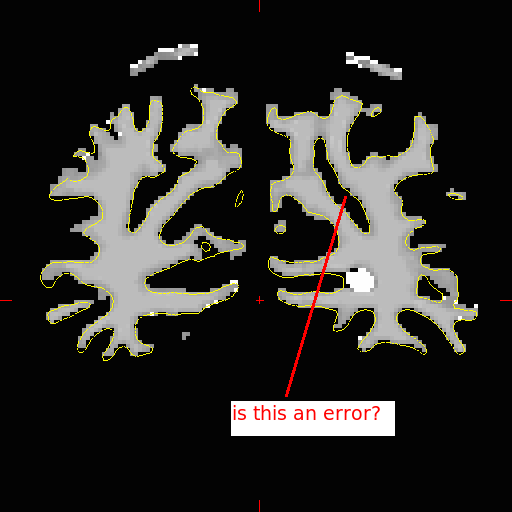
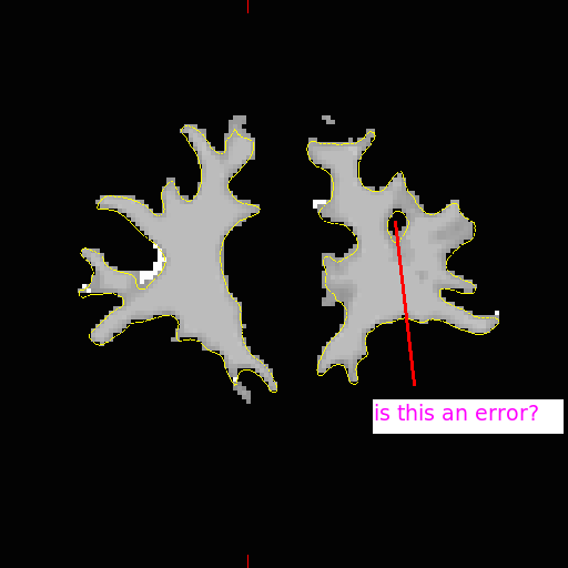
To make it more specific, I show you a slice of wm.mgh I have ( wmConfusion1 attached). Please look at the small yellow circle on the right. If I just follow the exercise, this looks like a lesion, as the yellow line cuts into it. but when I move up to next few slices, this hole opens up ( close to the green line ) giving me indications that it may NOT be a hole ( keeping in mind the inherent 3D nature of the MR image ). Also I am not sure whether there is an error in the image ( wmConfusion5 ) attched is an error is WM segmentation.
So my questions:
1. What are the things I have to look for, while trying to verify the WM segmentation ( after autorecon2 )? 2. Any thumb-rules laid out for this already? My googling was in fact in vain. 3. Any other tutorials/resources/books stating clearly what to verify, in different stages of reconstruction?
I am sorry to write a long email. Please be kind to help me in this regard.
Regards, Pradeep Reddy PhD Student, Simon Fraser Unviersity, Canada.

Hi Pradeep,
it's impossible to tell if it's an error from a single slice. Things that look like holes may actually just be sulci if you page through a couple of slices. You should also be looking at the intensity volume (e.g. norm.mgz) not just the wm.mgz. If the surface follows the gray/white boundary everywhere then you are all set. The problems occur if for example a lesion causes the topology correction to put the surface in the incorrect location in order to get the correct topology.
cheers, Bruce On Tue, 6 May 2008, Pradeep Reddy Ramana wrote:
Hello Everybody,
This is Pradeep Reddy, just started my PhD in Medical Image Analysis and hence a newbie to FreeSurfer.
I've passed the auto-recon2 stage in the reconstruction procedure. Now I am trying to verify WM segmentation produced by the recon-all/auto-recon2, before I move on to further processing. The excercise given in the freesurfer tutorial ( http://surfer.nmr.mgh.harvard.edu/fswiki/FsTutorial/WhiteMatterEdits ) gives me one example on how to detect if there were a lesion and outlines how to fix it. But now when I observe the outputs I have, I am quite confused as to how to detect inaccuracies in the segmentation of white matter.
To make it more specific, I show you a slice of wm.mgh I have ( wmConfusion1 attached). Please look at the small yellow circle on the right. If I just follow the exercise, this looks like a lesion, as the yellow line cuts into it. but when I move up to next few slices, this hole opens up ( close to the green line ) giving me indications that it may NOT be a hole ( keeping in mind the inherent 3D nature of the MR image ). Also I am not sure whether there is an error in the image ( wmConfusion5 ) attched is an error is WM segmentation.
So my questions:
- What are the things I have to look for, while trying to verify the
WM segmentation ( after autorecon2 )? 2. Any thumb-rules laid out for this already? My googling was in fact in vain. 3. Any other tutorials/resources/books stating clearly what to verify, in different stages of reconstruction?
I am sorry to write a long email. Please be kind to help me in this regard.
Regards, Pradeep Reddy PhD Student, Simon Fraser Unviersity, Canada.
freesurfer@nmr.mgh.harvard.edu
-
 Bruce Fischl
 -
 Pradeep Reddy Ramana