
Just as an update, it turns out that my segmentation volume had been slightly altered during the conversion from .nii to .mgz. I completely forgot to use the -rt nearest flag to prevent the labels from becoming distorted. That is why the segmentation looked so fuzzy with various regions labeled incorrectly.
Apparently, I should check the basics before overcomplicating the problem, haha. Thanks for the help everyone!
On Thu, Aug 1, 2013 at 9:36 AM, Mark Plantz < markplantz2016@u.northwestern.edu> wrote:
Sounds good. I also just realized that the segmentation values are varying from 0-255, which makes me think that the file is only designed for the grayscale/heat/etc. settings [where 0 is darkest regions and 255 is brightest regions]. Is that a valid assumption? Also, if this is the case, would it be impossible to create a .gca file using mri_ca_train with these files?
Thanks for the help.
MP
On Wed, Jul 31, 2013 at 8:43 PM, Douglas Greve greve@nmr.mgh.harvard.eduwrote:
Just make sure that you create a new file and do not repeat index numbers.
On 7/31/13 2:02 PM, Mark Plantz wrote:
That makes sense. Wouldn't that cause there to be some overlap between different regions, since the same index #'s used by the two programs (FreeSurfer & MRIcro) correspond to different brain regions?
On Tue, Jul 30, 2013 at 12:50 PM, Douglas Greve < greve@nmr.mgh.harvard.edu> wrote:
for every line in that file, you will need to create a line in the new LUT, something like
8 Frontal_Mid_R 0 255 0 0
This will make the right mid frontal green = (0,255,0)
On 7/30/13 9:55 AM, Mark Plantz wrote:
Hmm, so I received the text part of the LUT. Looks like this is completely different from the segmented regions that we would want. [attached]. Can't seem to understand how this text file could be related to the default FreeSurfer LUT. It looks like the labeling for an AAL map. This probably wont' work will it?
- Mark
On Tue, Jul 30, 2013 at 8:47 AM, Bruce Fischl < fischl@nmr.mgh.harvard.edu> wrote:
the UNC one? No, you should ask them On Tue, 30 Jul 2013, Mark Plantz wrote:
---------- Forwarded message ---------- From: Mark Plantz markplantz2016@u.northwestern.edu Date: Tue, Jul 30, 2013 at 8:39 AM Subject: Re: [Freesurfer] infant atlas segmentation To: Bruce Fischl fischl@nmr.mgh.harvard.edu
Just opened up the default FreeSurferColorLUT.txt file. So it looks like all I have to change is the #No. column of the file, to match the ones in the other LUT used. I actually just received the MRIcro .lut file from the UNC group. [attached]
Hopefully this will help with the conversion. However, I still have to figure out how to open the file. Any ideas?
On Tue, Jul 30, 2013 at 8:25 AM, Bruce Fischl < fischl@nmr.mgh.harvard.edu> wrote: Hi Mark
  creating a FreeSurfer LUT really isn't that hard. Have you
  looked at our standard one? Probably we have the names you
  already want and you just need to change our indices to match
  the ones in your segmentation file (and save it as a different
  LUT)

  cheers
  Bruce

  On Tue, 30 Jul 2013, Mark Plantz wrote:

        Turns out that the files had been labeled using a
        color LUT in a program
        called MRIcro. I am now installing this program
        simply to access the LUT.
        Will it be as simple as exchanging the index values
        from this LUT to a
        custom LUT in FreeSurfer?


        On Tue, Jul 30, 2013 at 12:35 AM, Douglas Greve
        <greve@nmr.mgh.harvard.edu>
        wrote:

              The problem is that you need to create a
        completely new LUT. The
              labels that get displayed with the standard
        LUT are going to be
              totally random with respect to what they
        should display.

              On 7/29/13 2:20 PM, Mark Plantz wrote:
              Hi Doug,
          Thanks for the reply. I will definitely go into
        the LUT and
        change those values to some specific region. My main
        concern is
        that I think all of these "out of bounds" labels are
        the
        symptoms of some larger problem. The segmentation
        looks very
        strange as compared to a typical segmentation (i.e.
        an
        'aseg.mgz' file). In fact, the segmentation appears
        to be
        labeling "left-<region" in the right hemisphere.
        Have you seen a
        segmentation file that looks like the once I
        attached before? I
        am used to seeing segmentation files such as the one
        attached to
        this e-mail.

        - Mark


        On Mon, Jul 29, 2013 at 12:28 PM, Douglas Greve
        <greve@nmr.mgh.harvard.edu> wrote:

              Mark, the only thing that is wrong with the
        out of
              bounds regions is that they are not
        represented in
              the LUT. Once you add an index corresponding
        to the
              index of the segmentation to the LUT, it will
        not
              show up as "out of bounds". The brain mask has
              nothing to do with it.
              best
              doug


              On 7/29/13 12:50 PM, Mark Plantz wrote:
              Sorry about that. I received the manual
              segmentations from a UNC medicalgroup<
http://www.med.unc.edu/bric/ideagroup/free-softwares/unc-infant-0-1-2 -atla ses>. I actually asked the main researcher about the out of bounds regions. He thinks that as long as the segmented file lines up with the input volumes, a few out of bounds regions might be OK. However, these seg volumes look very different from a typical .aseg volume that I am used to seeing after recon-all is run. Maybe the segmentation is simply incompatible with FreeSurfer?
Thanks for the help. These files are a nightmare! - MP On Mon, Jul 29, 2013 at 11:40 AM, Bruce Fischl <fischl@nmr.mgh.harvard.edu> wrote: can you cc the list please? Maybe we should start over. Where did you get the manual segmentations from? On Mon, 29 Jul 2013, Mark Plantz wrote: Ahh, right. So it looks like the red regions appear to be averaged around 248. I am not sure why that is the case or if it has any significance. I attached a screen shot for reference. On Mon, Jul 29, 2013 at 11:29 AM, Bruce Fischl <fischl@nmr.mgh.harvard.edu> wrote: it isn't a registration file. You should just specify it as a volume on the freeview command line, the same as brainmask.mgz On Mon, 29 Jul 2013, Mark Plantz wrote: I actually tried to view the brainmask.mgz file with the segmentation file [Freeview calls it 'registration file' I believe]. For some reason, the file will load without segmentation, but when I attempt to add the registration file, I get an error message: "Failed to load MRI ~/.../../../brainmask.mgz Could this potentially be part of the problem? Thanks MP On Mon, Jul 29, 2013 at 10:57 AM, Mark Plantz <markplantz2016@u.northwestern.edu> wrote: It looks like the values vary from 20-120 depending on which region I run the cursor over. However, aren't those the values that correspond to the brain.mgz regions? Is there another way to find the segmentation values? On Mon, Jul 29, 2013 at 10:37 AM, Bruce Fischl <fischl@nmr.mgh.harvard.edu> wrote: I mean the segmentation values that give you the "out of bounds" message On Mon, 29 Jul 2013, Mark Plantz wrote: Hi Bruce, Do you mean the brainmask.mgz values? I'm not sure what the segmentation #'s exactly are. Thanks! MP On Mon, Jul 29, 2013 at 10:17 AM, Bruce Fischl <fischl@nmr.mgh.harvard.edu> wrote: Hi Mark what #s are those segmentations? It should be trivial to just add them to the end of the LUT. I wouldn't let them be out of bounds as there might be code around that might ignore them then. cheers Bruce On Mon, 29 Jul 2013, Mark Plantz wrote: Hi Doug, Thanks for the reply. After doing some research, it looks like messing with the LUT might be a risky decision [and way out of my abilities!]. If I were to use this segmentation file to create a .gca atlas using the command mri_ca_train, do you think it would yield problems or be inaccurate? I guess what I am trying to figure out is if the color lookup values are even significant when using the segmentation file to create an atlas? Thanks for all of the help with this. Best, MP p.s. I attached the photo of the segmentation volume because this e-mail is sort of old On Wed, Jul 24, 2013 at 2:39 PM, Douglas N Greve <greve@nmr.mgh.harvard.edu> wrote: you need a new LUT. See my comment from a few emails ago. On 07/24/2013 03:29 PM, Mark Plantz wrote: I'm not actually quite sure what it means either. When I use the command line: tkmedit $Subject brain.mgz -segmentation <seg.mgz>, I can run the cursor over various regions and the name of the region should pop up. For some reason, the red regions are simply labeled as "out of bounds." I'll keep messing with tkmedit and let you know if I find out what the problem is. Thanks guys! MP On Wed, Jul 24, 2013 at 2:26 PM, Douglas N Greve <greve@nmr.mgh.harvard.edu <mailto:greve@nmr.mgh.harvard.edu>> wrote: what do you mean that they are out of bounds? On 07/24/2013 03:25 PM, Mark Plantz wrote: I guess the problem is that those regions should not be out of bounds. Maybe I need to create a new Lookup Table for the infant mri's? Is that possible to do? On Wed, Jul 24, 2013 at 2:21 PM, Douglas N Greve <greve@nmr.mgh.harvard.edu <mailto:greve@nmr.mgh.harvard.edu> <mailto:greve@nmr.mgh.harvard.edu <mailto:greve@nmr.mgh.harvard.edu>>> wrote: Hi Mark, please post to the list and not to me. thanks doug On 07/24/2013 03:21 PM, Mark Plantz wrote: I guess the problem is that those regions should not be out of bounds. Maybe I need to create a new Lookup Table for the infant mri's? Is that possible to do? On Wed, Jul 24, 2013 at 2:15 PM, Douglas N Greve <greve@nmr.mgh.harvard.edu <mailto:greve@nmr.mgh.harvard.edu> <mailto:greve@nmr.mgh.harvard.edu <mailto:greve@nmr.mgh.harvard.edu>> <mailto:greve@nmr.mgh.harvard.edu <mailto:greve@nmr.mgh.harvard.edu> <mailto:greve@nmr.mgh.harvard.edu <mailto:greve@nmr.mgh.harvard.edu>>>> wrote: what is the problem exactly? The fact that there are red regions or that that the red regions are labeled as "Out of Bounds"? If the latter, you will need to create a LUT that matches your regions. the out of bounds means that the index in the volume does not match an index in the LUT. doug On 07/24/2013 01:26 PM, Mark Plantz wrote: Finally got the registration to work. However, it looks like the out of bounds regions (in red) are still present (even though the regions are well within our boundaries). Is that expected since they were labeled in the original segmentation volume? Is there anyway to correct those? Thanks for all of the help! - Mark p.s. I attached a picture of the original brain.mgz file viewed with the corrected segmentation volume On Wed, Jul 24, 2013 at 12:22 PM, Mark Plantz <markplantz2016@u.northwestern.edu <mailto:markplantz2016@u.northwestern.edu> <mailto:markplantz2016@u.northwestern.edu <mailto:markplantz2016@u.northwestern.edu>> <mailto:markplantz2016@u.northwestern.edu <mailto:markplantz2016@u.northwestern.edu> <mailto:markplantz2016@u.northwestern.edu <mailto:markplantz2016@u.northwestern.edu>>> <mailto:markplantz2016@u.northwestern.edu <mailto:markplantz2016@u.northwestern.edu> <mailto:markplantz2016@u.northwestern.edu <mailto:markplantz2016@u.northwestern.edu>> <mailto:markplantz2016@u.northwestern.edu <mailto:markplantz2016@u.northwestern.edu> <mailto:markplantz2016@u.northwestern.edu <mailto:markplantz2016@u.northwestern.edu>>>>> wrote: Finally got the registration to work. However, it looks like the out of bound regions (in red) are still prevalent. Is that expected since they were present in the original segmentation volume? Would there be anyway to correct those? Thanks for all the help! - MP On Wed, Jul 24, 2013 at 11:11 AM, Mark Plantz <markplantz2016@u.northwestern.edu <mailto:markplantz2016@u.northwestern.edu> <mailto:markplantz2016@u.northwestern.edu <mailto:markplantz2016@u.northwestern.edu>> <mailto:markplantz2016@u.northwestern.edu <mailto:markplantz2016@u.northwestern.edu> <mailto:markplantz2016@u.northwestern.edu <mailto:markplantz2016@u.northwestern.edu>>> <mailto:markplantz2016@u.northwestern.edu <mailto:markplantz2016@u.northwestern.edu> <mailto:markplantz2016@u.northwestern.edu <mailto:markplantz2016@u.northwestern.edu>> <mailto:markplantz2016@u.northwestern.edu <mailto:markplantz2016@u.northwestern.edu> <mailto:markplantz2016@u.northwestern.edu <mailto:markplantz2016@u.northwestern.edu>>>>> wrote: Nevermind, it turned out to be a permissions issue. Thanks! On Wed, Jul 24, 2013 at 10:12 AM, Mark Plantz <markplantz2016@u.northwestern.edu <mailto:markplantz2016@u.northwestern.edu> <mailto:markplantz2016@u.northwestern.edu <mailto:markplantz2016@u.northwestern.edu>> <mailto:markplantz2016@u.northwestern.edu <mailto:markplantz2016@u.northwestern.edu> <mailto:markplantz2016@u.northwestern.edu <mailto:markplantz2016@u.northwestern.edu>>> <mailto:markplantz2016@u.northwestern.edu <mailto:markplantz2016@u.northwestern.edu>
Freesurfer mailing listFreesurfer@nmr.mgh.harvard.eduhttps://mail.nmr.mgh.harvard.edu/mailman/listinfo/freesurfer
Freesurfer mailing list Freesurfer@nmr.mgh.harvard.edu https://mail.nmr.mgh.harvard.edu/mailman/listinfo/freesurfer
The information in this e-mail is intended only for the person to whom it is addressed. If you believe this e-mail was sent to you in error and the e-mail contains patient information, please contact the Partners Compliance HelpLine at http://www.partners.org/complianceline . If the e-mail was sent to you in error but does not contain patient information, please contact the sender and properly dispose of the e-mail.

glad it worked out Bruce On Thu, 1 Aug 2013, Mark Plantz wrote:
Just as an update, it turns out that my segmentation volume had been slightly altered during the conversion from .nii to .mgz. I completely forgot to use the -rt nearest flag to prevent the labels from becoming distorted. That is why the segmentation looked so fuzzy with various regions labeled incorrectly. Apparently, I should check the basics before overcomplicating the problem, haha. Thanks for the help everyone!
On Thu, Aug 1, 2013 at 9:36 AM, Mark Plantz markplantz2016@u.northwestern.edu wrote: Sounds good. I also just realized that the segmentation values are varying from 0-255, which makes me think that the file is only designed for the grayscale/heat/etc. settings [where 0 is darkest regions and 255 is brightest regions]. Is that a valid assumption? Also, if this is the case, would it be impossible to create a .gca file using mri_ca_train with these files? Thanks for the help.
MP
On Wed, Jul 31, 2013 at 8:43 PM, Douglas Greve greve@nmr.mgh.harvard.edu wrote:
  Just make sure that you create a new file and do not
  repeat index numbers.


  On 7/31/13 2:02 PM, Mark Plantz wrote:
  That makes sense. Wouldn't that cause there to be
  some overlap between different regions, since the
  same index #'s used by the two programs (FreeSurfer
  & MRIcro) correspond to different brain regions?
On Tue, Jul 30, 2013 at 12:50 PM, Douglas Greve greve@nmr.mgh.harvard.edu wrote:
  for every line in that file, you will need to
  create a line in the new LUT, something like


  8 Frontal_Mid_R 0 255 0   0

  This will make the right mid frontal green =
  (0,255,0)


  On 7/30/13 9:55 AM, Mark Plantz wrote:
  Hmm, so I received the text part of the
  LUT. Looks like this is completely
  different from the segmented regions
  that we would want. [attached]. Can't
  seem to understand how this text file
  could be related to the default
  FreeSurfer LUT. It looks like the
  labeling for an AAL map. This probably
  wont' work will it?
- Mark
On Tue, Jul 30, 2013 at 8:47 AM, Bruce Fischl fischl@nmr.mgh.harvard.edu wrote: the UNC one? No, you should ask them On Tue, 30 Jul 2013, Mark Plantz wrote:
---------- Forwarded message ---------- From: Mark Plantz <markplantz2016@u.northwestern.edu> Date: Tue, Jul 30, 2013 at 8:39 AM Subject: Re: [Freesurfer] infant atlas segmentation To: Bruce Fischl <fischl@nmr.mgh.harvard.edu> Just opened up the default FreeSurferColorLUT.txt file. So it looks like all I have to change is the #No. column of the file, to match the ones in the other LUT used. I actually just received the MRIcro .lut file from the UNC group. [attached] Hopefully this will help with the conversion. However, I still have to figure out how to open the file. Any ideas? On Tue, Jul 30, 2013 at 8:25 AM, Bruce Fischl <fischl@nmr.mgh.harvard.edu> wrote: Hi Mark creating a FreeSurfer LUT really isn't that hard. Have you looked at our standard one? Probably we have the names you already want and you just need to change our indices to match the ones in your segmentation file (and save it as a different LUT) cheers Bruce On Tue, 30 Jul 2013, Mark Plantz wrote: Turns out that the files had been labeled using a color LUT in a program called MRIcro. I am now installing this program simply to access the LUT. Will it be as simple as exchanging the index values from this LUT to a custom LUT in FreeSurfer? On Tue, Jul 30, 2013 at 12:35 AM, Douglas Greve <greve@nmr.mgh.harvard.edu> wrote: The problem is that you need to create a completely new LUT. The labels that get displayed with the standard LUT are going to be totally random with respect to what they should display. On 7/29/13 2:20 PM, Mark Plantz wrote: Hi Doug, Thanks for the reply. I will definitely go into the LUT and change those values to some specific region. My main concern is that I think all of these "out of bounds" labels are the symptoms of some larger problem. The segmentation looks very strange as compared to a typical segmentation (i.e. an 'aseg.mgz' file). In fact, the segmentation appears to be labeling "left-<region" in the right hemisphere. Have you seen a segmentation file that looks like the once I attached before? I am used to seeing segmentation files such as the one attached to this e-mail. - Mark On Mon, Jul 29, 2013 at 12:28 PM, Douglas Greve <greve@nmr.mgh.harvard.edu> wrote: Mark, the only thing that is wrong with the out of bounds regions is that they are not represented in the LUT. Once you add an index corresponding to the index of the segmentation to the LUT, it will not show up as "out of bounds". The brain mask has nothing to do with it. best doug On 7/29/13 12:50 PM, Mark Plantz wrote: Sorry about that. I received the manual segmentations from a UNCmedicalgroup<http://www.med.unc.edu/bric/ideagroup/free-softwares/unc-infan t-0-1-2 -atla ses>. I actually asked the main researcher about the out of bounds regions. He thinks that as long as the segmented file lines up with the input volumes, a few out of bounds regions might be OK. However, these seg volumes look very different from a typical .aseg volume that I am used to seeing after recon-all is run. Maybe the segmentation is simply incompatible with FreeSurfer? Thanks for the help. These files are a nightmare! - MP On Mon, Jul 29, 2013 at 11:40 AM, Bruce Fischl <fischl@nmr.mgh.harvard.edu> wrote: can you cc the list please? Maybe we should start over. Where did you get the manual segmentations from? On Mon, 29 Jul 2013, Mark Plantz wrote: Ahh, right. So it looks like the red regions appear to be averaged around 248. I am not sure why that is the case or if it has any significance. I attached a screen shot for reference. On Mon, Jul 29, 2013 at 11:29 AM, Bruce Fischl <fischl@nmr.mgh.harvard.edu> wrote: it isn't a registration file. You should just specify it as a volume on the freeview command line, the same as brainmask.mgz On Mon, 29 Jul 2013, Mark Plantz wrote: I actually tried to view the brainmask.mgz file with the segmentation file [Freeview calls it 'registration file' I believe]. For some reason, the file will load without segmentation, but when I attempt to add the registration file, I get an error message: "Failed to load MRI ~/.../../../brainmask.mgz Could this potentially be part of the problem? Thanks MP On Mon, Jul 29, 2013 at 10:57 AM, Mark Plantz <markplantz2016@u.northwestern.edu> wrote: It looks like the values vary from 20-120 depending on which region I run the cursor over. However, aren't those the values that correspond to the brain.mgz regions? Is there another way to find the segmentation values? On Mon, Jul 29, 2013 at 10:37 AM, Bruce Fischl <fischl@nmr.mgh.harvard.edu> wrote: I mean the segmentation values that give you the "out of bounds" message On Mon, 29 Jul 2013, Mark Plantz wrote: Hi Bruce, Do you mean the brainmask.mgz values? I'm not sure what the segmentation #'s exactly are. Thanks! MP On Mon, Jul 29, 2013 at 10:17 AM, Bruce Fischl <fischl@nmr.mgh.harvard.edu> wrote: Hi Mark what #s are those segmentations? It should be trivial to just add them to the end of the LUT. I wouldn't let them be out of bounds as there might be code around that might ignore them then. cheers Bruce On Mon, 29 Jul 2013, Mark Plantz wrote: Hi Doug, Thanks for the reply. After doing some research, it looks like messing with the LUT might be a risky decision [and way out of my abilities!]. If I were to use this segmentation file to create a .gca atlas using the command mri_ca_train, do you think it would yield problems or be inaccurate? I guess what I am trying to figure out is if the color lookup values are even significant when using the segmentation file to create an atlas? Thanks for all of the help with this. Best, MP p.s. I attached the photo of the segmentation volume because this e-mail is sort of old On Wed, Jul 24, 2013 at 2:39 PM, Douglas N Greve <greve@nmr.mgh.harvard.edu> wrote: you need a new LUT. See my comment from a few emails ago. On 07/24/2013 03:29 PM, Mark Plantz wrote: I'm not actually quite sure what it means either. When I use the command line: tkmedit $Subject brain.mgz -segmentation <seg.mgz>, I can run the cursor over various regions and the name of the region should pop up. For some reason, the red regions are simply labeled as "out of bounds." I'll keep messing with tkmedit and let you know if I find out what the problem is. Thanks guys! MP On Wed, Jul 24, 2013 at 2:26 PM, Douglas N Greve <greve@nmr.mgh.harvard.edu <mailto:greve@nmr.mgh.harvard.edu>> wrote: what do you mean that they are out of bounds? On 07/24/2013 03:25 PM, Mark Plantz wrote: I guess the problem is that those regions should not be out of bounds. Maybe I need to create a new Lookup Table for the infant mri's? Is that possible to do? On Wed, Jul 24, 2013 at 2:21 PM, Douglas N Greve <greve@nmr.mgh.harvard.edu <mailto:greve@nmr.mgh.harvard.edu> <mailto:greve@nmr.mgh.harvard.edu <mailto:greve@nmr.mgh.harvard.edu>>> wrote: Hi Mark, please post to the list and not to me. thanks doug On 07/24/2013 03:21 PM, Mark Plantz wrote: I guess the problem is that those regions should not be out of bounds. Maybe I need to create a new Lookup Table for the infant mri's? Is that possible to do? On Wed, Jul 24, 2013 at 2:15 PM, Douglas N Greve <greve@nmr.mgh.harvard.edu <mailto:greve@nmr.mgh.harvard.edu> <mailto:greve@nmr.mgh.harvard.edu <mailto:greve@nmr.mgh.harvard.edu>> <mailto:greve@nmr.mgh.harvard.edu <mailto:greve@nmr.mgh.harvard.edu> <mailto:greve@nmr.mgh.harvard.edu <mailto:greve@nmr.mgh.harvard.edu>>>> wrote: what is the problem exactly? The fact that there are red regions or that that the red regions are labeled as "Out of Bounds"? If the latter, you will need to create a LUT that matches your regions. the out of bounds means that the index in the volume does not match an index in the LUT. doug On 07/24/2013 01:26 PM, Mark Plantz wrote: Finally got the registration to work. However, it looks like the out of bounds regions (in red) are still present (even though the regions are well within our boundaries). Is that expected since they were labeled in the original segmentation volume? Is there anyway to correct those? Thanks for all of the help! - Mark p.s. I attached a picture of the original brain.mgz file viewed with the corrected segmentation volume On Wed, Jul 24, 2013 at 12:22 PM, Mark Plantz <markplantz2016@u.northwestern.edu <mailto:markplantz2016@u.northwestern.edu> <mailto:markplantz2016@u.northwestern.edu <mailto:markplantz2016@u.northwestern.edu>> <mailto:markplantz2016@u.northwestern.edu <mailto:markplantz2016@u.northwestern.edu> <mailto:markplantz2016@u.northwestern.edu <mailto:markplantz2016@u.northwestern.edu>>> <mailto:markplantz2016@u.northwestern.edu <mailto:markplantz2016@u.northwestern.edu> <mailto:markplantz2016@u.northwestern.edu <mailto:markplantz2016@u.northwestern.edu>> <mailto:markplantz2016@u.northwestern.edu <mailto:markplantz2016@u.northwestern.edu> <mailto:markplantz2016@u.northwestern.edu <mailto:markplantz2016@u.northwestern.edu>>>>> wrote: Finally got the registration to work. However, it looks like theout of bound regions (in red) are still prevalent. Is that expected since they were present in the original segmentation volume? Would there be anyway to correct those?
Thanks for all the help!
- MP
On Wed, Jul 24, 2013 at 11:11 AM, Mark Plantz <markplantz2016@u.northwestern.edu mailto:markplantz2016@u.northwestern.edu <mailto:markplantz2016@u.northwestern.edu mailto:markplantz2016@u.northwestern.edu> <mailto:markplantz2016@u.northwestern.edu mailto:markplantz2016@u.northwestern.edu <mailto:markplantz2016@u.northwestern.edu mailto:markplantz2016@u.northwestern.edu>> <mailto:markplantz2016@u.northwestern.edu mailto:markplantz2016@u.northwestern.edu <mailto:markplantz2016@u.northwestern.edu mailto:markplantz2016@u.northwestern.edu>
<mailto:markplantz2016@u.northwestern.edu mailto:markplantz2016@u.northwestern.edu <mailto:markplantz2016@u.northwestern.edu mailto:markplantz2016@u.northwestern.edu>>>> wrote:
Nevermind, it turned out to be a permissions issue. Thanks!
On Wed, Jul 24, 2013 at 10:12 AM, Mark Plantz <markplantz2016@u.northwestern.edu mailto:markplantz2016@u.northwestern.edu <mailto:markplantz2016@u.northwestern.edu mailto:markplantz2016@u.northwestern.edu> <mailto:markplantz2016@u.northwestern.edu mailto:markplantz2016@u.northwestern.edu <mailto:markplantz2016@u.northwestern.edu mailto:markplantz2016@u.northwestern.edu>> <mailto:markplantz2016@u.northwestern.edu mailto:markplantz2016@u.northwestern.edu
Freesurfer mailing list Freesurfer@nmr.mgh.harvard.edu https://mail.nmr.mgh.harvard.edu/mailman/listinfo/freesurfer
Freesurfer mailing list Freesurfer@nmr.mgh.harvard.edu https://mail.nmr.mgh.harvard.edu/mailman/listinfo/freesurfer
The information in this e-mail is intended only for the person to whom it is addressed. If you believe this e-mail was sent to you in error and the e-mail contains patient information, please contact the Partners Compliance HelpLine at http://www.partners.org/complianceline . If the e-mail was sent to you in error but does not contain patient information, please contact the sender and properly dispose of the e-mail.
One last question: Is there any flag I can use with 'bbregister' that prevents the labels from changing in my segmentation volume? This is occuring because the volume that I am moving has labels. For some reason, the labels change ever so slightly in the output file.
On Thu, Aug 1, 2013 at 2:28 PM, Bruce Fischl fischl@nmr.mgh.harvard.eduwrote:
glad it worked out Bruce On Thu, 1 Aug 2013, Mark Plantz wrote:
Just as an update, it turns out that my segmentation volume had been
slightly altered during the conversion from .nii to .mgz. I completely forgot to use the -rt nearest flag to prevent the labels from becoming distorted. That is why the segmentation looked so fuzzy with various regions labeled incorrectly. Apparently, I should check the basics before overcomplicating the problem, haha. Thanks for the help everyone!
On Thu, Aug 1, 2013 at 9:36 AM, Mark Plantz <markplantz2016@u.**northwestern.edu markplantz2016@u.northwestern.edu> wrote: Sounds good. I also just realized that the segmentation values are varying from 0-255, which makes me think that the file is only designed for the grayscale/heat/etc. settings [where 0 is darkest regions and 255 is brightest regions]. Is that a valid assumption? Also, if this is the case, would it be impossible to create a .gca file using mri_ca_train with these files? Thanks for the help.
MP
On Wed, Jul 31, 2013 at 8:43 PM, Douglas Greve greve@nmr.mgh.harvard.edu wrote:
  Just make sure that you create a new file and do not
  repeat index numbers.


  On 7/31/13 2:02 PM, Mark Plantz wrote:
  That makes sense. Wouldn't that cause there to be
  some overlap between different regions, since the
  same index #'s used by the two programs (FreeSurfer
  & MRIcro) correspond to different brain regions?
On Tue, Jul 30, 2013 at 12:50 PM, Douglas Greve greve@nmr.mgh.harvard.edu wrote:
  for every line in that file, you will need to
  create a line in the new LUT, something like


  8 Frontal_Mid_R 0 255 0   0

  This will make the right mid frontal green =
  (0,255,0)


  On 7/30/13 9:55 AM, Mark Plantz wrote:
  Hmm, so I received the text part of the
  LUT. Looks like this is completely
  different from the segmented regions
  that we would want. [attached]. Can't
  seem to understand how this text file
  could be related to the default
  FreeSurfer LUT. It looks like the
  labeling for an AAL map. This probably
  wont' work will it?
- Mark
On Tue, Jul 30, 2013 at 8:47 AM, Bruce Fischl fischl@nmr.mgh.harvard.edu wrote: the UNC one? No, you should ask them On Tue, 30 Jul 2013, Mark Plantz wrote:
---------- Forwarded message ---------- From: Mark Plantz <markplantz2016@u.**northwestern.edu<markplantz2016@u.northwestern.edu>Date: Tue, Jul 30, 2013 at 8:39 AM Subject: Re: [Freesurfer] infant atlas segmentation To: Bruce Fischl <fischl@nmr.mgh.harvard.edu> Just opened up the default FreeSurferColorLUT.txt file. So it looks like all I have to change is the #No. column of the file, to match the ones in the other LUT used. I actually just received the MRIcro .lut file from the UNC group. [attached] Hopefully this will help with the conversion. However, I still have to figure out how to open the file. Any ideas? On Tue, Jul 30, 2013 at 8:25 AM, Bruce Fischl <fischl@nmr.mgh.harvard.edu> wrote: Hi Mark creating a FreeSurfer LUT really isn't that hard. Have you looked at our standard one? Probably we have the names you already want and you just need to change our indices to match the ones in your segmentation file (and save it as a different LUT) cheers Bruce On Tue, 30 Jul 2013, Mark Plantz wrote: Turns out that the files had been labeled using a color LUT in a program called MRIcro. I am now installing this program simply to access the LUT. Will it be as simple as exchanging the index values from this LUT to a custom LUT in FreeSurfer? On Tue, Jul 30, 2013 at 12:35 AM, Douglas Greve <greve@nmr.mgh.harvard.edu> wrote: The problem is that you need to create a completely new LUT. The labels that get displayed with the standard LUT are going to be totally random with respect to what they should display. On 7/29/13 2:20 PM, Mark Plantz wrote: Hi Doug, Thanks for the reply. I will definitely go into the LUT and change those values to some specific region. My main concern is that I think all of these "out of bounds" labels are the symptoms of some larger problem. The segmentation looks very strange as compared to a typical segmentation (i.e. an 'aseg.mgz' file). In fact, the segmentation appears to be labeling "left-<region" in the right hemisphere. Have you seen a segmentation file that looks like the once I attached before? I am used to seeing segmentation files such as the one attached to this e-mail. - Mark On Mon, Jul 29, 2013 at 12:28 PM, Douglas Greve <greve@nmr.mgh.harvard.edu> wrote: Mark, the only thing that is wrong with the out of bounds regions is that they are not represented in the LUT. Once you add an index corresponding to the index of the segmentation to the LUT, it will not show up as "out of bounds". The brain mask has nothing to do with it. best doug On 7/29/13 12:50 PM, Mark Plantz wrote: Sorry about that. I received the manual segmentations from a UNCmedicalgroup<http://www.**med.unc.edu/bric/ideagroup/**free-softwares/unc-infanhttp://www.med.unc.edu/bric/ideagroup/free-softwares/unc-infan t-0-1-2 -atla ses>. I actually asked the main researcher about the out of bounds regions. He thinks that as long as the segmented file lines up with the input volumes, a few out of bounds regions might be OK. However, these seg volumes look very different from a typical .aseg volume that I am used to seeing after recon-all is run. Maybe the segmentation is simply incompatible with FreeSurfer?
Thanks for the help. These files are a nightmare! - MP On Mon, Jul 29, 2013 at 11:40 AM, Bruce Fischl <fischl@nmr.mgh.harvard.edu> wrote: can you cc the list please? Maybe we should start over. Where did you get the manual segmentations from? On Mon, 29 Jul 2013, Mark Plantz wrote: Ahh, right. So it looks like the red regions appear to be averaged around 248. I am not sure why that is the case or if it has any significance. I attached a screen shot for reference. On Mon, Jul 29, 2013 at 11:29 AM, Bruce Fischl <fischl@nmr.mgh.harvard.edu> wrote: it isn't a registration file. You should just specify it as a volume on the freeview command line, the same as brainmask.mgz On Mon, 29 Jul 2013, Mark Plantz wrote: I actually tried to view the brainmask.mgz file with the segmentation file [Freeview calls it 'registration file' I believe]. For some reason, the file will load without segmentation, but when I attempt to add the registration file, I get an error message: "Failed to load MRI ~/.../../../brainmask.mgz Could this potentially be part of the problem? Thanks MP On Mon, Jul 29, 2013 at 10:57 AM, Mark Plantz <markplantz2016@u.**northwestern.edu<markplantz2016@u.northwestern.edu>wrote: It looks like the values vary from 20-120 depending on which region I run the cursor over. However, aren't those the values that correspond to the brain.mgz regions? Is there another way to find the segmentation values? On Mon, Jul 29, 2013 at 10:37 AM, Bruce Fischl <fischl@nmr.mgh.harvard.edu> wrote: I mean the segmentation values that give you the "out of bounds" message On Mon, 29 Jul 2013, Mark Plantz wrote: Hi Bruce, Do you mean the brainmask.mgz values? I'm not sure what the segmentation #'s exactly are. Thanks! MP On Mon, Jul 29, 2013 at 10:17 AM, Bruce Fischl <fischl@nmr.mgh.harvard.edu> wrote: Hi Mark what #s are those segmentations? It should be trivial to just add them to the end of the LUT. I wouldn't let them be out of bounds as there might be code around that might ignore them then. cheers Bruce On Mon, 29 Jul 2013, Mark Plantz wrote: Hi Doug, Thanks for the reply. After doing some research, it looks like messing with the LUT might be a risky decision [and way out of my abilities!]. If I were to use this segmentation file to create a .gca atlas using the command mri_ca_train, do you think it would yield problems or be inaccurate? I guess what I am trying to figure out is if the color lookup values are even significant when using the segmentation file to create an atlas? Thanks for all of the help with this. Best, MP p.s. I attached the photo of the segmentation volume because this e-mail is sort of old On Wed, Jul 24, 2013 at 2:39 PM, Douglas N Greve <greve@nmr.mgh.harvard.edu> wrote: you need a new LUT. See my comment from a few emails ago. On 07/24/2013 03:29 PM, Mark Plantz wrote: I'm not actually quite sure what it means either. When I use the command line: tkmedit $Subject brain.mgz -segmentation <seg.mgz>, I can run the cursor over various regions and the name of the region should pop up. For some reason, the red regions are simply labeled as "out of bounds." I'll keep messing with tkmedit and let you know if I find out what the problem is. Thanks guys! MP On Wed, Jul 24, 2013 at 2:26 PM, Douglas N Greve <greve@nmr.mgh.harvard.edu <mailto:greve@nmr.mgh.harvard.**edu <greve@nmr.mgh.harvard.edu>>> wrote: what do you mean that they are out of bounds? On 07/24/2013 03:25 PM, Mark Plantz wrote: I guess the problem is that those regions should not be out of bounds. Maybe I need to create a new Lookup Table for the infant mri's? Is that possible to do? On Wed, Jul 24, 2013 at 2:21 PM, Douglas N Greve <greve@nmr.mgh.harvard.edu <mailto:greve@nmr.mgh.harvard.**edu <greve@nmr.mgh.harvard.edu>> <mailto:greve@nmr.mgh.harvard.**edu <greve@nmr.mgh.harvard.edu> <mailto:greve@nmr.mgh.harvard.**edu <greve@nmr.mgh.harvard.edu>>>> wrote: Hi Mark, please post to the list and not to me. thanks doug On 07/24/2013 03:21 PM, Mark Plantz wrote: I guess the problem is that those regions should not be out of bounds. Maybe I need to create a new Lookup Table for the infant mri's? Is that possible to do? On Wed, Jul 24, 2013 at 2:15 PM, Douglas N Greve <greve@nmr.mgh.harvard.edu <mailto:greve@nmr.mgh.harvard.**edu <greve@nmr.mgh.harvard.edu>> <mailto:greve@nmr.mgh.harvard.**edu <greve@nmr.mgh.harvard.edu> <mailto:greve@nmr.mgh.harvard.**edu <greve@nmr.mgh.harvard.edu>>> <mailto:greve@nmr.mgh.harvard.**edu <greve@nmr.mgh.harvard.edu> <mailto:greve@nmr.mgh.harvard.**edu <greve@nmr.mgh.harvard.edu>> <mailto:greve@nmr.mgh.harvard.**edu <greve@nmr.mgh.harvard.edu> <mailto:greve@nmr.mgh.harvard.**edu <greve@nmr.mgh.harvard.edu>>>>> wrote: what is the problem exactly? The fact that there are red regions or that that the red regions are labeled as "Out of Bounds"? If the latter, you will need to create a LUT that matches your regions. the out of bounds means that the index in the volume does not match an index in the LUT. doug On 07/24/2013 01:26 PM, Mark Plantz wrote: Finally got the registration to work. However, it looks like the out of bounds regions (in red) are still present (even though the regions are well within our boundaries). Is that expected since they were labeled in the original segmentation volume? Is there anyway to correct those? Thanks for all of the help! - Mark p.s. I attached a picture of the original brain.mgz file viewed with the corrected segmentation volume On Wed, Jul 24, 2013 at 12:22 PM, Mark Plantz <markplantz2016@u.**northwestern.edu<markplantz2016@u.northwestern.edu> <mailto:markplantz2016@u.**northwestern.edu<markplantz2016@u.northwestern.edu><mailto:markplantz2016@u.**northwestern.edu<markplantz2016@u.northwestern.edu> <mailto:markplantz2016@u.**northwestern.edu<markplantz2016@u.northwestern.edu><mailto:markplantz2016@u.**northwestern.edu<markplantz2016@u.northwestern.edu> <mailto:markplantz2016@u.**northwestern.edu<markplantz2016@u.northwestern.edu><mailto:markplantz2016@u.**northwestern.edu<markplantz2016@u.northwestern.edu> <mailto:markplantz2016@u.**northwestern.edu<markplantz2016@u.northwestern.edu><mailto:markplantz2016@u.**northwestern.edu<markplantz2016@u.northwestern.edu> <mailto:markplantz2016@u.**northwestern.edu<markplantz2016@u.northwestern.edu><mailto:markplantz2016@u.**northwestern.edu<markplantz2016@u.northwestern.edu> <mailto:markplantz2016@u.**northwestern.edu<markplantz2016@u.northwestern.edu>
Hi Mark
you can use the transform that bbregister outputs and give it to mri_convert with -at <transform name>
cheers Bruce
On Thu, 1 Aug 2013, Mark Plantz wrote:
One last question: Is there any flag I can use with 'bbregister' that prevents the labels from changing in my segmentation volume? This is occuring because the volume that I am moving has labels. For some reason, the labels change ever so slightly in the output file.
On Thu, Aug 1, 2013 at 2:28 PM, Bruce Fischl fischl@nmr.mgh.harvard.edu wrote: glad it worked out Bruce On Thu, 1 Aug 2013, Mark Plantz wrote:
Just as an update, it turns out that my segmentation volume had been slightly altered during the conversion from .nii to .mgz. I completely forgot to use the -rt nearest flag to prevent the labels from becoming distorted. That is why the segmentation looked so fuzzy with various regions labeled incorrectly. Apparently, I should check the basics before overcomplicating the problem, haha. Thanks for the help everyone! On Thu, Aug 1, 2013 at 9:36 AM, Mark Plantz <markplantz2016@u.northwestern.edu> wrote: Sounds good. I also just realized that the segmentation values are varying from 0-255, which makes me think that the file is only designed for the grayscale/heat/etc. settings [where 0 is darkest regions and 255 is brightest regions]. Is that a valid assumption? Also, if this is the case, would it be impossible to create a .gca file using mri_ca_train with these files? Thanks for the help. MP On Wed, Jul 31, 2013 at 8:43 PM, Douglas Greve <greve@nmr.mgh.harvard.edu> wrote: Just make sure that you create a new file and do not repeat index numbers. On 7/31/13 2:02 PM, Mark Plantz wrote: That makes sense. Wouldn't that cause there to be some overlap between different regions, since the same index #'s used by the two programs (FreeSurfer & MRIcro) correspond to different brain regions? On Tue, Jul 30, 2013 at 12:50 PM, Douglas Greve <greve@nmr.mgh.harvard.edu> wrote: for every line in that file, you will need to create a line in the new LUT, something like 8 Frontal_Mid_R 0 255 0 0 This will make the right mid frontal green = (0,255,0) On 7/30/13 9:55 AM, Mark Plantz wrote: Hmm, so I received the text part of the LUT. Looks like this is completely different from the segmented regions that we would want. [attached]. Can't seem to understand how this text file could be related to the default FreeSurfer LUT. It looks like the labeling for an AAL map. This probably wont' work will it? - Mark On Tue, Jul 30, 2013 at 8:47 AM, Bruce Fischl <fischl@nmr.mgh.harvard.edu> wrote: the UNC one? No, you should ask them On Tue, 30 Jul 2013, Mark Plantz wrote: ---------- Forwarded message ---------- From: Mark Plantz <markplantz2016@u.northwestern.edu> Date: Tue, Jul 30, 2013 at 8:39 AM Subject: Re: [Freesurfer] infant atlas segmentation To: Bruce Fischl <fischl@nmr.mgh.harvard.edu> Just opened up the default FreeSurferColorLUT.txt file. So it looks like all I have to change is the #No. column of the file, to match the ones in the other LUT used. I actually just received the MRIcro .lut file from the UNC group. [attached] Hopefully this will help with the conversion. However, I still have to figure out how to open the file. Any ideas? On Tue, Jul 30, 2013 at 8:25 AM, Bruce Fischl <fischl@nmr.mgh.harvard.edu> wrote: Hi Mark creating a FreeSurfer LUT really isn't that hard. Have you looked at our standard one? Probably we have the names you already want and you just need to change our indices to match the ones in your segmentation file (and save it as a different LUT) cheers Bruce On Tue, 30 Jul 2013, Mark Plantz wrote: Turns out that the files had been labeled using a color LUT in a program called MRIcro. I am now installing this program simply to access the LUT. Will it be as simple as exchanging the index values from this LUT to a custom LUT in FreeSurfer? On Tue, Jul 30, 2013 at 12:35 AM, Douglas Greve <greve@nmr.mgh.harvard.edu> wrote: The problem is that you need to create a completely new LUT. The labels that get displayed with the standard LUT are going to be totally random with respect to what they should display. On 7/29/13 2:20 PM, Mark Plantz wrote: Hi Doug, Thanks for the reply. I will definitely go into the LUT and change those values to some specific region. My main concern is that I think all of these "out of bounds" labels are the symptoms of some larger problem. The segmentation looks very strange as compared to a typical segmentation (i.e. an 'aseg.mgz' file). In fact, the segmentation appears to be labeling "left-<region" in the right hemisphere. Have you seen a segmentation file that looks like the once I attached before? I am used to seeing segmentation files such as the one attached to this e-mail. - Mark On Mon, Jul 29, 2013 at 12:28 PM, Douglas Greve <greve@nmr.mgh.harvard.edu> wrote: Mark, the only thing that is wrong with the out of bounds regions is that they are not represented in the LUT. Once you add an index corresponding to the index of the segmentation to the LUT, it will not show up as "out of bounds". The brain mask has nothing to do with it. best doug On 7/29/13 12:50 PM, Mark Plantz wrote: Sorry about that. I received the manual segmentations from aUNCmedicalgroup<http://www.med.unc.edu/bric/ideagroup/free-softwares/unc-in fan t-0-1-2 -atla ses>. I actually asked the main researcher about the out of bounds regions. He thinks that as long as the segmented file lines up with the input volumes, a few out of bounds regions might be OK. However, these seg volumes look very different from a typical .aseg volume that I am used to seeing after recon-all is run. Maybe the segmentation is simply incompatible with FreeSurfer? Thanks for the help. These files are a nightmare! - MP On Mon, Jul 29, 2013 at 11:40 AM, Bruce Fischl <fischl@nmr.mgh.harvard.edu> wrote: can you cc the list please? Maybe we should start over. Where did you get the manual segmentations from? On Mon, 29 Jul 2013, Mark Plantz wrote: Ahh, right. So it looks like the red regions appear to be averaged around 248. I am not sure why that is the case or if it has any significance. I attached a screen shot for reference. On Mon, Jul 29, 2013 at 11:29 AM, Bruce Fischl <fischl@nmr.mgh.harvard.edu> wrote: it isn't a registration file. You should just specify it as a volume on the freeview command line, the same as brainmask.mgz On Mon, 29 Jul 2013, Mark Plantz wrote: I actually tried to view the brainmask.mgz file with the segmentation file [Freeview calls it 'registration file' I believe]. For some reason, the file will load without segmentation, but when I attempt to add the registration file, I get an error message: "Failed to load MRI ~/.../../../brainmask.mgz Could this potentially be part of the problem? Thanks MP On Mon, Jul 29, 2013 at 10:57 AM, Mark Plantz <markplantz2016@u.northwestern.edu> wrote: It looks like the values vary from 20-120 depending on which region I run the cursor over. However, aren't those the values that correspond to the brain.mgz regions? Is there another way to find the segmentation values? On Mon, Jul 29, 2013 at 10:37 AM, Bruce Fischl <fischl@nmr.mgh.harvard.edu> wrote: I mean the segmentation values that give you the "out of bounds" message On Mon, 29 Jul 2013, Mark Plantz wrote: Hi Bruce, Do you mean the brainmask.mgz values? I'm not sure what the segmentation #'s exactly are. Thanks! MP On Mon, Jul 29, 2013 at 10:17 AM, Bruce Fischl <fischl@nmr.mgh.harvard.edu> wrote: Hi Mark what #s are those segmentations? It should be trivial to just add them to the end of the LUT. I wouldn't let them be out of bounds as there might be code around that might ignore them then. cheers Bruce On Mon, 29 Jul 2013, Mark Plantz wrote: Hi Doug, Thanks for the reply. After doing some research, it looks like messing with the LUT might be a risky decision [and way out of my abilities!]. If I were to use this segmentation file to create a .gca atlas using the command mri_ca_train, do you think it would yield problems or be inaccurate? I guess what I am trying to figure out is if the color lookup values are even significant when using the segmentation file to create an atlas? Thanks for all of the help with this. Best, MP p.s. I attached the photo of the segmentation volume because this e-mail is sort of old On Wed, Jul 24, 2013 at 2:39 PM, Douglas N Greve <greve@nmr.mgh.harvard.edu> wrote: you need a new LUT. See my comment from a few emails ago. On 07/24/2013 03:29 PM, Mark Plantz wrote: I'm not actually quite sure what it means either. When I use the command line: tkmedit $Subject brain.mgz -segmentation <seg.mgz>, I can run the cursor over various regions and the name of the region should pop up. For some reason, the red regions are simply labeled as "out of bounds." I'll keep messing with tkmedit and let you know if I find out what the problem is. Thanks guys! MP On Wed, Jul 24, 2013 at 2:26 PM, Douglas N Greve <greve@nmr.mgh.harvard.edu <mailto:greve@nmr.mgh.harvard.edu>> wrote: what do you mean that they are out of bounds? On 07/24/2013 03:25 PM, Mark Plantz wrote: I guess the problem is that those regions should not be out of bounds. Maybe I need to create a new Lookup Table for the infant mri's? Is that possible to do? On Wed, Jul 24, 2013 at 2:21 PM, Douglas N Greve <greve@nmr.mgh.harvard.edu <mailto:greve@nmr.mgh.harvard.edu> <mailto:greve@nmr.mgh.harvard.edu <mailto:greve@nmr.mgh.harvard.edu>>> wrote: Hi Mark, please post to the list and not to me. thanks doug On 07/24/2013 03:21 PM, Mark Plantz wrote: I guess the problem is that those regions should not be out of bounds. Maybe I need to create a new Lookup Table for the infant mri's? Is that possible to do? On Wed, Jul 24, 2013 at 2:15 PM, Douglas N Greve <greve@nmr.mgh.harvard.edu <mailto:greve@nmr.mgh.harvard.edu> <mailto:greve@nmr.mgh.harvard.edu <mailto:greve@nmr.mgh.harvard.edu>> <mailto:greve@nmr.mgh.harvard.edu <mailto:greve@nmr.mgh.harvard.edu> <mailto:greve@nmr.mgh.harvard.edu <mailto:greve@nmr.mgh.harvard.edu>>>> wrote: what is the problem exactly? The fact that there are red regions or that that the red regions are labeled as "Out of Bounds"? If the latter, you will need to create a LUT that matches your regions. the out of bounds means that the index in the volume does not match an index in the LUT. doug On 07/24/2013 01:26 PM, Mark Plantz wrote: Finally got the registration to work. However, it looks like the out of bounds regions (in red) are still present (even though the regions are well within our boundaries). Is that expected since they were labeled in the original segmentation volume? Is there anyway to correct those? Thanks for all of the help! - Mark p.s. I attached a picture of the original brain.mgz file viewed with the corrected segmentation volume On Wed, Jul 24, 2013 at 12:22 PM, Mark Plantz <markplantz2016@u.northwestern.edu <mailto:markplantz2016@u.northwestern.edu> <mailto:markplantz2016@u.northwestern.edu <mailto:markplantz2016@u.northwestern.edu>> <mailto:markplantz2016@u.northwestern.edu <mailto:markplantz2016@u.northwestern.edu> <mailto:markplantz2016@u.northwestern.edu <mailto:markplantz2016@u.northwestern.edu>>> <mailto:markplantz2016@u.northwestern.edu <mailto:markplantz2016@u.northwestern.edu> <mailto:markplantz2016@u.northwestern.edu <mailto:markplantz2016@u.northwestern.edu>>
Thank you. Do you know how to access the transform that is outputted by bbregister. I am just obtaining an output volume <volume.mgz>>?
On Thu, Aug 1, 2013 at 3:56 PM, Bruce Fischl fischl@nmr.mgh.harvard.eduwrote:
Hi Mark
you can use the transform that bbregister outputs and give it to mri_convert with -at <transform name>
cheers
Bruce
On Thu, 1 Aug 2013, Mark Plantz wrote:
One last question: Is there any flag I can use with 'bbregister' that
prevents the labels from changing in my segmentation volume? This is occuring because the volume that I am moving has labels. For some reason, the labels change ever so slightly in the output file.
On Thu, Aug 1, 2013 at 2:28 PM, Bruce Fischl fischl@nmr.mgh.harvard.edu wrote: glad it worked out Bruce On Thu, 1 Aug 2013, Mark Plantz wrote:
        Just as an update, it turns out that my segmentation
        volume had been
        slightly altered during the conversion from .nii to
        .mgz. I completely
        forgot to use the -rt nearest flag to prevent the
        labels from becoming
        distorted. That is why the segmentation looked so
        fuzzy with various regions
        labeled incorrectly.
        Apparently, I should check the basics before
        overcomplicating the problem,
        haha. Thanks for the help everyone!


        On Thu, Aug 1, 2013 at 9:36 AM, Mark Plantz
        <markplantz2016@u.**northwestern.edu<markplantz2016@u.northwestern.edu>>
wrote: Sounds good. I also just realized that the segmentation values are varying from 0-255, which makes me think that the file is only designed for the grayscale/heat/etc. settings [where 0 is darkest regions and 255 is brightest regions]. Is that a valid assumption? Also, if this is the case, would it be impossible to create a .gca file using mri_ca_train with these files? Thanks for the help.
MP On Wed, Jul 31, 2013 at 8:43 PM, Douglas Greve <greve@nmr.mgh.harvard.edu> wrote: Just make sure that you create a new file and do not repeat index numbers. On 7/31/13 2:02 PM, Mark Plantz wrote: That makes sense. Wouldn't that cause there to be some overlap between different regions, since the same index #'s used by the two programs (FreeSurfer & MRIcro) correspond to different brain regions? On Tue, Jul 30, 2013 at 12:50 PM, Douglas Greve <greve@nmr.mgh.harvard.edu> wrote: for every line in that file, you will need to create a line in the new LUT, something like 8 Frontal_Mid_R 0 255 0 0 This will make the right mid frontal green = (0,255,0) On 7/30/13 9:55 AM, Mark Plantz wrote: Hmm, so I received the text part of the LUT. Looks like this is completely different from the segmented regions that we would want. [attached]. Can't seem to understand how this text file could be related to the default FreeSurfer LUT. It looks like the labeling for an AAL map. This probably wont' work will it? - Mark On Tue, Jul 30, 2013 at 8:47 AM, Bruce Fischl <fischl@nmr.mgh.harvard.edu> wrote: the UNC one? No, you should ask them On Tue, 30 Jul 2013, Mark Plantz wrote: ---------- Forwarded message ---------- From: Mark Plantz <markplantz2016@u.**northwestern.edu<markplantz2016@u.northwestern.edu>Date: Tue, Jul 30, 2013 at 8:39 AM Subject: Re: [Freesurfer] infant atlas segmentation To: Bruce Fischl <fischl@nmr.mgh.harvard.edu> Just opened up the default FreeSurferColorLUT.txt file. So it looks like all I have to change is the #No. column of the file, to match the ones in the other LUT used. I actually just received the MRIcro .lut file from the UNC group. [attached] Hopefully this will help with the conversion. However, I still have to figure out how to open the file. Any ideas? On Tue, Jul 30, 2013 at 8:25 AM, Bruce Fischl <fischl@nmr.mgh.harvard.edu> wrote: Hi Mark creating a FreeSurfer LUT really isn't that hard. Have you looked at our standard one? Probably we have the names you already want and you just need to change our indices to match the ones in your segmentation file (and save it as a different LUT) cheers Bruce On Tue, 30 Jul 2013, Mark Plantz wrote: Turns out that the files had been labeled using a color LUT in a program called MRIcro. I am now installing this program simply to access the LUT. Will it be as simple as exchanging the index values from this LUT to a custom LUT in FreeSurfer? On Tue, Jul 30, 2013 at 12:35 AM, Douglas Greve <greve@nmr.mgh.harvard.edu> wrote: The problem is that you need to create a completely new LUT. The labels that get displayed with the standard LUT are going to be totally random with respect to what they should display. On 7/29/13 2:20 PM, Mark Plantz wrote: Hi Doug, Thanks for the reply. I will definitely go into the LUT and change those values to some specific region. My main concern is that I think all of these "out of bounds" labels are the symptoms of some larger problem. The segmentation looks very strange as compared to a typical segmentation (i.e. an 'aseg.mgz' file). In fact, the segmentation appears to be labeling "left-<region" in the right hemisphere. Have you seen a segmentation file that looks like the once I attached before? I am used to seeing segmentation files such as the one attached to this e-mail. - Mark On Mon, Jul 29, 2013 at 12:28 PM, Douglas Greve <greve@nmr.mgh.harvard.edu> wrote: Mark, the only thing that is wrong with the out of bounds regions is that they are not represented in the LUT. Once you add an index corresponding to the index of the segmentation to the LUT, it will not show up as "out of bounds". The brain mask has nothing to do with it. best doug On 7/29/13 12:50 PM, Mark Plantz wrote: Sorry about that. I received the manual segmentations from aUNCmedicalgroup<http://www.**med.unc.edu/bric/ideagroup/**free-softwares/unc-inhttp://www.med.unc.edu/bric/ideagroup/free-softwares/unc-in fan t-0-1-2 -atla ses>. I actually asked the main researcher about the out of bounds regions. He thinks that as long as the segmented file lines up with the input volumes, a few out of bounds regions might be OK. However, these seg volumes look very different from a typical .aseg volume that I am used to seeing after recon-all is run. Maybe the segmentation is simply incompatible with FreeSurfer?
Thanks for the help. These files are a nightmare! - MP On Mon, Jul 29, 2013 at 11:40 AM, Bruce Fischl <fischl@nmr.mgh.harvard.edu> wrote: can you cc the list please? Maybe we should start over. Where did you get the manual segmentations from? On Mon, 29 Jul 2013, Mark Plantz wrote: Ahh, right. So it looks like the red regions appear to be averaged around 248. I am not sure why that is the case or if it has any significance. I attached a screen shot for reference. On Mon, Jul 29, 2013 at 11:29 AM, Bruce Fischl <fischl@nmr.mgh.harvard.edu> wrote: it isn't a registration file. You should just specify it as a volume on the freeview command line, the same as brainmask.mgz On Mon, 29 Jul 2013, Mark Plantz wrote: I actually tried to view the brainmask.mgz file with the segmentation file [Freeview calls it 'registration file' I believe]. For some reason, the file will load without segmentation, but when I attempt to add the registration file, I get an error message: "Failed to load MRI ~/.../../../brainmask.mgz Could this potentially be part of the problem? Thanks MP On Mon, Jul 29, 2013 at 10:57 AM, Mark Plantz <markplantz2016@u.**northwestern.edu<markplantz2016@u.northwestern.edu>wrote: It looks like the values vary from 20-120 depending on which region I run the cursor over. However, aren't those the values that correspond to the brain.mgz regions? Is there another way to find the segmentation values? On Mon, Jul 29, 2013 at 10:37 AM, Bruce Fischl <fischl@nmr.mgh.harvard.edu> wrote: I mean the segmentation values that give you the "out of bounds" message On Mon, 29 Jul 2013, Mark Plantz wrote: Hi Bruce, Do you mean the brainmask.mgz values? I'm not sure what the segmentation #'s exactly are. Thanks! MP On Mon, Jul 29, 2013 at 10:17 AM, Bruce Fischl <fischl@nmr.mgh.harvard.edu> wrote: Hi Mark what #s are those segmentations? It should be trivial to just add them to the end of the LUT. I wouldn't let them be out of bounds as there might be code around that might ignore them then. cheers Bruce On Mon, 29 Jul 2013, Mark Plantz wrote: Hi Doug, Thanks for the reply. After doing some research, it looks like messing with the LUT might be a risky decision [and way out of my abilities!]. If I were to use this segmentation file to create a .gca atlas using the command mri_ca_train, do you think it would yield problems or be inaccurate? I guess what I am trying to figure out is if the color lookup values are even significant when using the segmentation file to create an atlas? Thanks for all of the help with this. Best, MP p.s. I attached the photo of the segmentation volume because this e-mail is sort of old On Wed, Jul 24, 2013 at 2:39 PM, Douglas N Greve <greve@nmr.mgh.harvard.edu> wrote: you need a new LUT. See my comment from a few emails ago. On 07/24/2013 03:29 PM, Mark Plantz wrote: I'm not actually quite sure what it means either. When I use the command line: tkmedit $Subject brain.mgz -segmentation <seg.mgz>, I can run the cursor over various regions and the name of the region should pop up. For some reason, the red regions are simply labeled as "out of bounds." I'll keep messing with tkmedit and let you know if I find out what the problem is. Thanks guys! MP On Wed, Jul 24, 2013 at 2:26 PM, Douglas N Greve <greve@nmr.mgh.harvard.edu <mailto:greve@nmr.mgh.harvard.**edu<greve@nmr.mgh.harvard.edu>wrote: what do you mean that they are out of bounds? On 07/24/2013 03:25 PM, Mark Plantz wrote: I guess the problem is that those regions should not be out of bounds. Maybe I need to create a new Lookup Table for the infant mri's? Is that possible to do? On Wed, Jul 24, 2013 at 2:21 PM, Douglas N Greve <greve@nmr.mgh.harvard.edu <mailto:greve@nmr.mgh.harvard.**edu<greve@nmr.mgh.harvard.edu><mailto:greve@nmr.mgh.harvard.**edu<greve@nmr.mgh.harvard.edu> <mailto:greve@nmr.mgh.harvard.**edu<greve@nmr.mgh.harvard.edu>wrote: Hi Mark, please post to the list and not to me. thanks doug On 07/24/2013 03:21 PM, Mark Plantz wrote: I guess the problem is that those regions should not be out of bounds. Maybe I need to create a new Lookup Table for...
[Message clipped]
Sorry, you can ignore that last e-mail. It looks like it cannot read the transform file because c_(r,a,s) are not equal to zero:
INFO: Reading transformation from file register.dat... Reading transform with LTAreadEx() INFO: This REGISTER_DAT transform is valid only for volumes between COR types with c_(r,a,s) = 0. INFO: Applying transformation from file register.dat... --------------------------------- INFO: Transform Matrix (linear_cor_to_cor) -1.000 0.024 -0.002 -0.300; -0.009 -0.279 0.960 -21.552; -0.023 -0.960 -0.279 -14.628; 0.000 0.000 0.000 1.000; --------------------------------- Applying LTAtransformInterp (resample_type 1) INFO: Transform dst volume info is not used (valid flag = 0). LTAtransform: unknown linear transform
Is there a way to manually change these values to equal zero?
On Mon, Aug 5, 2013 at 9:22 AM, Mark Plantz < markplantz2016@u.northwestern.edu> wrote:
Thank you. Do you know how to access the transform that is outputted by bbregister. I am just obtaining an output volume <volume.mgz>>?
On Thu, Aug 1, 2013 at 3:56 PM, Bruce Fischl fischl@nmr.mgh.harvard.eduwrote:
Hi Mark
you can use the transform that bbregister outputs and give it to mri_convert with -at <transform name>
cheers
Bruce
On Thu, 1 Aug 2013, Mark Plantz wrote:
One last question: Is there any flag I can use with 'bbregister' that
prevents the labels from changing in my segmentation volume? This is occuring because the volume that I am moving has labels. For some reason, the labels change ever so slightly in the output file.
On Thu, Aug 1, 2013 at 2:28 PM, Bruce Fischl <fischl@nmr.mgh.harvard.edu
wrote: glad it worked out Bruce On Thu, 1 Aug 2013, Mark Plantz wrote:
        Just as an update, it turns out that my segmentation
        volume had been
        slightly altered during the conversion from .nii to
        .mgz. I completely
        forgot to use the -rt nearest flag to prevent the
        labels from becoming
        distorted. That is why the segmentation looked so
        fuzzy with various regions
        labeled incorrectly.
        Apparently, I should check the basics before
        overcomplicating the problem,
        haha. Thanks for the help everyone!


        On Thu, Aug 1, 2013 at 9:36 AM, Mark Plantz
        <markplantz2016@u.**northwestern.edu<markplantz2016@u.northwestern.edu>>
wrote: Sounds good. I also just realized that the segmentation values are varying from 0-255, which makes me think that the file is only designed for the grayscale/heat/etc. settings [where 0 is darkest regions and 255 is brightest regions]. Is that a valid assumption? Also, if this is the case, would it be impossible to create a .gca file using mri_ca_train with these files? Thanks for the help.
MP On Wed, Jul 31, 2013 at 8:43 PM, Douglas Greve <greve@nmr.mgh.harvard.edu> wrote: Just make sure that you create a new file and do not repeat index numbers. On 7/31/13 2:02 PM, Mark Plantz wrote: That makes sense. Wouldn't that cause there to be some overlap between different regions, since the same index #'s used by the two programs (FreeSurfer & MRIcro) correspond to different brain regions? On Tue, Jul 30, 2013 at 12:50 PM, Douglas Greve <greve@nmr.mgh.harvard.edu> wrote: for every line in that file, you will need to create a line in the new LUT, something like 8 Frontal_Mid_R 0 255 0 0 This will make the right mid frontal green = (0,255,0) On 7/30/13 9:55 AM, Mark Plantz wrote: Hmm, so I received the text part of the LUT. Looks like this is completely different from the segmented regions that we would want. [attached]. Can't seem to understand how this text file could be related to the default FreeSurfer LUT. It looks like the labeling for an AAL map. This probably wont' work will it? - Mark On Tue, Jul 30, 2013 at 8:47 AM, Bruce Fischl <fischl@nmr.mgh.harvard.edu> wrote: the UNC one? No, you should ask them On Tue, 30 Jul 2013, Mark Plantz wrote: ---------- Forwarded message ---------- From: Mark Plantz <markplantz2016@u.**northwestern.edu<markplantz2016@u.northwestern.edu>Date: Tue, Jul 30, 2013 at 8:39 AM Subject: Re: [Freesurfer] infant atlas segmentation To: Bruce Fischl <fischl@nmr.mgh.harvard.edu> Just opened up the default FreeSurferColorLUT.txt file. So it looks like all I have to change is the #No. column of the file, to match the ones in the other LUT used. I actually just received the MRIcro .lut file from the UNC group. [attached] Hopefully this will help with the conversion. However, I still have to figure out how to open the file. Any ideas? On Tue, Jul 30, 2013 at 8:25 AM, Bruce Fischl <fischl@nmr.mgh.harvard.edu> wrote: Hi Mark creating a FreeSurfer LUT really isn't that hard. Have you looked at our standard one? Probably we have the names you already want and you just need to change our indices to match the ones in your segmentation file (and save it as a different LUT) cheers Bruce On Tue, 30 Jul 2013, Mark Plantz wrote: Turns out that the files had been labeled using a color LUT in a program called MRIcro. I am now installing this program simply to access the LUT. Will it be as simple as exchanging the index values from this LUT to a custom LUT in FreeSurfer? On Tue, Jul 30, 2013 at 12:35 AM, Douglas Greve <greve@nmr.mgh.harvard.edu> wrote: The problem is that you need to create a completely new LUT. The labels that get displayed with the standard LUT are going to be totally random with respect to what they should display. On 7/29/13 2:20 PM, Mark Plantz wrote: Hi Doug, Thanks for the reply. I will definitely go into the LUT and change those values to some specific region. My main concern is that I think all of these "out of bounds" labels are the symptoms of some larger problem. The segmentation looks very strange as compared to a typical segmentation (i.e. an 'aseg.mgz' file). In fact, the segmentation appears to be labeling "left-<region" in the right hemisphere. Have you seen a segmentation file that looks like the once I attached before? I am used to seeing segmentation files such as the one attached to this e-mail. - Mark On Mon, Jul 29, 2013 at 12:28 PM, Douglas Greve <greve@nmr.mgh.harvard.edu> wrote: Mark, the only thing that is wrong with the out of bounds regions is that they are not represented in the LUT. Once you add an index corresponding to the index of the segmentation to the LUT, it will not show up as "out of bounds". The brain mask has nothing to do with it. best doug On 7/29/13 12:50 PM, Mark Plantz wrote: Sorry about that. I received the manual segmentations from aUNCmedicalgroup<http://www.**med.unc.edu/bric/ideagroup/**free-softwares/unc-inhttp://www.med.unc.edu/bric/ideagroup/free-softwares/unc-in fan t-0-1-2 -atla ses>. I actually asked the main researcher about the out of bounds regions. He thinks that as long as the segmented file lines up with the input volumes, a few out of bounds regions might be OK. However, these seg volumes look very different from a typical .aseg volume that I am used to seeing after recon-all is run. Maybe the segmentation is simply incompatible with FreeSurfer?
Thanks for the help. These files are a nightmare! - MP On Mon, Jul 29, 2013 at 11:40 AM, Bruce Fischl <fischl@nmr.mgh.harvard.edu> wrote: can you cc the list please? Maybe we should start over. Where did you get the manual segmentations from? On Mon, 29 Jul 2013, Mark Plantz wrote: Ahh, right. So it looks like the red regions appear to be averaged around 248. I am not sure why that is the case or if it has any significance. I attached a screen shot for reference. On Mon, Jul 29, 2013 at 11:29 AM, Bruce Fischl <fischl@nmr.mgh.harvard.edu> wrote: it isn't a registration file. You should just specify it as a volume on the freeview command line, the same as brainmask.mgz On Mon, 29 Jul 2013, Mark Plantz wrote: I actually tried to view the brainmask.mgz file with the segmentation file [Freeview calls it 'registration file' I believe]. For some reason, the file will load without segmentation, but when I attempt to add the registration file, I get an error message: "Failed to load MRI ~/.../../../brainmask.mgz Could this potentially be part of the problem? Thanks MP On Mon, Jul 29, 2013 at 10:57 AM, Mark Plantz <markplantz2016@u.**northwestern.edu<markplantz2016@u.northwestern.edu>wrote: It looks like the values vary from 20-120 depending on which region I run the cursor over. However, aren't those the values that correspond to the brain.mgz regions? Is there another way to find the segmentation values? On Mon, Jul 29, 2013 at 10:37 AM, Bruce Fischl <fischl@nmr.mgh.harvard.edu> wrote: I mean the segmentation values that give you the "out of bounds" message On Mon, 29 Jul 2013, Mark Plantz wrote: Hi Bruce, Do you mean the brainmask.mgz values? I'm not sure what the segmentation #'s exactly are. Thanks! MP On Mon, Jul 29, 2013 at 10:17 AM, Bruce Fischl <fischl@nmr.mgh.harvard.edu> wrote: Hi Mark what #s are those segmentations? It should be trivial to just add them to the end of the LUT. I wouldn't let them be out of bounds as there might be code around that might ignore them then. cheers Bruce On Mon, 29 Jul 2013, Mark Plantz wrote: Hi Doug, Thanks for the reply. After doing some research, it looks like messing with the LUT might be a risky decision [and way out of my abilities!]. If I were to use this segmentation file to create a .gca atlas using the command mri_ca_train, do you think it would yield problems or be inaccurate? I guess what I am trying to figure out is if the color lookup values are even significant when using the segmentation file to create an atlas? Thanks for all of the help with this. Best, MP p.s. I attached the photo of the segmentation volume because this e-mail is sort of old On Wed, Jul 24, 2013 at 2:39 PM, Douglas N Greve <greve@nmr.mgh.harvard.edu> wrote: you need a new LUT. See my comment from a few emails ago. On 07/24/2013 03:29 PM, Mark Plantz wrote: I'm not actually quite sure what it means either. When I use the command line: tkmedit $Subject brain.mgz -segmentation <seg.mgz>, I can run the cursor over various regions and the name of the region should pop up. For some reason, the red regions are simply labeled as "out of bounds." I'll keep messing with tkmedit and let you know if I find out what the problem is. Thanks guys! MP On Wed, Jul 24, 2013 at 2:26 PM, Douglas N Greve <greve@nmr.mgh.harvard.edu <mailto:greve@nmr.mgh.harvard.**edu<greve@nmr.mgh.harvard.edu>wrote: what do you mean that they are out of bounds? On 07/24/2013 03:25 PM, Mark Plantz wrote: I guess the problem is that those regions should not be out of bounds. Maybe I need to create a new Lookup Table for the infant mri's? Is that possible to do? On Wed, Jul 24, 2013 at 2:21 PM, Douglas N Greve <greve@nmr.mgh.harvard.edu <mailto:greve@nmr.mgh.harvard.**edu<greve@nmr.mgh.harvard.edu><mailto:greve@nmr.mgh.harvard.**edu<greve@nmr.mgh.harvard.edu> <mailto:greve@nmr.mgh.harvard.**edu<greve@nmr.mgh.harvard.edu>wrote: Hi Mark, please post to the list and not to me. thanks doug On 07/24/2013 03:21 PM, Mark Plantz wrote: I guess the problem is that those regions should not be out of bounds. Maybe I need to create a new Lookup Table for...
[Message clipped]

The file that you pass it with the --reg option is the registration file (eg, --reg register.dat). This can be used for input to mri_vol2vol or mri_label2vol. mri_label2vol is appropriate for a segmetation
doug
On 8/5/13 10:22 AM, Mark Plantz wrote:
Thank you. Do you know how to access the transform that is outputted by bbregister. I am just obtaining an output volume <volume.mgz>>?
On Thu, Aug 1, 2013 at 3:56 PM, Bruce Fischl <fischl@nmr.mgh.harvard.edu mailto:fischl@nmr.mgh.harvard.edu> wrote:
Hi Mark you can use the transform that bbregister outputs and give it to mri_convert with -at <transform name> cheers Bruce On Thu, 1 Aug 2013, Mark Plantz wrote: One last question: Is there any flag I can use with 'bbregister' that prevents the labels from changing in my segmentation volume? This is occuring because the volume that I am moving has labels. For some reason, the labels change ever so slightly in the output file. On Thu, Aug 1, 2013 at 2:28 PM, Bruce Fischl <fischl@nmr.mgh.harvard.edu <mailto:fischl@nmr.mgh.harvard.edu>> wrote: glad it worked out Bruce On Thu, 1 Aug 2013, Mark Plantz wrote: Just as an update, it turns out that my segmentation volume had been slightly altered during the conversion from .nii to .mgz. I completely forgot to use the -rt nearest flag to prevent the labels from becoming distorted. That is why the segmentation looked so fuzzy with various regions labeled incorrectly. Apparently, I should check the basics before overcomplicating the problem, haha. Thanks for the help everyone! On Thu, Aug 1, 2013 at 9:36 AM, Mark Plantz <markplantz2016@u.northwestern.edu <mailto:markplantz2016@u.northwestern.edu>> wrote: Sounds good. I also just realized that the segmentation values are varying from 0-255, which makes me think that the file is only designed for the grayscale/heat/etc. settings [where 0 is darkest regions and 255 is brightest regions]. Is that a valid assumption? Also, if this is the case, would it be impossible to create a .gca file using mri_ca_train with these files? Thanks for the help. MP On Wed, Jul 31, 2013 at 8:43 PM, Douglas Greve <greve@nmr.mgh.harvard.edu <mailto:greve@nmr.mgh.harvard.edu>> wrote: Just make sure that you create a new file and do not repeat index numbers. On 7/31/13 2:02 PM, Mark Plantz wrote: That makes sense. Wouldn't that cause there to be some overlap between different regions, since the same index #'s used by the two programs (FreeSurfer & MRIcro) correspond to different brain regions? On Tue, Jul 30, 2013 at 12:50 PM, Douglas Greve <greve@nmr.mgh.harvard.edu <mailto:greve@nmr.mgh.harvard.edu>> wrote: for every line in that file, you will need to create a line in the new LUT, something like 8 Frontal_Mid_R 0 255 0 0 This will make the right mid frontal green = (0,255,0) On 7/30/13 9:55 AM, Mark Plantz wrote: Hmm, so I received the text part of the LUT. Looks like this is completely different from the segmented regions that we would want. [attached]. Can't seem to understand how this text file could be related to the default FreeSurfer LUT. It looks like the labeling for an AAL map. This probably wont' work will it? - Mark On Tue, Jul 30, 2013 at 8:47 AM, Bruce Fischl <fischl@nmr.mgh.harvard.edu <mailto:fischl@nmr.mgh.harvard.edu>> wrote: the UNC one? No, you should ask them On Tue, 30 Jul 2013, Mark Plantz wrote: ---------- Forwarded message ---------- From: Mark Plantz <markplantz2016@u.northwestern.edu <mailto:markplantz2016@u.northwestern.edu>> Date: Tue, Jul 30, 2013 at 8:39 AM Subject: Re: [Freesurfer] infant atlas segmentation To: Bruce Fischl <fischl@nmr.mgh.harvard.edu <mailto:fischl@nmr.mgh.harvard.edu>> Just opened up the default FreeSurferColorLUT.txt file. So it looks like all I have to change is the #No. column of the file, to match the ones in the other LUT used. I actually just received the MRIcro .lut file from the UNC group. [attached] Hopefully this will help with the conversion. However, I still have to figure out how to open the file. Any ideas? On Tue, Jul 30, 2013 at 8:25 AM, Bruce Fischl <fischl@nmr.mgh.harvard.edu <mailto:fischl@nmr.mgh.harvard.edu>> wrote: Hi Mark creating a FreeSurfer LUT really isn't that hard. Have you looked at our standard one? Probably we have the names you already want and you just need to change our indices to match the ones in your segmentation file (and save it as a different LUT) cheers Bruce On Tue, 30 Jul 2013, Mark Plantz wrote: Turns out that the files had been labeled using a color LUT in a program called MRIcro. I am now installing this program simply to access the LUT. Will it be as simple as exchanging the index values from this LUT to a custom LUT in FreeSurfer? On Tue, Jul 30, 2013 at 12:35 AM, Douglas Greve <greve@nmr.mgh.harvard.edu <mailto:greve@nmr.mgh.harvard.edu>> wrote: The problem is that you need to create a completely new LUT. The labels that get displayed with the standard LUT are going to be totally random with respect to what they should display. On 7/29/13 2:20 PM, Mark Plantz wrote: Hi Doug, Thanks for the reply. I will definitely go into the LUT and change those values to some specific region. My main concern is that I think all of these "out of bounds" labels are the symptoms of some larger problem. The segmentation looks very strange as compared to a typical segmentation (i.e. an 'aseg.mgz' file). In fact, the segmentation appears to be labeling "left-<region" in the right hemisphere. Have you seen a segmentation file that looks like the once I attached before? I am used to seeing segmentation files such as the one attached to this e-mail. - Mark On Mon, Jul 29, 2013 at 12:28 PM, Douglas Greve <greve@nmr.mgh.harvard.edu <mailto:greve@nmr.mgh.harvard.edu>> wrote: Mark, the only thing that is wrong with the out of bounds regions is that they are not represented in the LUT. Once you add an index corresponding to the index of the segmentation to the LUT, it will not show up as "out of bounds". The brain mask has nothing to do with it. best doug On 7/29/13 12:50 PM, Mark Plantz wrote: Sorry about that. I received the manual segmentations from aUNCmedicalgroup<http://www.med.unc.edu/bric/ideagroup/free-softwares/unc-in fan t-0-1-2 -atla ses>. I actually asked the main researcher about the out of bounds regions. He thinks that as long as the segmented file lines up with the input volumes, a few out of bounds regions might be OK. However, these seg volumes look very different from a typical .aseg volume that I am used to seeing after recon-all is run. Maybe the segmentation is simply incompatible with FreeSurfer? Thanks for the help. These files are a nightmare! - MP On Mon, Jul 29, 2013 at 11:40 AM, Bruce Fischl <fischl@nmr.mgh.harvard.edu <mailto:fischl@nmr.mgh.harvard.edu>> wrote: can you cc the list please? Maybe we should start over. Where did you get the manual segmentations from? On Mon, 29 Jul 2013, Mark Plantz wrote: Ahh, right. So it looks like the red regions appear to be averaged around 248. I am not sure why that is the case or if it has any significance. I attached a screen shot for reference. On Mon, Jul 29, 2013 at 11:29 AM, Bruce Fischl <fischl@nmr.mgh.harvard.edu <mailto:fischl@nmr.mgh.harvard.edu>> wrote: it isn't a registration file. You should just specify it as a volume on the freeview command line, the same as brainmask.mgz On Mon, 29 Jul 2013, Mark Plantz wrote: I actually tried to view the brainmask.mgz file with the segmentation file [Freeview calls it 'registration file' I believe]. For some reason, the file will load without segmentation, but when I attempt to add the registration file, I get an error message: "Failed to load MRI ~/.../../../brainmask.mgz Could this potentially be part of the problem? Thanks MP On Mon, Jul 29, 2013 at 10:57 AM, Mark Plantz <markplantz2016@u.northwestern.edu <mailto:markplantz2016@u.northwestern.edu>> wrote: It looks like the values vary from 20-120 depending on which region I run the cursor over. However, aren't those the values that correspond to the brain.mgz regions? Is there another way to find the segmentation values? On Mon, Jul 29, 2013 at 10:37 AM, Bruce Fischl <fischl@nmr.mgh.harvard.edu <mailto:fischl@nmr.mgh.harvard.edu>> wrote: I mean the segmentation values that give you the "out of bounds" message On Mon, 29 Jul 2013, Mark Plantz wrote: Hi Bruce, Do you mean the brainmask.mgz values? I'm not sure what the segmentation #'s exactly are. Thanks! MP On Mon, Jul 29, 2013 at 10:17 AM, Bruce Fischl <fischl@nmr.mgh.harvard.edu <mailto:fischl@nmr.mgh.harvard.edu>> wrote: Hi Mark what #s are those segmentations? It should be trivial to just add them to the end of the LUT. I wouldn't let them be out of bounds as there might be code around that might ignore them then. cheers Bruce On Mon, 29 Jul 2013, Mark Plantz wrote: Hi Doug, Thanks for the reply. After doing some research, it looks like messing with the LUT might be a risky decision [and way out of my abilities!]. If I were to use this segmentation file to create a .gca atlas using the command mri_ca_train, do you think it would yield problems or be inaccurate? I guess what I am trying to figure out is if the color lookup values are even significant when using the segmentation file to create an atlas? Thanks for all of the help with this. Best, MP p.s. I attached the photo of the segmentation volume because this e-mail is sort of old On Wed, Jul 24, 2013 at 2:39 PM, Douglas N Greve <greve@nmr.mgh.harvard.edu <mailto:greve@nmr.mgh.harvard.edu>> wrote: you need a new LUT. See my comment from a few emails ago. On 07/24/2013 03:29 PM, Mark Plantz wrote: I'm not actually quite sure what it means either. When I use the command line: tkmedit $Subject brain.mgz -segmentation <seg.mgz>, I can run the cursor over various regions and the name of the region should pop up. For some reason, the red regions are simply labeled as "out of bounds." I'll keep messing with tkmedit and let you know if I find out what the problem is. Thanks guys! MP On Wed, Jul 24, 2013 at 2:26 PM, Douglas N Greve <greve@nmr.mgh.harvard.edu <mailto:greve@nmr.mgh.harvard.edu> <mailto:greve@nmr.mgh.harvard.edu <mailto:greve@nmr.mgh.harvard.edu>>> wrote: what do you mean that they are out of bounds? On 07/24/2013 03:25 PM, Mark Plantz wrote: I guess the problem is that those regions should not be out of bounds. Maybe I need to create a new Lookup Table for the infant mri's? Is that possible to do? On Wed, Jul 24, 2013 at 2:21 PM, Douglas N Greve <greve@nmr.mgh.harvard.edu <mailto:greve@nmr.mgh.harvard.edu> <mailto:greve@nmr.mgh.harvard.edu <mailto:greve@nmr.mgh.harvard.edu>> <mailto:greve@nmr.mgh.harvard.edu <mailto:greve@nmr.mgh.harvard.edu> <mailto:greve@nmr.mgh.harvard.edu <mailto:greve@nmr.mgh.harvard.edu>>>> wrote: Hi Mark, please post to the list and not to me. thanks doug On 07/24/2013 03:21 PM, Mark Plantz wrote: I guess the problem is that those regions should not be out of bounds. Maybe I need to create a new Lookup Table for ... [Message clipped]
Freesurfer mailing list Freesurfer@nmr.mgh.harvard.edu https://mail.nmr.mgh.harvard.edu/mailman/listinfo/freesurfer
Thanks Doug. mri_label2vol worked well, except it looks like all of the files are inverted by 90 degrees. They appear inverted in both tkmedit and 'FreeView.' I read that inversions are common in "fslview" but not in tkmedit. Any idea what could be causing this problem?
My command line is:
*mri_label2vol --seg /Volumes/Wong_Lab/CochlearImplant/UNC_NiFTI/infant-1yr-mgz/seg.mgz --temp /Users/IngvalsonLab/Desktop/temp.mgz --o seg_new.mgz --reg register.dat* * * I didn't encounter any errors with mri_label2vol nor bbregister [which was used to generate register.dat and temp.mgz].
Any ideas what could be causing this problem?
I can attach more images if this one doesn't portray the inversion well.
Thanks a lot
MP
On Mon, Aug 5, 2013 at 10:12 AM, Douglas Greve greve@nmr.mgh.harvard.eduwrote:
The file that you pass it with the --reg option is the registration file (eg, --reg register.dat). This can be used for input to mri_vol2vol or mri_label2vol. mri_label2vol is appropriate for a segmetation
doug
On 8/5/13 10:22 AM, Mark Plantz wrote:
Thank you. Do you know how to access the transform that is outputted by bbregister. I am just obtaining an output volume <volume.mgz>>?
On Thu, Aug 1, 2013 at 3:56 PM, Bruce Fischl fischl@nmr.mgh.harvard.eduwrote:
Hi Mark
you can use the transform that bbregister outputs and give it to mri_convert with -at <transform name>
cheers
Bruce
On Thu, 1 Aug 2013, Mark Plantz wrote:
One last question: Is there any flag I can use with 'bbregister' that
prevents the labels from changing in my segmentation volume? This is occuring because the volume that I am moving has labels. For some reason, the labels change ever so slightly in the output file.
On Thu, Aug 1, 2013 at 2:28 PM, Bruce Fischl <fischl@nmr.mgh.harvard.edu
wrote: glad it worked out Bruce On Thu, 1 Aug 2013, Mark Plantz wrote:
        Just as an update, it turns out that my segmentation
        volume had been
        slightly altered during the conversion from .nii to
        .mgz. I completely
        forgot to use the -rt nearest flag to prevent the
        labels from becoming
        distorted. That is why the segmentation looked so
        fuzzy with various regions
        labeled incorrectly.
        Apparently, I should check the basics before
        overcomplicating the problem,
        haha. Thanks for the help everyone!


        On Thu, Aug 1, 2013 at 9:36 AM, Mark Plantz
        <markplantz2016@u.northwestern.edu> wrote:
              Sounds good. I also just realized that the
        segmentation values
              are varying from 0-255, which makes me think
        that the file is
              only designed for the grayscale/heat/etc.
        settings [where 0 is
              darkest regions and 255 is brightest regions].
        Is that a valid
              assumption? Also, if this is the case, would
        it be impossible to
              create a .gca file using mri_ca_train with
        these files?
        Thanks for the help.

        MP


        On Wed, Jul 31, 2013 at 8:43 PM, Douglas Greve
        <greve@nmr.mgh.harvard.edu> wrote:

              Just make sure that you create a new file and
        do not
              repeat index numbers.


              On 7/31/13 2:02 PM, Mark Plantz wrote:
              That makes sense. Wouldn't that cause there to
        be
              some overlap between different regions, since
        the
              same index #'s used by the two programs
        (FreeSurfer
              & MRIcro) correspond to different brain
        regions?


        On Tue, Jul 30, 2013 at 12:50 PM, Douglas Greve
        <greve@nmr.mgh.harvard.edu> wrote:

              for every line in that file, you will need to
              create a line in the new LUT, something like


              8 Frontal_Mid_R 0 255 0   0

              This will make the right mid frontal green =
              (0,255,0)


              On 7/30/13 9:55 AM, Mark Plantz wrote:
              Hmm, so I received the text part of the
              LUT. Looks like this is completely
              different from the segmented regions
              that we would want. [attached]. Can't
              seem to understand how this text file
              could be related to the default
              FreeSurfer LUT. It looks like the
              labeling for an AAL map. This probably
              wont' work will it?
        - Mark


        On Tue, Jul 30, 2013 at 8:47 AM, Bruce Fischl
        <fischl@nmr.mgh.harvard.edu> wrote:
              the UNC one? No, you should ask
              them
              On Tue, 30 Jul 2013, Mark Plantz
              wrote:


              ---------- Forwarded message
              ----------
              From: Mark Plantz
              <markplantz2016@u.northwestern.edu>
              Date: Tue, Jul 30, 2013 at
              8:39 AM
              Subject: Re: [Freesurfer]
              infant atlas segmentation
              To: Bruce Fischl
              <fischl@nmr.mgh.harvard.edu>


              Just opened up the default
              FreeSurferColorLUT.txt file.
              So it looks like all
              I have to change is the #No.
              column of the file, to match
              the ones in the
              other LUT used.
              I actually just received the
              MRIcro .lut file from the
              UNC group. [attached]

              Hopefully this will help
              with the conversion.
              However, I still have to
              figure out how to open the
              file. Any ideas?


              On Tue, Jul 30, 2013 at 8:25
              AM, Bruce Fischl
              <fischl@nmr.mgh.harvard.edu>
              wrote:
                    Hi Mark

                    creating a FreeSurfer
              LUT really isn't that hard.
              Have you
                    looked at our standard
              one? Probably we have the
              names you
                    already want and you
              just need to change our
              indices to match
                    the ones in your
              segmentation file (and save
              it as a different
                    LUT)

                    cheers
                    Bruce

                    On Tue, 30 Jul 2013,
              Mark Plantz wrote:

                          Turns out that
              the files had been labeled
              using a
                          color LUT in a
              program
                          called MRIcro. I
              am now installing this
              program
                          simply to access
              the LUT.
                          Will it be as
              simple as exchanging the
              index values
                          from this LUT to
              a
                          custom LUT in
              FreeSurfer?


                          On Tue, Jul 30,
              2013 at 12:35 AM, Douglas
              Greve

              <greve@nmr.mgh.harvard.edu>
                          wrote:

                                The
              problem is that you need to
              create a
                          completely new
              LUT. The
                                labels
              that get displayed with the
              standard
                          LUT are going to
              be
                                totally
              random with respect to what
              they
                          should display.

                                On 7/29/13
              2:20 PM, Mark Plantz wrote:
                                Hi Doug,
                            Thanks for the
              reply. I will definitely go
              into
                          the LUT and
                          change those
              values to some specific
              region. My main
                          concern is
                          that I think all
              of these "out of bounds"
              labels are
                          the
                          symptoms of some
              larger problem. The
              segmentation
                          looks very
                          strange as
              compared to a typical
              segmentation (i.e.
                          an
                          'aseg.mgz'
              file). In fact, the
              segmentation appears
                          to be
                          labeling
              "left-<region" in the right
              hemisphere.
                          Have you seen a
                          segmentation
              file that looks like the
              once I
                          attached before?
              I
                          am used to
              seeing segmentation files
              such as the one
                          attached to
                          this e-mail.

                          - Mark


                          On Mon, Jul 29,
              2013 at 12:28 PM, Douglas
              Greve

              <greve@nmr.mgh.harvard.edu>
              wrote:

                                Mark, the
              only thing that is wrong
              with the
                          out of
                                bounds
              regions is that they are not
                          represented in
                                the LUT.
              Once you add an index
              corresponding
                          to the
                                index of
              the segmentation to the LUT,
              it will
                          not
                                show up as
              "out of bounds". The brain
              mask has
                                nothing to
              do with it.
                                best
                                doug


                                On 7/29/13
              12:50 PM, Mark Plantz wrote:
                                Sorry
              about that. I received the
              manual

              segmentations from aUNCmedicalgroup<
http://www.med.unc.edu/bric/ideagroup/free-softwares/unc-in fan t-0-1-2 -atla ses>. I actually asked the main researcher about the out of bounds regions. He thinks that as long as the segmented file lines up with the input volumes, a few out of bounds regions might be OK. However, these seg volumes look very different from a typical .aseg volume that I am used to seeing after recon-all is run. Maybe the segmentation is simply incompatible with FreeSurfer?
Thanks for the help. These files are a nightmare! - MP On Mon, Jul 29, 2013 at 11:40 AM, Bruce Fischl <fischl@nmr.mgh.harvard.edu> wrote: can you cc the list please? Maybe we should start over. Where did you get the manual segmentations from? On Mon, 29 Jul 2013, Mark Plantz wrote: Ahh, right. So it looks like the red regions appear to be averaged around 248. I am not sure why that is the case or if it has any significance. I attached a screen shot for reference. On Mon, Jul 29, 2013 at 11:29 AM, Bruce Fischl <fischl@nmr.mgh.harvard.edu> wrote: it isn't a registration file. You should just specify it as a volume on the freeview command line, the same as brainmask.mgz On Mon, 29 Jul 2013, Mark Plantz wrote: I actually tried to view the brainmask.mgz file with the segmentation file [Freeview calls it 'registration file' I believe]. For some reason, the file will load without segmentation, but when I attempt to add the registration file, I get an error message: "Failed to load MRI ~/.../../../brainmask.mgz Could this potentially be part of the problem? Thanks MP On Mon, Jul 29, 2013 at 10:57 AM, Mark Plantz <markplantz2016@u.northwestern.edu> wrote: It looks like the values vary from 20-120 depending on which region I run the cursor over. However, aren't those the values that correspond to the brain.mgz regions? Is there another way to find the segmentation values? On Mon, Jul 29, 2013 at 10:37 AM, Bruce Fischl <fischl@nmr.mgh.harvard.edu> wrote: I mean the segmentation values that give you the "out of bounds" message On Mon, 29 Jul 2013, Mark Plantz wrote: Hi Bruce, Do you mean the brainmask.mgz values? I'm not sure what the segmentation #'s exactly are. Thanks! MP On Mon, Jul 29, 2013 at 10:17 AM, Bruce Fischl <fischl@nmr.mgh.harvard.edu> wrote: Hi Mark what #s are those segmentations? It should be trivial to just add them to the end of the LUT. I wouldn't let them be out of bounds as there might be code around that might ignore them then. cheers Bruce On Mon, 29 Jul 2013, Mark Plantz wrote: Hi Doug, Thanks for the reply. After doing some research, it looks like messing with the LUT might be a risky decision [and way out of my abilities!]. If I were to use this segmentation file to create a .gca atlas using the command mri_ca_train, do you think it would yield problems or be inaccurate? I guess what I am trying to figure out is if the color lookup values are even significant when using the segmentation file to create an atlas? Thanks for all of the help with this. Best, MP p.s. I attached the photo of the segmentation volume because this e-mail is sort of old On Wed, Jul 24, 2013 at 2:39 PM, Douglas N Greve <greve@nmr.mgh.harvard.edu> wrote: you need a new LUT. See my comment from a few emails ago. On 07/24/2013 03:29 PM, Mark Plantz wrote: I'm not actually quite sure what it means either. When I use the command line: tkmedit $Subject brain.mgz -segmentation <seg.mgz>, I can run the cursor over various regions and the name of the region should pop up. For some reason, the red regions are simply labeled as "out of bounds." I'll keep messing with tkmedit and let you know if I find out what the problem is. Thanks guys! MP O...
[Message clipped] _______________________________________________ Freesurfer mailing list Freesurfer@nmr.mgh.harvard.edu https://mail.nmr.mgh.harvard.edu/mailman/listinfo/freesurfer
The information in this e-mail is intended only for the person to whom it is addressed. If you believe this e-mail was sent to you in error and the e-mail contains patient information, please contact the Partners Compliance HelpLine at http://www.partners.org/complianceline . If the e-mail was sent to you in error but does not contain patient information, please contact the sender and properly dispose of the e-mail.
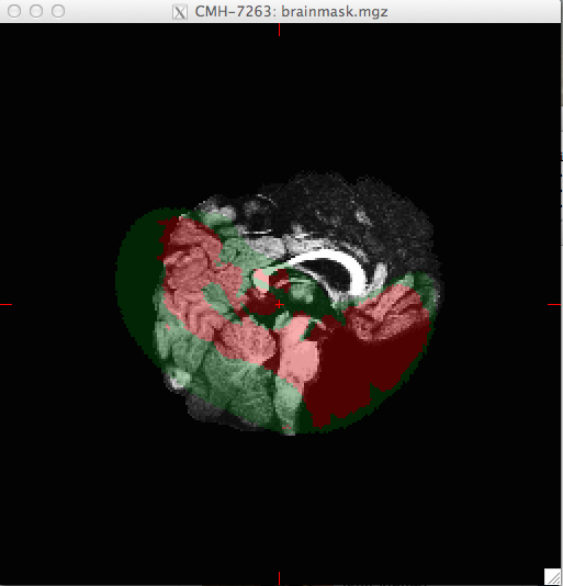
did you look at the registration with tkregister2? When bbregister finished, it would have printed out the command line to run (it is also at the end of the log file). If that looks ok, please send your bbregister command. doug
On 8/5/13 1:47 PM, Mark Plantz wrote:
Thanks Doug. mri_label2vol worked well, except it looks like all of the files are inverted by 90 degrees. They appear inverted in both tkmedit and 'FreeView.' I read that inversions are common in "fslview" but not in tkmedit. Any idea what could be causing this problem?
My command line is:
*mri_label2vol --seg /Volumes/Wong_Lab/CochlearImplant/UNC_NiFTI/infant-1yr-mgz/seg.mgz --temp /Users/IngvalsonLab/Desktop/temp.mgz --o seg_new.mgz --reg register.dat*
I didn't encounter any errors with mri_label2vol nor bbregister [which was used to generate register.dat and temp.mgz].
Any ideas what could be causing this problem?
I can attach more images if this one doesn't portray the inversion well.
Thanks a lot
MP
On Mon, Aug 5, 2013 at 10:12 AM, Douglas Greve <greve@nmr.mgh.harvard.edu mailto:greve@nmr.mgh.harvard.edu> wrote:
The file that you pass it with the --reg option is the registration file (eg, --reg register.dat). This can be used for input to mri_vol2vol or mri_label2vol. mri_label2vol is appropriate for a segmetation doug On 8/5/13 10:22 AM, Mark Plantz wrote:Thank you. Do you know how to access the transform that is outputted by bbregister. I am just obtaining an output volume <volume.mgz>>? On Thu, Aug 1, 2013 at 3:56 PM, Bruce Fischl <fischl@nmr.mgh.harvard.edu <mailto:fischl@nmr.mgh.harvard.edu>> wrote: Hi Mark you can use the transform that bbregister outputs and give it to mri_convert with -at <transform name> cheers Bruce On Thu, 1 Aug 2013, Mark Plantz wrote: One last question: Is there any flag I can use with 'bbregister' that prevents the labels from changing in my segmentation volume? This is occuring because the volume that I am moving has labels. For some reason, the labels change ever so slightly in the output file. On Thu, Aug 1, 2013 at 2:28 PM, Bruce Fischl <fischl@nmr.mgh.harvard.edu <mailto:fischl@nmr.mgh.harvard.edu>> wrote: glad it worked out Bruce On Thu, 1 Aug 2013, Mark Plantz wrote: Just as an update, it turns out that my segmentation volume had been slightly altered during the conversion from .nii to .mgz. I completely forgot to use the -rt nearest flag to prevent the labels from becoming distorted. That is why the segmentation looked so fuzzy with various regions labeled incorrectly. Apparently, I should check the basics before overcomplicating the problem, haha. Thanks for the help everyone! On Thu, Aug 1, 2013 at 9:36 AM, Mark Plantz <markplantz2016@u.northwestern.edu <mailto:markplantz2016@u.northwestern.edu>> wrote: Sounds good. I also just realized that the segmentation values are varying from 0-255, which makes me think that the file is only designed for the grayscale/heat/etc. settings [where 0 is darkest regions and 255 is brightest regions]. Is that a valid assumption? Also, if this is the case, would it be impossible to create a .gca file using mri_ca_train with these files? Thanks for the help. MP On Wed, Jul 31, 2013 at 8:43 PM, Douglas Greve <greve@nmr.mgh.harvard.edu <mailto:greve@nmr.mgh.harvard.edu>> wrote: Just make sure that you create a new file and do not repeat index numbers. On 7/31/13 2:02 PM, Mark Plantz wrote: That makes sense. Wouldn't that cause there to be some overlap between different regions, since the same index #'s used by the two programs (FreeSurfer & MRIcro) correspond to different brain regions? On Tue, Jul 30, 2013 at 12:50 PM, Douglas Greve <greve@nmr.mgh.harvard.edu <mailto:greve@nmr.mgh.harvard.edu>> wrote: for every line in that file, you will need to create a line in the new LUT, something like 8 Frontal_Mid_R 0 255 0 0 This will make the right mid frontal green = (0,255,0) On 7/30/13 9:55 AM, Mark Plantz wrote: Hmm, so I received the text part of the LUT. Looks like this is completely different from the segmented regions that we would want. [attached]. Can't seem to understand how this text file could be related to the default FreeSurfer LUT. It looks like the labeling for an AAL map. This probably wont' work will it? - Mark On Tue, Jul 30, 2013 at 8:47 AM, Bruce Fischl <fischl@nmr.mgh.harvard.edu <mailto:fischl@nmr.mgh.harvard.edu>> wrote: the UNC one? No, you should ask them On Tue, 30 Jul 2013, Mark Plantz wrote: ---------- Forwarded message ---------- From: Mark Plantz <markplantz2016@u.northwestern.edu <mailto:markplantz2016@u.northwestern.edu>> Date: Tue, Jul 30, 2013 at 8:39 AM Subject: Re: [Freesurfer] infant atlas segmentation To: Bruce Fischl <fischl@nmr.mgh.harvard.edu <mailto:fischl@nmr.mgh.harvard.edu>> Just opened up the default FreeSurferColorLUT.txt file. So it looks like all I have to change is the #No. column of the file, to match the ones in the other LUT used. I actually just received the MRIcro .lut file from the UNC group. [attached] Hopefully this will help with the conversion. However, I still have to figure out how to open the file. Any ideas? On Tue, Jul 30, 2013 at 8:25 AM, Bruce Fischl <fischl@nmr.mgh.harvard.edu <mailto:fischl@nmr.mgh.harvard.edu>> wrote: Hi Mark creating a FreeSurfer LUT really isn't that hard. Have you looked at our standard one? Probably we have the names you already want and you just need to change our indices to match the ones in your segmentation file (and save it as a different LUT) cheers Bruce On Tue, 30 Jul 2013, Mark Plantz wrote: Turns out that the files had been labeled using a color LUT in a program called MRIcro. I am now installing this program simply to access the LUT. Will it be as simple as exchanging the index values from this LUT to a custom LUT in FreeSurfer? On Tue, Jul 30, 2013 at 12:35 AM, Douglas Greve <greve@nmr.mgh.harvard.edu <mailto:greve@nmr.mgh.harvard.edu>> wrote: The problem is that you need to create a completely new LUT. The labels that get displayed with the standard LUT are going to be totally random with respect to what they should display. On 7/29/13 2:20 PM, Mark Plantz wrote: Hi Doug, Thanks for the reply. I will definitely go into the LUT and change those values to some specific region. My main concern is that I think all of these "out of bounds" labels are the symptoms of some larger problem. The segmentation looks very strange as compared to a typical segmentation (i.e. an 'aseg.mgz' file). In fact, the segmentation appears to be labeling "left-<region" in the right hemisphere. Have you seen a segmentation file that looks like the once I attached before? I am used to seeing segmentation files such as the one attached to this e-mail. - Mark On Mon, Jul 29, 2013 at 12:28 PM, Douglas Greve <greve@nmr.mgh.harvard.edu <mailto:greve@nmr.mgh.harvard.edu>> wrote: Mark, the only thing that is wrong with the out of bounds regions is that they are not represented in the LUT. Once you add an index corresponding to the index of the segmentation to the LUT, it will not show up as "out of bounds". The brain mask has nothing to do with it. best doug On 7/29/13 12:50 PM, Mark Plantz wrote: Sorry about that. I received the manual segmentations from aUNCmedicalgroup<http://www.med.unc.edu/bric/ideagroup/free-softwares/unc-in fan t-0-1-2 -atla ses>. I actually asked the main researcher about the out of bounds regions. He thinks that as long as the segmented file lines up with the input volumes, a few out of bounds regions might be OK. However, these seg volumes look very different from a typical .aseg volume that I am used to seeing after recon-all is run. Maybe the segmentation is simply incompatible with FreeSurfer? Thanks for the help. These files are a nightmare! - MP On Mon, Jul 29, 2013 at 11:40 AM, Bruce Fischl <fischl@nmr.mgh.harvard.edu <mailto:fischl@nmr.mgh.harvard.edu>> wrote: can you cc the list please? Maybe we should start over. Where did you get the manual segmentations from? On Mon, 29 Jul 2013, Mark Plantz wrote: Ahh, right. So it looks like the red regions appear to be averaged around 248. I am not sure why that is the case or if it has any significance. I attached a screen shot for reference. On Mon, Jul 29, 2013 at 11:29 AM, Bruce Fischl <fischl@nmr.mgh.harvard.edu <mailto:fischl@nmr.mgh.harvard.edu>> wrote: it isn't a registration file. You should just specify it as a volume on the freeview command line, the same as brainmask.mgz On Mon, 29 Jul 2013, Mark Plantz wrote: I actually tried to view the brainmask.mgz file with the segmentation file [Freeview calls it 'registration file' I believe]. For some reason, the file will load without segmentation, but when I attempt to add the registration file, I get an error message: "Failed to load MRI ~/.../../../brainmask.mgz Could this potentially be part of the problem? Thanks MP On Mon, Jul 29, 2013 at 10:57 AM, Mark Plantz <markplantz2016@u.northwestern.edu <mailto:markplantz2016@u.northwestern.edu>> wrote: It looks like the values vary from 20-120 depending on which region I run the cursor over. However, aren't those the values that correspond to the brain.mgz regions? Is there another way to find the segmentation values? On Mon, Jul 29, 2013 at 10:37 AM, Bruce Fischl <fischl@nmr.mgh.harvard.edu <mailto:fischl@nmr.mgh.harvard.edu>> wrote: I mean the segmentation values that give you the "out of bounds" message On Mon, 29 Jul 2013, Mark Plantz wrote: Hi Bruce, Do you mean the brainmask.mgz values? I'm not sure what the segmentation #'s exactly are. Thanks! MP On Mon, Jul 29, 2013 at 10:17 AM, Bruce Fischl <fischl@nmr.mgh.harvard.edu <mailto:fischl@nmr.mgh.harvard.edu>> wrote: Hi Mark what #s are those segmentations? It should be trivial to just add them to the end of the LUT. I wouldn't let them be out of bounds as there might be code around that might ignore them then. cheers Bruce On Mon, 29 Jul 2013, Mark Plantz wrote: Hi Doug, Thanks for the reply. After doing some research, it looks like messing with the LUT might be a risky decision [and way out of my abilities!]. If I were to use this segmentation file to create a .gca atlas using the command mri_ca_train, do you think it would yield problems or be inaccurate? I guess what I am trying to figure out is if the color lookup values are even significant when using the segmentation file to create an atlas? Thanks for all of the help with this. Best, MP p.s. I attached the photo of the segmentation volume because this e-mail is sort of old On Wed, Jul 24, 2013 at 2:39 PM, Douglas N Greve <greve@nmr.mgh.harvard.edu <mailto:greve@nmr.mgh.harvard.edu>> wrote: you need a new LUT. See my comment from a few emails ago. On 07/24/2013 03:29 PM, Mark Plantz wrote: I'm not actually quite sure what it means either. When I use the command line: tkmedit $Subject brain.mgz -segmentation <seg.mgz>, I can run the cursor over various regions and the name of the region should pop up. For some reason, the red regions are simply labeled as "out of bounds." I'll keep messing with tkmedit and let you know if I find out what the problem is. Thanks guys! MP O... [Message clipped] _______________________________________________ Freesurfer mailing list Freesurfer@nmr.mgh.harvard.edu <mailto:Freesurfer@nmr.mgh.harvard.edu> https://mail.nmr.mgh.harvard.edu/mailman/listinfo/freesurfer The information in this e-mail is intended only for the person to whom it is addressed. If you believe this e-mail was sent to you in error and the e-mail contains patient information, please contact the Partners Compliance HelpLine at http://www.partners.org/complianceline . If the e-mail was sent to you in error but does not contain patient information, please contact the sender and properly dispose of the e-mail.
freesurfer@nmr.mgh.harvard.edu
-
 Bruce Fischl
 -
 Douglas Greve
 -
 Mark Plantz