
16 Aug
2011
16 Aug
'11
3:05 p.m.
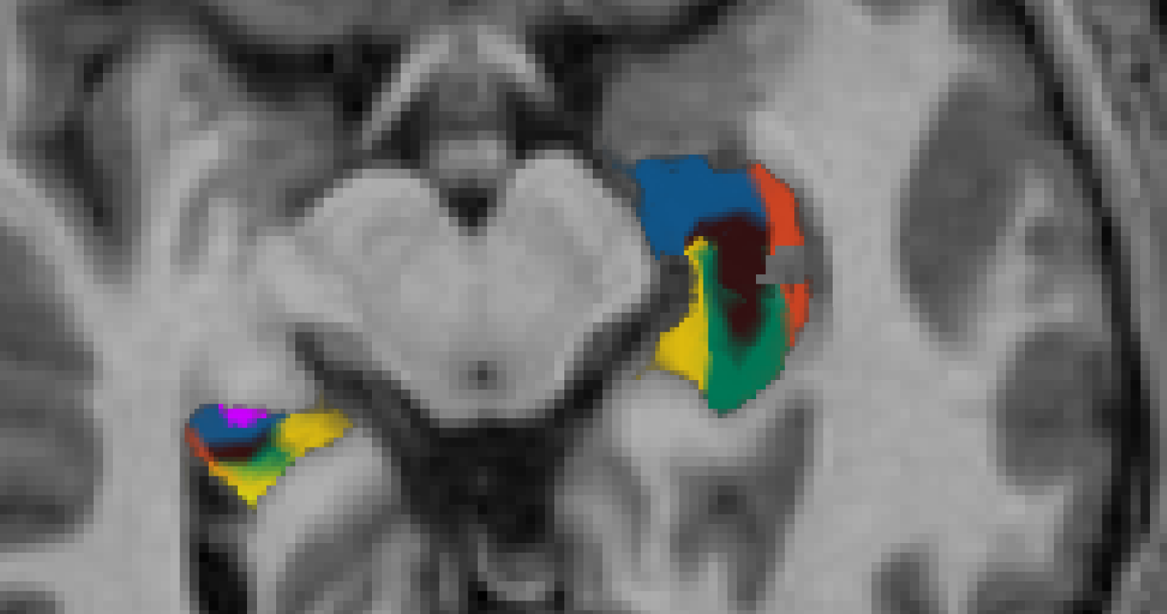
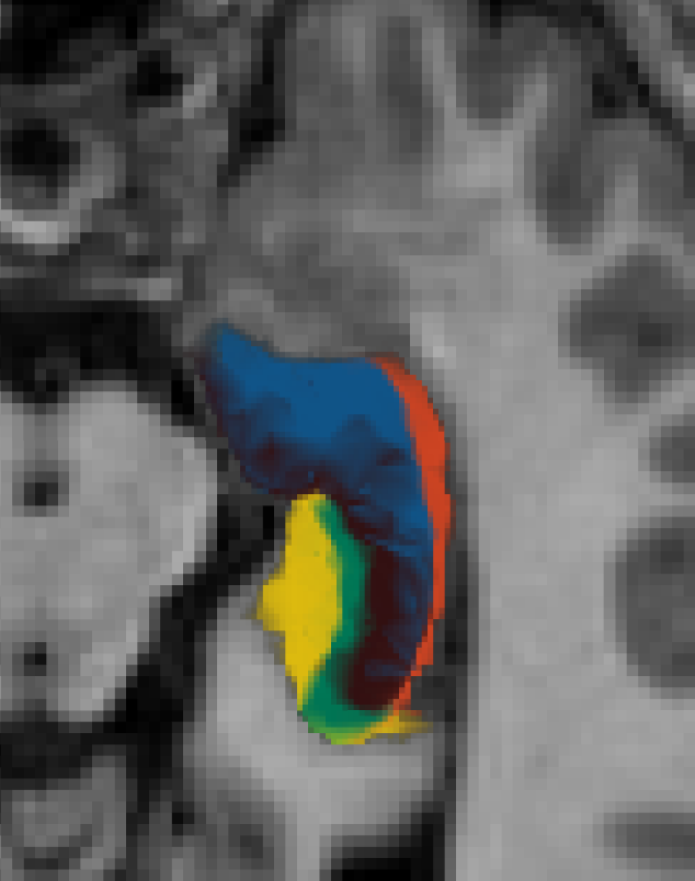
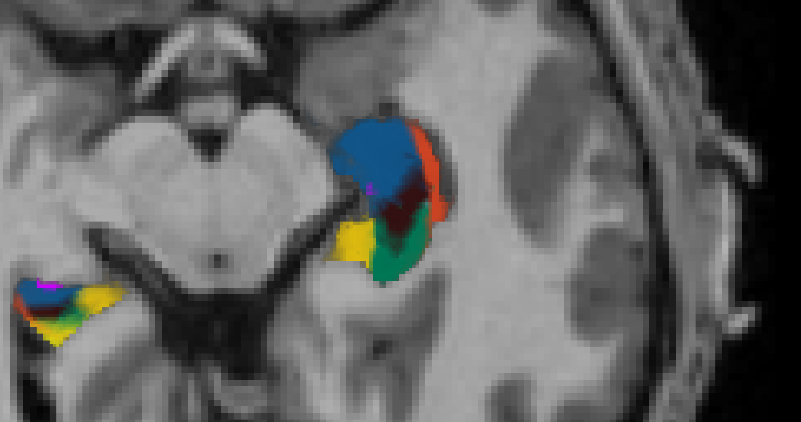
Hi, I've been inspecting the hippocampal subfield segmentation in my subjects. So far, the subfields segmentation appear to be consistent across my images.
1. Is there any criterion used in defining proper subfield segmentation (any landmarks I should look out during QC)?
2. What are the corrective measures when the subfield segmentation fails? I attach examples of 2 subjects that have two T1-weighted scans taken in tandem in the same session (of which one two images appears to have malfunctioned segmentation)
Thanks, Newfei
5454
Age (days ago)
5454
Last active (days ago)
freesurfer@nmr.mgh.harvard.edu
0 comments
1 participants
participants (1)
-
 New Fei Ho