
Hi All,
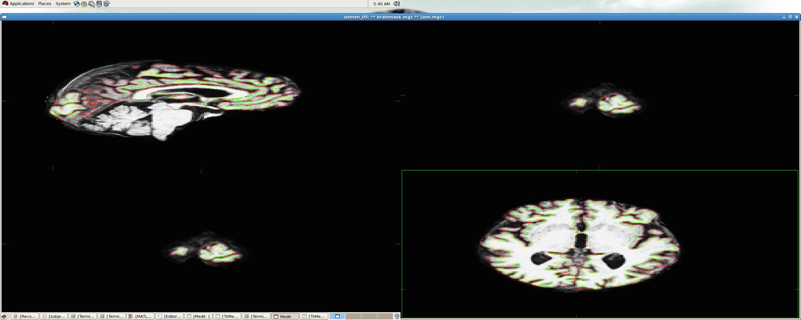
I am working in ALS dementia patients where there is ventricle enlargement and more CSF filling when compared with controls. Since i am new to freesurfer and my knowledge of anatomy is limited i would like to clarify the following. Pial surface in right and left cerebral cortex looks suspicious to me, the brain extraction step left out some non brain regions, i tried to edit them but since some of the non brain regions are close to brain regions i didn't do too much editing for the fear of removing brain regions. I have attached a screen shot image in one of our subjects, kindly tell me whether the pial surface looks ok if not any suggestions to correct this problem.
Thanks in advance
venkat
===================================
P Please consider the environment before printing this e-mail
Cleveland Clinic is ranked one of the top hospitals in America by U.S.News & World Report (2009). Visit us online at http://www.clevelandclinic.org for a complete listing of our services, staff and locations.
Confidentiality Note: This message is intended for use only by the individual or entity to which it is addressed and may contain information that is privileged, confidential, and exempt from disclosure under applicable law. If the reader of this message is not the intended recipient or the employee or agent responsible for delivering the message to the intended recipient, you are hereby notified that any dissemination, distribution or copying of this communication is strictly prohibited. If you have received this communication in error, please contact the sender immediately and destroy the material in its entirety, whether electronic or hard copy. Thank you.

Hi Venkat,
the image looks ok, but it's hard to tell from a single slice or two and not being able to change the window levels. If you want to ftp a subject to us we're happy to take a look and let you know what we think.
cheers, Bruce On Fri, 12 Mar 2010, Rajagopalan, Venkateswaran wrote:
Hi All,
I am working in ALS dementia patients where there is ventricle enlargement and more CSF filling when compared with controls. Since i am new to freesurfer and my knowledge of anatomy is limited i would like to clarify the following. Pial surface in right and left cerebral cortex looks suspicious to me, the brain extraction step left out some non brain regions, i tried to edit them but since some of the non brain regions are close to brain regions i didn't do too much editing for the fear of removing brain regions. I have attached a screen shot image in one of our subjects, kindly tell me whether the pial surface looks ok if not any suggestions to correct this problem.
Thanks in advance
venkat
===================================
P Please consider the environment before printing this e-mail
Cleveland Clinic is ranked one of the top hospitals in America by U.S.News & World Report (2009). Visit us online at http://www.clevelandclinic.org for a complete listing of our services, staff and locations.
Confidentiality Note: This message is intended for use only by the individual or entity to which it is addressed and may contain information that is privileged, confidential, and exempt from disclosure under applicable law. If the reader of this message is not the intended recipient or the employee or agent responsible for delivering the message to the intended recipient, you are hereby notified that any dissemination, distribution or copying of this communication is strictly prohibited. If you have received this communication in error, please contact the sender immediately and destroy the material in its entirety, whether electronic or hard copy. Thank you.
Thanks Bruce, i don't know much about ftps, do you have a ftp address (FSL they give us some ftp address which you can use to send your images) which you can send it to me and i can upload my images. I am very sorry.
Thanks a lot
venkat
________________________________
From: freesurfer-bounces@nmr.mgh.harvard.edu on behalf of Bruce Fischl Sent: Sat 3/13/2010 12:35 PM To: Rajagopalan, Venkateswaran Cc: freesurfer@nmr.mgh.harvard.edu Subject: Re: [Freesurfer] Pial surface- clarification
Hi Venkat,
the image looks ok, but it's hard to tell from a single slice or two and not being able to change the window levels. If you want to ftp a subject to us we're happy to take a look and let you know what we think.
cheers, Bruce On Fri, 12 Mar 2010, Rajagopalan, Venkateswaran wrote:
Hi All,
I am working in ALS dementia patients where there is ventricle enlargement and more CSF filling when compared with controls. Since i am new to freesurfer and my knowledge of anatomy is limited i would like to clarify the following. Pial surface in right and left cerebral cortex looks suspicious to me, the brain extraction step left out some non brain regions, i tried to edit them but since some of the non brain regions are close to brain regions i didn't do too much editing for the fear of removing brain regions. I have attached a screen shot image in one of our subjects, kindly tell me whether the pial surface looks ok if not any suggestions to correct this problem.
Thanks in advance
venkat
===================================
P Please consider the environment before printing this e-mail
Cleveland Clinic is ranked one of the top hospitals in America by U.S.News & World Report (2009). Visit us online at http://www.clevelandclinic.org http://www.clevelandclinic.org/ for a complete listing of our services, staff and locations.
Confidentiality Note: This message is intended for use only by the individual or entity to which it is addressed and may contain information that is privileged, confidential, and exempt from disclosure under applicable law. If the reader of this message is not the intended recipient or the employee or agent responsible for delivering the message to the intended recipient, you are hereby notified that any dissemination, distribution or copying of this communication is strictly prohibited. If you have received this communication in error, please contact the sender immediately and destroy the material in its entirety, whether electronic or hard copy. Thank you.
_______________________________________________ Freesurfer mailing list Freesurfer@nmr.mgh.harvard.edu https://mail.nmr.mgh.harvard.edu/mailman/listinfo/freesurfer
The information in this e-mail is intended only for the person to whom it is addressed. If you believe this e-mail was sent to you in error and the e-mail contains patient information, please contact the Partners Compliance HelpLine at http://www.partners.org/complianceline . If the e-mail was sent to you in error but does not contain patient information, please contact the sender and properly dispose of the e-mail.
===================================
P Please consider the environment before printing this e-mail
Cleveland Clinic is ranked one of the top hospitals in America by U.S.News & World Report (2009). Visit us online at http://www.clevelandclinic.org for a complete listing of our services, staff and locations.
Confidentiality Note: This message is intended for use only by the individual or entity to which it is addressed and may contain information that is privileged, confidential, and exempt from disclosure under applicable law. If the reader of this message is not the intended recipient or the employee or agent responsible for delivering the message to the intended recipient, you are hereby notified that any dissemination, distribution or copying of this communication is strictly prohibited. If you have received this communication in error, please contact the sender immediately and destroy the material in its entirety, whether electronic or hard copy. Thank you.
freesurfer@nmr.mgh.harvard.edu
-
 Bruce Fischl
 -
 Rajagopalan, Venkateswaran