
External Email - Use Caution
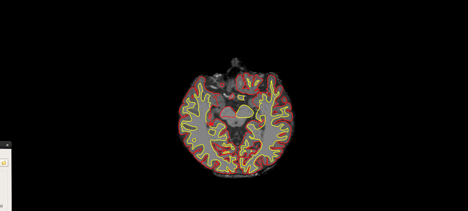
Dear experts, When I see the images through freeview to edit, upper brainstem and optic chiasm were always included in wm or brainmask.mgz, as an attached picture below. Left side seems to be recognized as white matter, and right side as cortex. My concern is that this could affect the final value of e-TIV after recon-all process, so I need to edit these areas manually. I'd appreciated your help. Thanks so much.
Kyung

External Email - Use Caution
Hi, This won't affect the eTIV as that is derived purely from the affine registration. See Buckner et al. (2004) NeuroImage 23:724-738. for more details.
-Niels
On Mon, Nov 26, 2018 at 6:46 AM 박경일 ideopki@gmail.com wrote:
External Email - Use CautionDear experts, When I see the images through freeview to edit, upper brainstem and optic chiasm were always included in wm or brainmask.mgz, as an attached picture below. Left side seems to be recognized as white matter, and right side as cortex. My concern is that this could affect the final value of e-TIV after recon-all process, so I need to edit these areas manually. I'd appreciated your help. Thanks so much.
Kyung _______________________________________________ Freesurfer mailing list Freesurfer@nmr.mgh.harvard.edu https://mail.nmr.mgh.harvard.edu/mailman/listinfo/freesurfer

Hi Kyung
eTIV is estimated from the determinant of the Talairach transform, so won't be affected much by bits of things like the chiasm being left around.
cheers Bruce
On Mon, 26 Nov 2018, 박경일 wrote:
External Email - Use Caution
Dear experts,When I see the images through freeview to edit, upper brainstem and optic chiasm were always included in wm or brainmask.mgz, as an attached picture below. Left side seems to be recognized as white matter, and right side as cortex. My concern is that this could affect the final value of e-TIV after recon-all process, so I need to edit these areas manually. I'd appreciated your help. Thanks so much.
Kyung
freesurfer@nmr.mgh.harvard.edu
-
 Bruce Fischl
 -
 Niels Bergsland
 -
 박경일
박경일