Fwd: Correnting image bias before segmentation and parcelation - due to frontal and occipital shading.

Dear Freesurfer Team,
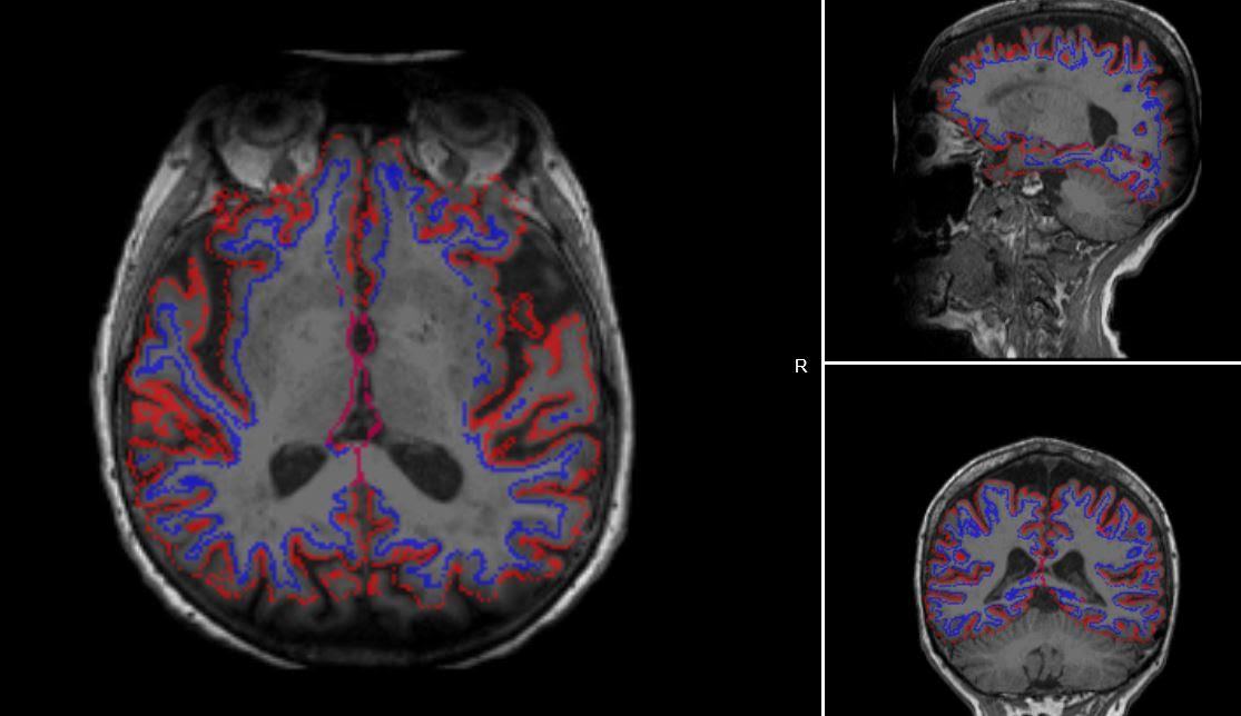
I have about 200 T1 coronal scans that were obtained with some shading in the frontal and occipital regions. Not sure if it can be seen from the attached picture, but the image fades toward the top and bottom (on the axial view). The segmentation left out a large section of the frontal and occipital, probably because the signal loss brings those voxels below the threshold for white and gray matter.
I have tried to run FSL bias correction and got a small improvement, but I wonder if there is any way these faded slides can be corrected so that the segmentation is capable of seeing these as proper white and gray matter.
In the attached picture, the blue is the wm surface and the red is the gray matter (wm and pial surfaces)
The image shown is actually the T1.mgz from freesufer that has already gone through the nu_correct step in recon_all, the original image looks very similar...
Thanks
Gabriel
On Sun, Mar 18, 2018 at 2:28 AM, Gamaliz gliza002@fiu.edu wrote:
Dear Freesurfer Team,
I have about 200 T1 coronal scans that were obtained with some shading in the frontal and occipital regions. Not sure if it can be seen from the attached picture, but the image fades toward the top and bottom (on the axial view). The segmentation left out a large section of the frontal and occipital, probably because the signal loss brings those voxels below the threshold for white and gray matter.
I have tried to run FSL bias correction and got a small improvement, but I wonder if there is any way these faded slides can be corrected so that the segmentation is capable of seeing these as proper white and gray matter.
In the attached picture, the blue is the wm surface and the red is the gray matter (wm and pial surfaces)
The image shown is actually the T1.mgz from freesufer that has already gone through the nu_correct step in recon_all, the original image looks very similar...
Thanks
Gabriel
-- gAbE

Hi Gabriel
you can try adding control points in the white matter in those regions, but typically when there is so much signal loss there is very little anatomical SNR left to build models with.
good luck Bruce On Sun, 18 Mar 2018, Gamaliz wrote:
[IMAGE]
On Sun, Mar 18, 2018 at 2:28 AM, Gamaliz gliza002@fiu.edu wrote:
Dear Freesurfer Team,
I have about 200 T1 coronal scans that were obtained with some shading in the frontal and occipital regions. Not sure if it can be seen from the attached picture, but the image fades toward the top and bottom (on the axial view). The segmentation left out a large section of the frontal and occipital, probably because the signal loss brings those voxels below the threshold for white and gray matter.
I have tried to run FSL bias correction and got a small improvement, but I wonder if there is any way these faded slides can be corrected so that the segmentation is capable of seeing these as proper white and gray matter.
In the attached picture, the blue is the wm surface and the red is the gray matter (wm and pial surfaces)
The image shown is actually the T1.mgz from freesufer that has already gone through the nu_correct step in recon_all, the original image looks very similar...
Thanks
Gabriel
-- gAbE
-- gAbE
Before I try that I will try increasing the intensity of the shaded voxels. I already tried running nu_correct, and does improve a little bit. The shading is a gradient (from darken into brighter, I will see if I can find the gradient in the out of brain pixels (like the dark areas of the image) and create a function that models it, and then apply it to the back and front sections of the scan. By brightening those pixels I might be able include the white and gray matter in the segmentation. Has anyone tried something like this before? I understand that there might be a bias, but if I get it to improve the segmentation and specially the ICV, it will be an improvement.
Gabriel
On Sun, Mar 18, 2018 at 3:19 PM, Bruce Fischl fischl@nmr.mgh.harvard.edu wrote:
Hi Gabriel
you can try adding control points in the white matter in those regions, but typically when there is so much signal loss there is very little anatomical SNR left to build models with.
good luck Bruce On Sun, 18 Mar 2018, Gamaliz wrote:
[IMAGE]
On Sun, Mar 18, 2018 at 2:28 AM, Gamaliz gliza002@fiu.edu wrote:
Dear Freesurfer Team,
I have about 200 T1 coronal scans that were obtained with some shading in the frontal and occipital regions. Not sure if it can be seen from the attached picture, but the image fades toward the top and bottom (on the axial view). The segmentation left out a large section of the frontal and occipital, probably because the signal loss brings those voxels below the threshold for white and gray matter.
I have tried to run FSL bias correction and got a small improvement, but I wonder if there is any way these faded slides can be corrected so that the segmentation is capable of seeing these as proper white and gray matter.
In the attached picture, the blue is the wm surface and the red is the gray matter (wm and pial surfaces)
The image shown is actually the T1.mgz from freesufer that has already gone through the nu_correct step in recon_all, the original image looks very similar...
Thanks
Gabriel
-- gAbE
-- gAbE
The information in this e-mail is intended only for the person to whom it is addressed. If you believe this e-mail was sent to you in error and the e-mail contains patient information, please contact the Partners Compliance HelpLine at http://www.partners.org/complianceline . If the e-mail was sent to you in error but does not contain patient information, please contact the sender and properly dispose of the e-mail.
Hi Gabriel
if you put control points in the wm of the darker regions it will do exactly that - increase the intensity of those regions
cheers Bruce On Wed, 21 Mar 2018, Gamaliz wrote:
Before I try that I will try increasing the intensity of the shaded voxels. I already tried running nu_correct, and does improve a little bit. The shading is a gradient (from darken into brighter, I will see if I can find the gradient in the out of brain pixels (like the dark areas of the image) and create a function that models it, and then apply it to the back and front sections of the scan. By brightening those pixels I might be able include the white and gray matter in the segmentation. Has anyone tried something like this before? I understand that there might be a bias, but if I get it to improve the segmentation and specially the ICV, it will be an improvement.
Gabriel
On Sun, Mar 18, 2018 at 3:19 PM, Bruce Fischl fischl@nmr.mgh.harvard.edu wrote: Hi Gabriel
  you can try adding control points in the white matter in those regions, but typically
  when there is so much signal loss there is very little anatomical SNR left to build
  models with.

  good luck
  Bruce
  On Sun, 18 Mar 2018, Gamaliz wrote:

        [IMAGE]


        On Sun, Mar 18, 2018 at 2:28 AM, Gamaliz <gliza002@fiu.edu> wrote:

        Dear Freesurfer Team,

        I have about 200 T1 coronal scans that were obtained with some shading in
        the frontal and
        occipital regions. Not sure if it can be seen from the attached picture, but
        the image fades
        toward the top and bottom (on the axial view). The segmentation left out a
        large section of
        the frontal and occipital, probably because the signal loss brings those
        voxels below the
        threshold for white and gray matter.

        I have tried to run FSL bias correction and got a small improvement, but I
        wonder if there is
        any way these faded slides can be corrected so that the segmentation is
        capable of seeing
        these as proper white and gray matter.

        In the attached picture, the blue is the wm surface and the red is the gray
        matter (wm and
        pial surfaces)

        The image shown is actually the T1.mgz from freesufer that has already gone
        through the
        nu_correct step in recon_all, the original image looks very similar...

        Thanks

        Gabriel

        --
        gAbE


        --
        gAbE


  The information in this e-mail is intended only for the person to whom it is
  addressed. If you believe this e-mail was sent to you in error and the e-mail
  contains patient information, please contact the Partners Compliance HelpLine at
  http://www.partners.org/complianceline . If the e-mail was sent to you in error
  but does not contain patient information, please contact the sender and properly
  dispose of the e-mail.
-- gAbE
freesurfer@nmr.mgh.harvard.edu
-
 Bruce Fischl
 -
 Gamaliz