
Hello All,
We have processed retinotopy data and piped the phase maps into freesurfer stream. As can be seen on the attached reconstructed image, there are patches of cortex (some reds) within the calcarine sulcus that are wrongly assigned to the upper bank. Any ideas on how to correct for these mis-identified patches in FreeSurfer ? I have already checked using tkregister the registration of the functional data to the freesurfer anatomy and that looks fine.
Please let me know
Thanks
Ri

This is the polar angle map? What does it look like inflated? Do you acquire data with the wedge going in both directions? Even if you do, there can be some delay errors that creep in.
doug
Ritobrato Datta wrote:
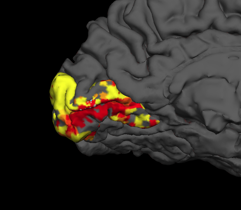
Hello All,
We have processed retinotopy data and piped the phase maps into freesurfer stream. As can be seen on the attached reconstructed image, there are patches of cortex (some reds) within the calcarine sulcus that are wrongly assigned to the upper bank. Any ideas on how to correct for these mis-identified patches in FreeSurfer ? I have already checked using tkregister the registration of the functional data to the freesurfer anatomy and that looks fine.
Please let me know
Thanks
Ri
Freesurfer mailing list Freesurfer@nmr.mgh.harvard.edu https://mail.nmr.mgh.harvard.edu/mailman/listinfo/freesurfer
freesurfer@nmr.mgh.harvard.edu
-
 Douglas N Greve
 -
 Ritobrato Datta