
External Email - Use Caution
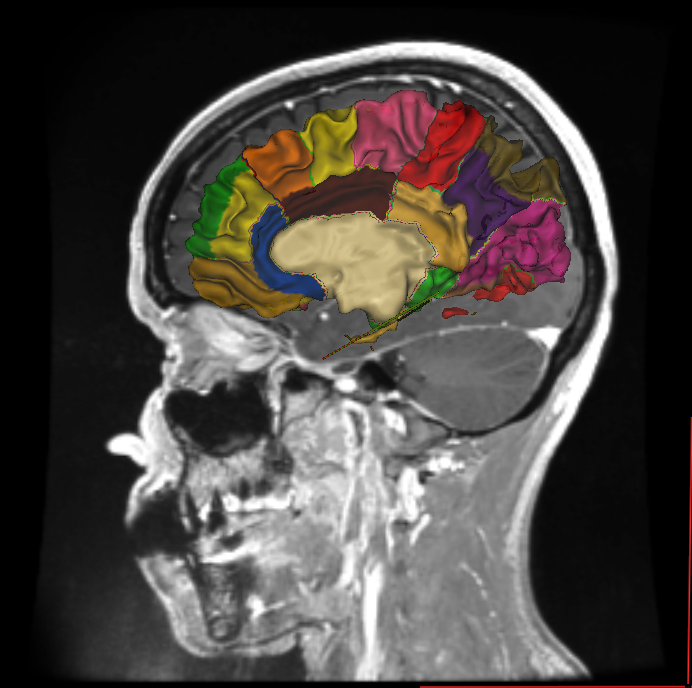
Dear all, Some of my images have been taken in patients with gadolinium, so some regions are highlighted because of the contrast effect. The problem is that, in many cases, this produces a bad parcelation of the brain. For instance, in the example you will find attached, the insula is smaller than it should be (parcels blue, dark read and yellow must be included in the insula).
Is there any way to avoid this effect or to correct these high intensities without using manually correction?
Thanks in advance, Maria.

Hi Maria
yes, potentially. What acqusitions do you have? Do you have a highres T2? You can erase the bright dura that is causing this effect manually if not
cheers Bruce On Wed, 29 May 2019, María Martínez Rodrigo wrote:
External Email - Use Caution
Dear all, Some of my images have been taken in patients with gadolinium, so some regions are highlighted because of the contrast effect. The problem is that, in many cases, this produces a bad parcelation of the brain. For instance, in the example you will find attached, the insula is smaller than it should be (parcels blue, dark read and yellow must be included in the insula).
Is there any way to avoid this effect or to correct these high intensities without using manually correction?
Thanks in advance, Maria.
External Email - Use Caution
Thanks for your answer. For the most of the patients I just have T1 images. Isn't there any alternative to manually correction for this cases?
Best regards, Maria
El mié., 29 may. 2019 a las 15:39, Bruce Fischl (fischl@nmr.mgh.harvard.edu) escribió:
Hi Maria
yes, potentially. What acqusitions do you have? Do you have a highres T2? You can erase the bright dura that is causing this effect manually if not
cheers Bruce On Wed, 29 May 2019, María Martínez Rodrigo wrote:
External Email - Use CautionDear all, Some of my images have been taken in patients with gadolinium, so some
regions are highlighted
because of the contrast effect. The problem is that, in many cases, this
produces a bad parcelation
of the brain. For instance, in the example you will find attached, the
insula is smaller than it
should be (parcels blue, dark read and yellow must be included in the
insula).
Is there any way to avoid this effect or to correct these high
intensities without using manually
correction?
Thanks in advance, Maria.
Freesurfer mailing list Freesurfer@nmr.mgh.harvard.edu https://mail.nmr.mgh.harvard.edu/mailman/listinfo/freesurfer
you can try the graph cuts skull strip to see if it removes more, or play with the watershed parameters cheers Bruce On Wed, 29 May 2019, María Martínez Rodrigo wrote:
External Email - Use Caution
Thanks for your answer. For the most of the patients I just have T1 images. Isn't there any alternative to manually correction for this cases?
Best regards, Maria
El mié., 29 may. 2019 a las 15:39, Bruce Fischl (fischl@nmr.mgh.harvard.edu) escribió: Hi Maria
  yes, potentially. What acqusitions do you have? Do you have a
  highres T2?
  You can erase the bright dura that is causing this effect
  manually if not

  cheers
  Bruce
  On
  Wed, 29 May 2019, María Martínez Rodrigo wrote:

  >
  >         External Email - Use Caution
  >
  > Dear all,
  > Some of my images have been taken in patients with gadolinium,
  so some regions are highlighted
  > because of the contrast effect. The problem is that, in many
  cases, this produces a bad parcelation
  > of the brain. For instance, in the example you will find
  attached, the insula is smaller than it
  > should be (parcels blue, dark read and yellow must be included
  in the insula).
  >
  > Is there any way to avoid this effect or to correct these high
  intensities without using manually
  > correction?
  >
  > Thanks in advance,
  > Maria.
  >
  >_______________________________________________
  Freesurfer mailing list
  Freesurfer@nmr.mgh.harvard.edu
  https://mail.nmr.mgh.harvard.edu/mailman/listinfo/freesurfer
freesurfer@nmr.mgh.harvard.edu
-
 Bruce Fischl
 -
 María Martínez Rodrigo