

Can you send us the recon-all.log?
On Sep 3, 2009, at 4:29 AM, "Zheng Hui" zheng.hui@duke-nus.edu.sg wrote:
Dear Bruce,
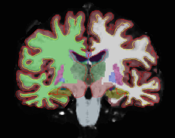
In the attached image at the ventricle area, you can see the pial and white surfaces go into the ventricle, following the white/CSF boundary rather than going through the medial separation. And by looking at the wm.mgz, I can see the ventricles were not filled by the program although the ventricles were labelled correctly in aseg.mgz. Hope I describe the problem clearer this time. I have attached the image again.
Thanks, Zheng Hui
Hi Zheng,
sorry, I don't see what you mean in the image. If you want to upload a dataset that has this problem I'll take a look.
cheers, Bruce On Wed, 2 Sep 2009, Hui Zheng wrote:
Hi freesurfer,
We are reprocessing our data from FS version 3.05 with the latest
4.50. We
found there are about 20% of the subjects have problem at the
ventricle as
shown in the attached image. I have tried the method suggested by
Doug to
remove the wm.mgz and rerun from autorecon2-cp. However, for two
subjects I
have tried, the method did not help. It will be tedious to
manually fill in
all the ventricles in wm.mgz. Any other work around?
Thanks, Zheng Hui
<aseg.jpg> _______________________________________________ Freesurfer mailing list Freesurfer@nmr.mgh.harvard.edu https://mail.nmr.mgh.harvard.edu/mailman/listinfo/freesurfer

HI Bruce, Zheng Hui and all.
I just wanted to add that we have also seen the same issues in our data that was been updated from 3.5. However, I noticed this issue started to occur around version 4.20 with another data set of ours.
Many of us had assumed that this was the new way Freesurfer segments the area around the medial wall. Is this the wrong assumption?
Thanks,
Judith
On Thu, Sep 3, 2009 at 7:41 PM, Zheng Hui zheng.hui@duke-nus.edu.sg wrote:
Hi Bruce,
I have attached the recon-all.log file for one of the subjects. By comparing the log file with other successful subjects, I can see some differences at the mri_edit_with_aseg:
Failed subject: mri_edit_wm_with_aseg wm.seg.mgz brain.mgz aseg.mgz wm.asegedit.mgz
MRIboundingBox: unsupported type 1 reading wm segmentation from wm.seg.mgz... MRIboundingBox: unsupported type 1 auto filling took 1.68 minutes 0 voxels added to wm to prevent paths from MTL structures to cortex 0 additional wm voxels added SEG EDIT: 0 voxels turned on, 1215 voxels turned off. writing edited volume to wm.asegedit.mgz....
Succeeded subject: mri_edit_wm_with_aseg -keep-in wm.seg.mgz brain.mgz aseg.mgz wm.asegedit.mgz
preserving editing changes in input volume... auto filling took 1.03 minutes reading wm segmentation from wm.seg.mgz... 515 voxels added to wm to prevent paths from MTL structures to cortex 2705 additional wm voxels added 0 additional wm voxels added SEG EDIT: 69225 voxels turned on, 29249 voxels turned off. propagating editing to output volume from wm.seg.mgz 115,126,128 old 109 new 109 115,126,128 old 109 new 109 writing edited volume to wm.asegedit.mgz....
Zheng Hui
-----Original Message----- From: Bruce Fischl [mailto:fischl@nmr.mgh.harvard.edufischl@nmr.mgh.harvard.edu ] Sent: Thu 9/3/2009 11:25 PM To: Zheng Hui Cc: Freesurfer@nmr.mgh.harvard.edu Subject: Re: [Freesurfer] Wrong boundaries at ventricle.
Can you send us the recon-all.log?
On Sep 3, 2009, at 4:29 AM, "Zheng Hui" zheng.hui@duke-nus.edu.sg wrote:
Dear Bruce,
In the attached image at the ventricle area, you can see the pial and white surfaces go into the ventricle, following the white/CSF boundary rather than going through the medial separation. And by looking at the wm.mgz, I can see the ventricles were not filled by the program although the ventricles were labelled correctly in aseg.mgz. Hope I describe the problem clearer this time. I have attached the image again.
Thanks, Zheng Hui
Hi Zheng,
sorry, I don't see what you mean in the image. If you want to upload a dataset that has this problem I'll take a look.
cheers, Bruce On Wed, 2 Sep 2009, Hui Zheng wrote:
Hi freesurfer,
We are reprocessing our data from FS version 3.05 with the latest
4.50. We
found there are about 20% of the subjects have problem at the
ventricle as
shown in the attached image. I have tried the method suggested by
Doug to
remove the wm.mgz and rerun from autorecon2-cp. However, for two
subjects I
have tried, the method did not help. It will be tedious to
manually fill in
all the ventricles in wm.mgz. Any other work around?
Thanks, Zheng Hui
<aseg.jpg> _______________________________________________ Freesurfer mailing list Freesurfer@nmr.mgh.harvard.edu https://mail.nmr.mgh.harvard.edu/mailman/listinfo/freesurfer
Freesurfer mailing list Freesurfer@nmr.mgh.harvard.edu https://mail.nmr.mgh.harvard.edu/mailman/listinfo/freesurfer
thanks Judith,
this is a bug that should be pretty easy to fix if someone can send us a sample set that it occurs on.
cheers, Bruce
On Fri, 4 Sep 2009, Judith Segall wrote:
HI Bruce, Zheng Hui and all.
I just wanted to add that we have also seen the same issues in our data that was been updated from 3.5. However, I noticed this issue started to occur around version 4.20 with another data set of ours.
Many of us had assumed that this was the new way Freesurfer segments the area around the medial wall. Is this the wrong assumption?
Thanks,
Judith
On Thu, Sep 3, 2009 at 7:41 PM, Zheng Hui zheng.hui@duke-nus.edu.sg wrote:
Hi Bruce,
I have attached the recon-all.log file for one of the subjects. By comparing the log file with other successful subjects, I can see some differences at the mri_edit_with_aseg:
Failed subject: mri_edit_wm_with_aseg wm.seg.mgz brain.mgz aseg.mgz wm.asegedit.mgz
MRIboundingBox: unsupported type 1 reading wm segmentation from wm.seg.mgz... MRIboundingBox: unsupported type 1 auto filling took 1.68 minutes 0 voxels added to wm to prevent paths from MTL structures to cortex 0 additional wm voxels added SEG EDIT: 0 voxels turned on, 1215 voxels turned off. writing edited volume to wm.asegedit.mgz....
Succeeded subject: mri_edit_wm_with_aseg -keep-in wm.seg.mgz brain.mgz aseg.mgz wm.asegedit.mgz
preserving editing changes in input volume... auto filling took 1.03 minutes reading wm segmentation from wm.seg.mgz... 515 voxels added to wm to prevent paths from MTL structures to cortex 2705 additional wm voxels added 0 additional wm voxels added SEG EDIT: 69225 voxels turned on, 29249 voxels turned off. propagating editing to output volume from wm.seg.mgz 115,126,128 old 109 new 109 115,126,128 old 109 new 109 writing edited volume to wm.asegedit.mgz....
Zheng Hui
-----Original Message----- From: Bruce Fischl [mailto:fischl@nmr.mgh.harvard.edufischl@nmr.mgh.harvard.edu ] Sent: Thu 9/3/2009 11:25 PM To: Zheng Hui Cc: Freesurfer@nmr.mgh.harvard.edu Subject: Re: [Freesurfer] Wrong boundaries at ventricle.
Can you send us the recon-all.log?
On Sep 3, 2009, at 4:29 AM, "Zheng Hui" zheng.hui@duke-nus.edu.sg wrote:
Dear Bruce,
In the attached image at the ventricle area, you can see the pial and white surfaces go into the ventricle, following the white/CSF boundary rather than going through the medial separation. And by looking at the wm.mgz, I can see the ventricles were not filled by the program although the ventricles were labelled correctly in aseg.mgz. Hope I describe the problem clearer this time. I have attached the image again.
Thanks, Zheng Hui
Hi Zheng,
sorry, I don't see what you mean in the image. If you want to upload a dataset that has this problem I'll take a look.
cheers, Bruce On Wed, 2 Sep 2009, Hui Zheng wrote:
Hi freesurfer,
We are reprocessing our data from FS version 3.05 with the latest
4.50. We
found there are about 20% of the subjects have problem at the
ventricle as
shown in the attached image. I have tried the method suggested by
Doug to
remove the wm.mgz and rerun from autorecon2-cp. However, for two
subjects I
have tried, the method did not help. It will be tedious to
manually fill in
all the ventricles in wm.mgz. Any other work around?
Thanks, Zheng Hui
<aseg.jpg> _______________________________________________ Freesurfer mailing list Freesurfer@nmr.mgh.harvard.edu https://mail.nmr.mgh.harvard.edu/mailman/listinfo/freesurfer
Freesurfer mailing list Freesurfer@nmr.mgh.harvard.edu https://mail.nmr.mgh.harvard.edu/mailman/listinfo/freesurfer

Zheng,
Can you send me the following files to our file drop? wm.mgz, wm.seg.mgz, brain.mgz and aseg.mgz
The file drop is https://www.nmr.mgh.harvard.edu/facility/filedrop/index.html
Also, were edits made to wm.mgz? I see from the log that it is being treated as if there were (although I need to check if they were handled properly in the script).
Nick
On Fri, 2009-09-04 at 09:41 +0800, Zheng Hui wrote:
Hi Bruce,
I have attached the recon-all.log file for one of the subjects. By comparing the log file with other successful subjects, I can see some differences at the mri_edit_with_aseg:
Failed subject: mri_edit_wm_with_aseg wm.seg.mgz brain.mgz aseg.mgz wm.asegedit.mgz
MRIboundingBox: unsupported type 1 reading wm segmentation from wm.seg.mgz... MRIboundingBox: unsupported type 1 auto filling took 1.68 minutes 0 voxels added to wm to prevent paths from MTL structures to cortex 0 additional wm voxels added SEG EDIT: 0 voxels turned on, 1215 voxels turned off. writing edited volume to wm.asegedit.mgz....
Succeeded subject: mri_edit_wm_with_aseg -keep-in wm.seg.mgz brain.mgz aseg.mgz wm.asegedit.mgz
preserving editing changes in input volume... auto filling took 1.03 minutes reading wm segmentation from wm.seg.mgz... 515 voxels added to wm to prevent paths from MTL structures to cortex 2705 additional wm voxels added 0 additional wm voxels added SEG EDIT: 69225 voxels turned on, 29249 voxels turned off. propagating editing to output volume from wm.seg.mgz 115,126,128 old 109 new 109 115,126,128 old 109 new 109 writing edited volume to wm.asegedit.mgz....
Zheng Hui
-----Original Message----- From: Bruce Fischl [mailto:fischl@nmr.mgh.harvard.edu] Sent: Thu 9/3/2009 11:25 PM To: Zheng Hui Cc: Freesurfer@nmr.mgh.harvard.edu Subject: Re: [Freesurfer] Wrong boundaries at ventricle.
Can you send us the recon-all.log?
On Sep 3, 2009, at 4:29 AM, "Zheng Hui" zheng.hui@duke-nus.edu.sg wrote:
Dear Bruce,
In the attached image at the ventricle area, you can see the pial and white surfaces go into the ventricle, following the white/CSF boundary rather than going through the medial separation. And by looking at the wm.mgz, I can see the ventricles were not filled by the program although the ventricles were labelled correctly in aseg.mgz. Hope I describe the problem clearer this time. I have attached the image again.
Thanks, Zheng Hui
Hi Zheng,
sorry, I don't see what you mean in the image. If you want to upload
a
dataset that has this problem I'll take a look.
cheers, Bruce On Wed, 2 Sep 2009, Hui Zheng wrote:
Hi freesurfer,
We are reprocessing our data from FS version 3.05 with the latest
4.50. We
found there are about 20% of the subjects have problem at the
ventricle as
shown in the attached image. I have tried the method suggested by
Doug to
remove the wm.mgz and rerun from autorecon2-cp. However, for two
subjects I
have tried, the method did not help. It will be tedious to
manually fill in
all the ventricles in wm.mgz. Any other work around?
Thanks, Zheng Hui
<aseg.jpg> _______________________________________________ Freesurfer mailing list Freesurfer@nmr.mgh.harvard.edu https://mail.nmr.mgh.harvard.edu/mailman/listinfo/freesurfer
Freesurfer mailing list Freesurfer@nmr.mgh.harvard.edu https://mail.nmr.mgh.harvard.edu/mailman/listinfo/freesurfer
freesurfer@nmr.mgh.harvard.edu
-
 Bruce Fischl
 -
 Judith Segall
 -
 Nick Schmansky
 -
 Zheng Hui