
13 Jan
2009
13 Jan
'09
4:54 a.m.
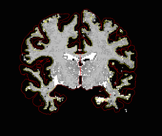
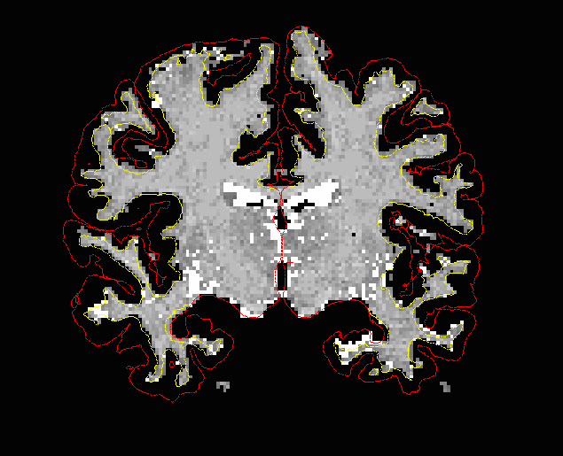
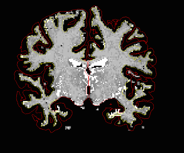
Hi all, I have a question relating to some quite poor data I have been analyzing. It seems that not all of the gray matter is included in the segmentation, especially in the lower part of the left temporal lobes (see attached image). It looks like the white matter has been correctly segmented in the area (i have also included 3 wm-images from the slices around the problematic area). I am not sure however, whether the tissue seen outside the pial surface is in fact gray matter. I have had some problems during scull stripping on these subjects. I would appreciate any opinions and tips on this.
Regards, Martin Ystad
6392
Age (days ago)
6392
Last active (days ago)
freesurfer@nmr.mgh.harvard.edu
0 comments
1 participants
participants (1)
-
 Martin Ystad