
I was wondering how the calculations of the wm volume were performed as well. The six values below are volume values from aseg I believe. I assume mris_wm_volume does more than sub-divide these regions into the wm parcellations, but probably also includes partial volume estimations along the wm/gm surface, is this correct? I basically have the same question for the gm volume values in aparc.stats; do they include partial volume calculations from the gm/pial and gm/white borders? Which binary is used to create these values for gm in aparc.stats... is it mri_segstats (with --pv)? I was assuming it would be an mris (surface) calculation as well. I am asking because if you use tkmedit to view the ?h.surface files and load the aparc+aseg or wmparc segmentation volumes in also, the surfaces and the segmentations don't seem to line up. Is there a way to create a segmentation volume that includes partial-volumed voxels at the gray/white and gray/pial boundries? Thanks for your help.
Chris Bell
the interior voxels are only included in the volume if they are one of the following in the aseg.mgz:
case Left_Cerebral_Cortex: case Right_Cerebral_Cortex: case Left_Cerebral_White_Matter: case Right_Cerebral_White_Matter: case Left_WM_hypointensities: case Right_WM_hypointensities:
cheers, Bruce
On Tue, 15 Jul 2008, Marin Richardson wrote:
OK, so aparc.stats is better for cortical gray matter. I want to look at the parcellated white matter volumes as well - is the only/best option to use the wm volumes from wmparc.stats?
Also, when using mris_wm_volume to calculate total white matter, I saw the result defined as the volume within the white matter surface minus non-WM structures? Can you tell me which regions are and are not included?
Marin
Doug Greve <[EMAIL PROTECTED]> 7/15/2008 12:02 PM >>>
It is better to use the aparc as it is derived from surface measurements, which have sub-voxel resolution. The wmparc.stats are generated by counting 1mm^3 voxels.
doug
Marin Richardson wrote:
  Hi all,

   I noticed that the gray volumes for parcellated cortical regions in
   wmparc.stats are different from those for the same region in
aparc.stats (e.g. ctx-lh-middletemporal = 8419 in wmparc.stats, middletemporal = 9466 in lh.aparc.stats). Why are the values different and which one is a better measure to use?
   Thanks,
   Marin

When the volume is created, a decision has to be made at each voxel as to what to classify it as. This is used when the aseg.mgz and ribbon.mgz are created. aparc+aseg.mgz inherit these decisions from these files. When using mri_segstats with --pv, it will take the volume of a 1mm3 voxel and divide it up so that it can contribute to more than one region. But there's not a way to put the partial volume information into the segmentation itself. Not sure why that do not appear alingned, but we've been recently looking into this for other reasons. Can you send a pic.?
doug
On Wed, 16 Jul 2008, bell0368@umn.edu wrote:
I was wondering how the calculations of the wm volume were performed as well. The six values below are volume values from aseg I believe. I assume mris_wm_volume does more than sub-divide these regions into the wm parcellations, but probably also includes partial volume estimations along the wm/gm surface, is this correct? I basically have the same question for the gm volume values in aparc.stats; do they include partial volume calculations from the gm/pial and gm/white borders? Which binary is used to create these values for gm in aparc.stats... is it mri_segstats (with --pv)? I was assuming it would be an mris (surface) calculation as well. I am asking because if you use tkmedit to view the ?h.surface files and load the aparc+aseg or wmparc segmentation volumes in also, the surfaces and the segmentations don't seem to line up. Is there a way to create a segmentation volume that includes partial-volumed voxels at the gray/white and gray/pial boundries? Thanks for your help.
Chris Bell
the interior voxels are only included in the volume if they are one of the following in the aseg.mgz:
case Left_Cerebral_Cortex: case Right_Cerebral_Cortex: case Left_Cerebral_White_Matter: case Right_Cerebral_White_Matter: case Left_WM_hypointensities: case Right_WM_hypointensities:
cheers, Bruce
On Tue, 15 Jul 2008, Marin Richardson wrote:
OK, so aparc.stats is better for cortical gray matter. I want to look at the parcellated white matter volumes as well - is the only/best option to use the wm volumes from wmparc.stats?
Also, when using mris_wm_volume to calculate total white matter, I saw the result defined as the volume within the white matter surface minus non-WM structures? Can you tell me which regions are and are not included?
Marin
Doug Greve <[EMAIL PROTECTED]> 7/15/2008 12:02 PM >>>It is better to use the aparc as it is derived from surface measurements, which have sub-voxel resolution. The wmparc.stats are generated by counting 1mm^3 voxels.
doug
Marin Richardson wrote:
  Hi all,

   I noticed that the gray volumes for parcellated cortical regions
in wmparc.stats are different from those for the same region inaparc.stats (e.g. ctx-lh-middletemporal = 8419 in wmparc.stats, middletemporal = 9466 in lh.aparc.stats). Why are the values different and which one is a better measure to use?
   Thanks,
   Marin
Freesurfer mailing list Freesurfer@nmr.mgh.harvard.edu https://mail.nmr.mgh.harvard.edu/mailman/listinfo/freesurfer
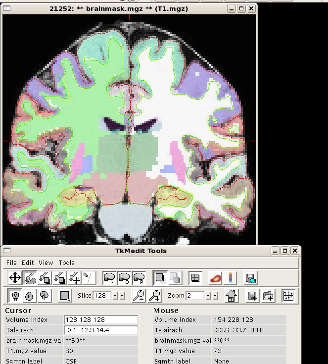
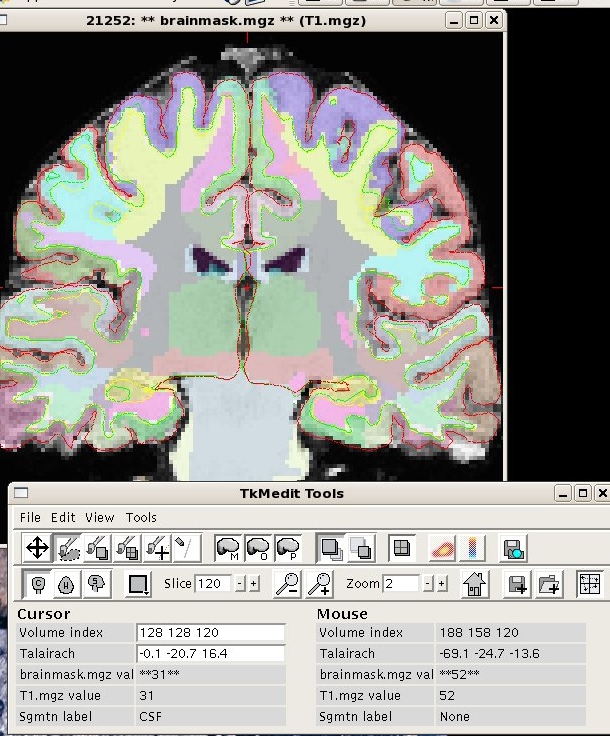
I have sent two images created with the following commands.
tkmedit $subjid brainmask.mgz lh.white -aux T1.mgz -aux-surface rh.white -segmentation (aparc+aseg.mgz and wmparc.mgz) $FREESURFER_HOME/FreesurferColorLUT.txt
The wm segmenations go through the "main surface" and the "orig surface" into the gray matter, for example in the rh-supramarginal and rh-postcentral. From your last response I am unsure as to which pipeline creates the wm parcellations, the volume-based pipeline (.i.e aseg.mgz) or the surface-based pipeline (i.e. ribbon.mgz) or some combination of the two. It seems to me that the gm/wm parcellations are created using the surface pipeline and then transferred to the closest class-matched voxels from Left-Cerebral-Cortex and Left-Cerebral-White-Matter aseg segmentations. I guess the question is how are we getting from the surface parcellations back to volume space; is there a binary? In a related vein, the value from aseg.stats "surface-based-volume mm3 lh-cerebral-white-matter" seems to be created with mri_segstats or is it from mris_wm_volume? Thanks for all your help!
Chris Bell
On Jul 17 2008, Doug Greve wrote:
When the volume is created, a decision has to be made at each voxel as to what to classify it as. This is used when the aseg.mgz and ribbon.mgz are created. aparc+aseg.mgz inherit these decisions from these files. When using mri_segstats with --pv, it will take the volume of a 1mm3 voxel and divide it up so that it can contribute to more than one region. But there's not a way to put the partial volume information into the segmentation itself. Not sure why that do not appear alingned, but we've been recently looking into this for other reasons. Can you send a pic.?
doug
On Wed, 16 Jul 2008, bell0368@umn.edu wrote:
I was wondering how the calculations of the wm volume were performed as well. The six values below are volume values from aseg I believe. I assume mris_wm_volume does more than sub-divide these regions into the wm parcellations, but probably also includes partial volume estimations along the wm/gm surface, is this correct? I basically have the same question for the gm volume values in aparc.stats; do they include partial volume calculations from the gm/pial and gm/white borders? Which binary is used to create these values for gm in aparc.stats... is it mri_segstats (with --pv)? I was assuming it would be an mris (surface) calculation as well. I am asking because if you use tkmedit to view the ?h.surface files and load the aparc+aseg or wmparc segmentation volumes in also, the surfaces and the segmentations don't seem to line up. Is there a way to create a segmentation volume that includes partial-volumed voxels at the gray/white and gray/pial boundries? Thanks for your help.
Chris Bell
the interior voxels are only included in the volume if they are one of the following in the aseg.mgz:
case Left_Cerebral_Cortex: case Right_Cerebral_Cortex: case Left_Cerebral_White_Matter: case Right_Cerebral_White_Matter: case Left_WM_hypointensities: case Right_WM_hypointensities:
cheers, Bruce
On Tue, 15 Jul 2008, Marin Richardson wrote:
OK, so aparc.stats is better for cortical gray matter. I want to look at the parcellated white matter volumes as well - is the only/best option to use the wm volumes from wmparc.stats?
Also, when using mris_wm_volume to calculate total white matter, I saw the result defined as the volume within the white matter surface minus non-WM structures? Can you tell me which regions are and are not included?
Marin
Doug Greve <[EMAIL PROTECTED]> 7/15/2008 12:02 PM >>>It is better to use the aparc as it is derived from surface measurements, which have sub-voxel resolution. The wmparc.stats are generated by counting 1mm^3 voxels.
doug
Marin Richardson wrote:
  Hi all,

   I noticed that the gray volumes for parcellated cortical regions
in wmparc.stats are different from those for the same region in aparc.stats (e.g. ctx-lh-middletemporal = 8419 in wmparc.stats, middletemporal = 9466 in lh.aparc.stats). Why are the values different and which one is a better measure to use?
   Thanks,
   Marin
Freesurfer mailing list Freesurfer@nmr.mgh.harvard.edu https://mail.nmr.mgh.harvard.edu/mailman/listinfo/freesurfer

Hi Chris, Doug, please correct me if I'm wrong, but the original gm/wm segmentation contained in aseg.mgz is based on the 3D volume-based tissue segmentation. This original segmentation then forms the basis for all subsequent additional segmentations of either GM or WM in the 3D volume. That is, aparc+aseg.mgz and wmparc.mgz make "use" of the surface based parcellation, but only to decide the most appropriate label to assign to GM/WM as defined in the already existing volume-based segmentation that is contained in aseg.mgz.
Thus, since the GM/WM in aparc+aseg.mgz and wmparc.mgz is still based on the 3D volume segmentation, there is no apriori reason that their spatial extent will correspond precisely to the more accurate gray and white mattes surfaces that come out of the surface-based processing stream. (Although certainly we would hope that the discrepancies don't become too large).
cheers, Mike H.
On Thu, 2008-07-17 at 12:00 -0500, bell0368@umn.edu wrote:
I have sent two images created with the following commands.
tkmedit $subjid brainmask.mgz lh.white -aux T1.mgz -aux-surface rh.white -segmentation (aparc+aseg.mgz and wmparc.mgz) $FREESURFER_HOME/FreesurferColorLUT.txt
The wm segmenations go through the "main surface" and the "orig surface" into the gray matter, for example in the rh-supramarginal and rh-postcentral. From your last response I am unsure as to which pipeline creates the wm parcellations, the volume-based pipeline (.i.e aseg.mgz) or the surface-based pipeline (i.e. ribbon.mgz) or some combination of the two. It seems to me that the gm/wm parcellations are created using the surface pipeline and then transferred to the closest class-matched voxels from Left-Cerebral-Cortex and Left-Cerebral-White-Matter aseg segmentations. I guess the question is how are we getting from the surface parcellations back to volume space; is there a binary? In a related vein, the value from aseg.stats "surface-based-volume mm3 lh-cerebral-white-matter" seems to be created with mri_segstats or is it from mris_wm_volume? Thanks for all your help!
Chris Bell
On Jul 17 2008, Doug Greve wrote:
When the volume is created, a decision has to be made at each voxel as to what to classify it as. This is used when the aseg.mgz and ribbon.mgz are created. aparc+aseg.mgz inherit these decisions from these files. When using mri_segstats with --pv, it will take the volume of a 1mm3 voxel and divide it up so that it can contribute to more than one region. But there's not a way to put the partial volume information into the segmentation itself. Not sure why that do not appear alingned, but we've been recently looking into this for other reasons. Can you send a pic.?
doug
On Wed, 16 Jul 2008, bell0368@umn.edu wrote:
I was wondering how the calculations of the wm volume were performed as well. The six values below are volume values from aseg I believe. I assume mris_wm_volume does more than sub-divide these regions into the wm parcellations, but probably also includes partial volume estimations along the wm/gm surface, is this correct? I basically have the same question for the gm volume values in aparc.stats; do they include partial volume calculations from the gm/pial and gm/white borders? Which binary is used to create these values for gm in aparc.stats... is it mri_segstats (with --pv)? I was assuming it would be an mris (surface) calculation as well. I am asking because if you use tkmedit to view the ?h.surface files and load the aparc+aseg or wmparc segmentation volumes in also, the surfaces and the segmentations don't seem to line up. Is there a way to create a segmentation volume that includes partial-volumed voxels at the gray/white and gray/pial boundries? Thanks for your help.
Chris Bell
the interior voxels are only included in the volume if they are one of the following in the aseg.mgz:
case Left_Cerebral_Cortex: case Right_Cerebral_Cortex: case Left_Cerebral_White_Matter: case Right_Cerebral_White_Matter: case Left_WM_hypointensities: case Right_WM_hypointensities:
cheers, Bruce
On Tue, 15 Jul 2008, Marin Richardson wrote:
OK, so aparc.stats is better for cortical gray matter. I want to look at the parcellated white matter volumes as well - is the only/best option to use the wm volumes from wmparc.stats?
Also, when using mris_wm_volume to calculate total white matter, I saw the result defined as the volume within the white matter surface minus non-WM structures? Can you tell me which regions are and are not included?
Marin
Doug Greve <[EMAIL PROTECTED]> 7/15/2008 12:02 PM >>>It is better to use the aparc as it is derived from surface measurements, which have sub-voxel resolution. The wmparc.stats are generated by counting 1mm^3 voxels.
doug
Marin Richardson wrote:
 Hi all,

  I noticed that the gray volumes for parcellated cortical regions
in wmparc.stats are different from those for the same region in aparc.stats (e.g. ctx-lh-middletemporal = 8419 in wmparc.stats, middletemporal = 9466 in lh.aparc.stats). Why are the values different and which one is a better measure to use?
  Thanks,
  Marin
Freesurfer mailing list Freesurfer@nmr.mgh.harvard.edu https://mail.nmr.mgh.harvard.edu/mailman/listinfo/freesurfer
_______________________________________________ Freesurfer mailing list Freesurfer@nmr.mgh.harvard.edu https://mail.nmr.mgh.harvard.edu/mailman/listinfo/freesurfer
Actually, the construction of aparc+aseg and wmparc do use info about where the surface is to refine the aseg cortical boundaries. This information is stored in ribbon.mgz (and it overrides the aseg definition). At some point, we will use the surfaces to directly refine the aseg boundaries.
doug
Michael Harms wrote:
Hi Chris, Doug, please correct me if I'm wrong, but the original gm/wm segmentation contained in aseg.mgz is based on the 3D volume-based tissue segmentation. This original segmentation then forms the basis for all subsequent additional segmentations of either GM or WM in the 3D volume. That is, aparc+aseg.mgz and wmparc.mgz make "use" of the surface based parcellation, but only to decide the most appropriate label to assign to GM/WM as defined in the already existing volume-based segmentation that is contained in aseg.mgz.
Thus, since the GM/WM in aparc+aseg.mgz and wmparc.mgz is still based on the 3D volume segmentation, there is no apriori reason that their spatial extent will correspond precisely to the more accurate gray and white mattes surfaces that come out of the surface-based processing stream. (Although certainly we would hope that the discrepancies don't become too large).
cheers, Mike H.
On Thu, 2008-07-17 at 12:00 -0500, bell0368@umn.edu wrote:
I have sent two images created with the following commands.
tkmedit $subjid brainmask.mgz lh.white -aux T1.mgz -aux-surface rh.white -segmentation (aparc+aseg.mgz and wmparc.mgz) $FREESURFER_HOME/FreesurferColorLUT.txt
The wm segmenations go through the "main surface" and the "orig surface" into the gray matter, for example in the rh-supramarginal and rh-postcentral. From your last response I am unsure as to which pipeline creates the wm parcellations, the volume-based pipeline (.i.e aseg.mgz) or the surface-based pipeline (i.e. ribbon.mgz) or some combination of the two. It seems to me that the gm/wm parcellations are created using the surface pipeline and then transferred to the closest class-matched voxels from Left-Cerebral-Cortex and Left-Cerebral-White-Matter aseg segmentations. I guess the question is how are we getting from the surface parcellations back to volume space; is there a binary? In a related vein, the value from aseg.stats "surface-based-volume mm3 lh-cerebral-white-matter" seems to be created with mri_segstats or is it from mris_wm_volume? Thanks for all your help!
Chris Bell
On Jul 17 2008, Doug Greve wrote:
When the volume is created, a decision has to be made at each voxel as to what to classify it as. This is used when the aseg.mgz and ribbon.mgz are created. aparc+aseg.mgz inherit these decisions from these files. When using mri_segstats with --pv, it will take the volume of a 1mm3 voxel and divide it up so that it can contribute to more than one region. But there's not a way to put the partial volume information into the segmentation itself. Not sure why that do not appear alingned, but we've been recently looking into this for other reasons. Can you send a pic.?
doug
On Wed, 16 Jul 2008, bell0368@umn.edu wrote:
I was wondering how the calculations of the wm volume were performed as well. The six values below are volume values from aseg I believe. I assume mris_wm_volume does more than sub-divide these regions into the wm parcellations, but probably also includes partial volume estimations along the wm/gm surface, is this correct? I basically have the same question for the gm volume values in aparc.stats; do they include partial volume calculations from the gm/pial and gm/white borders? Which binary is used to create these values for gm in aparc.stats... is it mri_segstats (with --pv)? I was assuming it would be an mris (surface) calculation as well. I am asking because if you use tkmedit to view the ?h.surface files and load the aparc+aseg or wmparc segmentation volumes in also, the surfaces and the segmentations don't seem to line up. Is there a way to create a segmentation volume that includes partial-volumed voxels at the gray/white and gray/pial boundries? Thanks for your help.
Chris Bell
the interior voxels are only included in the volume if they are one of the following in the aseg.mgz:
case Left_Cerebral_Cortex: case Right_Cerebral_Cortex: case Left_Cerebral_White_Matter: case Right_Cerebral_White_Matter: case Left_WM_hypointensities: case Right_WM_hypointensities:
cheers, Bruce
On Tue, 15 Jul 2008, Marin Richardson wrote:
OK, so aparc.stats is better for cortical gray matter. I want to look at the parcellated white matter volumes as well - is the only/best option to use the wm volumes from wmparc.stats?
Also, when using mris_wm_volume to calculate total white matter, I saw the result defined as the volume within the white matter surface minus non-WM structures? Can you tell me which regions are and are not included?
Marin
Doug Greve <[EMAIL PROTECTED]> 7/15/2008 12:02 PM >>>It is better to use the aparc as it is derived from surface measurements, which have sub-voxel resolution. The wmparc.stats are generated by counting 1mm^3 voxels.
doug
Marin Richardson wrote:
  Hi all,

   I noticed that the gray volumes for parcellated cortical regions
in wmparc.stats are different from those for the same region in aparc.stats (e.g. ctx-lh-middletemporal = 8419 in wmparc.stats, middletemporal = 9466 in lh.aparc.stats). Why are the values different and which one is a better measure to use?
   Thanks,
   Marin
Freesurfer mailing list Freesurfer@nmr.mgh.harvard.edu https://mail.nmr.mgh.harvard.edu/mailman/listinfo/freesurfer
_______________________________________________ Freesurfer mailing list Freesurfer@nmr.mgh.harvard.edu https://mail.nmr.mgh.harvard.edu/mailman/listinfo/freesurfer
Freesurfer mailing list Freesurfer@nmr.mgh.harvard.edu https://mail.nmr.mgh.harvard.edu/mailman/listinfo/freesurfer
Hi Doug, I was comparing the aseg.mgz and aparc+aseg.mgz segmentations (FS v4.0.2), and recalled a previous thread on this issue in which you corrected me on an aspect of how the aparc+aseg.mgz is affected by the surfaces.
From what I can tell looking at this again, voxels labeled as "None" in
the aseg.mgz are also still ALWAYS labelled as "None" in the aparc +aseg.mgz, even if those voxels are clearly interior to the final pial surface. However, voxels interior to the white surface that are labeled as "Cortex" in the aseg.mgz get converted to "None" in aparc+aseg.mgz. That is, it appears that a voxel must be in ribbon.mgz to be eligible for one of the "ctx" parcellation labels in aparc+aseg.mgz, but not necessarily all voxels in the ribbon will be labelled as "ctx" (some will remain labeled as "None", apparently propagated from aseg.mgz).
Do I have this summary correct?
thanks, Mike H.
On Tue, 2008-07-22 at 14:54 -0400, Doug Greve wrote:
Actually, the construction of aparc+aseg and wmparc do use info about where the surface is to refine the aseg cortical boundaries. This information is stored in ribbon.mgz (and it overrides the aseg definition). At some point, we will use the surfaces to directly refine the aseg boundaries.
doug
Michael Harms wrote:
Hi Chris, Doug, please correct me if I'm wrong, but the original gm/wm segmentation contained in aseg.mgz is based on the 3D volume-based tissue segmentation. This original segmentation then forms the basis for all subsequent additional segmentations of either GM or WM in the 3D volume. That is, aparc+aseg.mgz and wmparc.mgz make "use" of the surface based parcellation, but only to decide the most appropriate label to assign to GM/WM as defined in the already existing volume-based segmentation that is contained in aseg.mgz.
Thus, since the GM/WM in aparc+aseg.mgz and wmparc.mgz is still based on the 3D volume segmentation, there is no apriori reason that their spatial extent will correspond precisely to the more accurate gray and white mattes surfaces that come out of the surface-based processing stream. (Although certainly we would hope that the discrepancies don't become too large).
cheers, Mike H.
On Thu, 2008-07-17 at 12:00 -0500, bell0368@umn.edu wrote:
I have sent two images created with the following commands.
tkmedit $subjid brainmask.mgz lh.white -aux T1.mgz -aux-surface rh.white -segmentation (aparc+aseg.mgz and wmparc.mgz) $FREESURFER_HOME/FreesurferColorLUT.txt
The wm segmenations go through the "main surface" and the "orig surface" into the gray matter, for example in the rh-supramarginal and rh-postcentral. From your last response I am unsure as to which pipeline creates the wm parcellations, the volume-based pipeline (.i.e aseg.mgz) or the surface-based pipeline (i.e. ribbon.mgz) or some combination of the two. It seems to me that the gm/wm parcellations are created using the surface pipeline and then transferred to the closest class-matched voxels from Left-Cerebral-Cortex and Left-Cerebral-White-Matter aseg segmentations. I guess the question is how are we getting from the surface parcellations back to volume space; is there a binary? In a related vein, the value from aseg.stats "surface-based-volume mm3 lh-cerebral-white-matter" seems to be created with mri_segstats or is it from mris_wm_volume? Thanks for all your help!
Chris Bell
On Jul 17 2008, Doug Greve wrote:
When the volume is created, a decision has to be made at each voxel as to what to classify it as. This is used when the aseg.mgz and ribbon.mgz are created. aparc+aseg.mgz inherit these decisions from these files. When using mri_segstats with --pv, it will take the volume of a 1mm3 voxel and divide it up so that it can contribute to more than one region. But there's not a way to put the partial volume information into the segmentation itself. Not sure why that do not appear alingned, but we've been recently looking into this for other reasons. Can you send a pic.?
doug
On Wed, 16 Jul 2008, bell0368@umn.edu wrote:
I was wondering how the calculations of the wm volume were performed as well. The six values below are volume values from aseg I believe. I assume mris_wm_volume does more than sub-divide these regions into the wm parcellations, but probably also includes partial volume estimations along the wm/gm surface, is this correct? I basically have the same question for the gm volume values in aparc.stats; do they include partial volume calculations from the gm/pial and gm/white borders? Which binary is used to create these values for gm in aparc.stats... is it mri_segstats (with --pv)? I was assuming it would be an mris (surface) calculation as well. I am asking because if you use tkmedit to view the ?h.surface files and load the aparc+aseg or wmparc segmentation volumes in also, the surfaces and the segmentations don't seem to line up. Is there a way to create a segmentation volume that includes partial-volumed voxels at the gray/white and gray/pial boundries? Thanks for your help.
Chris Bell
the interior voxels are only included in the volume if they are one of the following in the aseg.mgz:
case Left_Cerebral_Cortex: case Right_Cerebral_Cortex: case Left_Cerebral_White_Matter: case Right_Cerebral_White_Matter: case Left_WM_hypointensities: case Right_WM_hypointensities:
cheers, Bruce
On Tue, 15 Jul 2008, Marin Richardson wrote:
OK, so aparc.stats is better for cortical gray matter. I want to look at the parcellated white matter volumes as well - is the only/best option to use the wm volumes from wmparc.stats?
Also, when using mris_wm_volume to calculate total white matter, I saw the result defined as the volume within the white matter surface minus non-WM structures? Can you tell me which regions are and are not included?
Marin
Doug Greve <[EMAIL PROTECTED]> 7/15/2008 12:02 PM >>>It is better to use the aparc as it is derived from surface measurements, which have sub-voxel resolution. The wmparc.stats are generated by counting 1mm^3 voxels.
doug
Marin Richardson wrote:
 Hi all,

  I noticed that the gray volumes for parcellated cortical regions
in wmparc.stats are different from those for the same region in aparc.stats (e.g. ctx-lh-middletemporal = 8419 in wmparc.stats, middletemporal = 9466 in lh.aparc.stats). Why are the values different and which one is a better measure to use?
  Thanks,
  Marin
Freesurfer mailing list Freesurfer@nmr.mgh.harvard.edu https://mail.nmr.mgh.harvard.edu/mailman/listinfo/freesurfer
_______________________________________________ Freesurfer mailing list Freesurfer@nmr.mgh.harvard.edu https://mail.nmr.mgh.harvard.edu/mailman/listinfo/freesurfer
Freesurfer mailing list Freesurfer@nmr.mgh.harvard.edu https://mail.nmr.mgh.harvard.edu/mailman/listinfo/freesurfer
-- Douglas N. Greve, Ph.D. MGH-NMR Center greve@nmr.mgh.harvard.edu Phone Number: 617-724-2358 Fax: 617-726-7422
In order to help us help you, please follow the steps in: surfer.nmr.mgh.harvard.edu/fswiki/BugReporting
I've gone through a few iterations re-working this code over the last month or so. The aparc+aseg (and wmparc) should respect the ribbon. So, voxels within the white surface but labeled as Cortex in aseg should be labeled as WhiteMatter in aparc+aseg. Voxels labeled as "None" should also be re-labeled as Cortex or WhiteMatter if they fall into those places on the ribbon.
There was a bug that was fixed in two stages, one on 8/6/08 and the other on 8/28/08. I'm not sure when our last stable version was cut, but we'll have a new version with these fixes in a few weeks.
doug
Michael Harms wrote:
Hi Doug, I was comparing the aseg.mgz and aparc+aseg.mgz segmentations (FS v4.0.2), and recalled a previous thread on this issue in which you corrected me on an aspect of how the aparc+aseg.mgz is affected by the surfaces.
From what I can tell looking at this again, voxels labeled as "None" in
the aseg.mgz are also still ALWAYS labelled as "None" in the aparc +aseg.mgz, even if those voxels are clearly interior to the final pial surface. However, voxels interior to the white surface that are labeled as "Cortex" in the aseg.mgz get converted to "None" in aparc+aseg.mgz. That is, it appears that a voxel must be in ribbon.mgz to be eligible for one of the "ctx" parcellation labels in aparc+aseg.mgz, but not necessarily all voxels in the ribbon will be labelled as "ctx" (some will remain labeled as "None", apparently propagated from aseg.mgz).
Do I have this summary correct?
thanks, Mike H.
On Tue, 2008-07-22 at 14:54 -0400, Doug Greve wrote:
Actually, the construction of aparc+aseg and wmparc do use info about where the surface is to refine the aseg cortical boundaries. This information is stored in ribbon.mgz (and it overrides the aseg definition). At some point, we will use the surfaces to directly refine the aseg boundaries.
doug
Michael Harms wrote:
Hi Chris, Doug, please correct me if I'm wrong, but the original gm/wm segmentation contained in aseg.mgz is based on the 3D volume-based tissue segmentation. This original segmentation then forms the basis for all subsequent additional segmentations of either GM or WM in the 3D volume. That is, aparc+aseg.mgz and wmparc.mgz make "use" of the surface based parcellation, but only to decide the most appropriate label to assign to GM/WM as defined in the already existing volume-based segmentation that is contained in aseg.mgz.
Thus, since the GM/WM in aparc+aseg.mgz and wmparc.mgz is still based on the 3D volume segmentation, there is no apriori reason that their spatial extent will correspond precisely to the more accurate gray and white mattes surfaces that come out of the surface-based processing stream. (Although certainly we would hope that the discrepancies don't become too large).
cheers, Mike H.
On Thu, 2008-07-17 at 12:00 -0500, bell0368@umn.edu wrote:
I have sent two images created with the following commands.
tkmedit $subjid brainmask.mgz lh.white -aux T1.mgz -aux-surface rh.white -segmentation (aparc+aseg.mgz and wmparc.mgz) $FREESURFER_HOME/FreesurferColorLUT.txt
The wm segmenations go through the "main surface" and the "orig surface" into the gray matter, for example in the rh-supramarginal and rh-postcentral. From your last response I am unsure as to which pipeline creates the wm parcellations, the volume-based pipeline (.i.e aseg.mgz) or the surface-based pipeline (i.e. ribbon.mgz) or some combination of the two. It seems to me that the gm/wm parcellations are created using the surface pipeline and then transferred to the closest class-matched voxels from Left-Cerebral-Cortex and Left-Cerebral-White-Matter aseg segmentations. I guess the question is how are we getting from the surface parcellations back to volume space; is there a binary? In a related vein, the value from aseg.stats "surface-based-volume mm3 lh-cerebral-white-matter" seems to be created with mri_segstats or is it from mris_wm_volume? Thanks for all your help!
Chris Bell
On Jul 17 2008, Doug Greve wrote:
When the volume is created, a decision has to be made at each voxel as to what to classify it as. This is used when the aseg.mgz and ribbon.mgz are created. aparc+aseg.mgz inherit these decisions from these files. When using mri_segstats with --pv, it will take the volume of a 1mm3 voxel and divide it up so that it can contribute to more than one region. But there's not a way to put the partial volume information into the segmentation itself. Not sure why that do not appear alingned, but we've been recently looking into this for other reasons. Can you send a pic.?
doug
On Wed, 16 Jul 2008, bell0368@umn.edu wrote:
I was wondering how the calculations of the wm volume were performed as well. The six values below are volume values from aseg I believe. I assume mris_wm_volume does more than sub-divide these regions into the wm parcellations, but probably also includes partial volume estimations along the wm/gm surface, is this correct? I basically have the same question for the gm volume values in aparc.stats; do they include partial volume calculations from the gm/pial and gm/white borders? Which binary is used to create these values for gm in aparc.stats... is it mri_segstats (with --pv)? I was assuming it would be an mris (surface) calculation as well. I am asking because if you use tkmedit to view the ?h.surface files and load the aparc+aseg or wmparc segmentation volumes in also, the surfaces and the segmentations don't seem to line up. Is there a way to create a segmentation volume that includes partial-volumed voxels at the gray/white and gray/pial boundries? Thanks for your help.
Chris Bell
the interior voxels are only included in the volume if they are one of the following in the aseg.mgz:
case Left_Cerebral_Cortex: case Right_Cerebral_Cortex: case Left_Cerebral_White_Matter: case Right_Cerebral_White_Matter: case Left_WM_hypointensities: case Right_WM_hypointensities:
cheers, Bruce
On Tue, 15 Jul 2008, Marin Richardson wrote:
OK, so aparc.stats is better for cortical gray matter. I want to look at the parcellated white matter volumes as well - is the only/best option to use the wm volumes from wmparc.stats?
Also, when using mris_wm_volume to calculate total white matter, I saw the result defined as the volume within the white matter surface minus non-WM structures? Can you tell me which regions are and are not included?
Marin
Doug Greve <[EMAIL PROTECTED]> 7/15/2008 12:02 PM >>>It is better to use the aparc as it is derived from surface measurements, which have sub-voxel resolution. The wmparc.stats are generated by counting 1mm^3 voxels.
doug
Marin Richardson wrote:
  Hi all,

   I noticed that the gray volumes for parcellated cortical regions
in wmparc.stats are different from those for the same region in aparc.stats (e.g. ctx-lh-middletemporal = 8419 in wmparc.stats, middletemporal = 9466 in lh.aparc.stats). Why are the values different and which one is a better measure to use?
   Thanks,
   Marin
Freesurfer mailing list Freesurfer@nmr.mgh.harvard.edu https://mail.nmr.mgh.harvard.edu/mailman/listinfo/freesurfer
_______________________________________________ Freesurfer mailing list Freesurfer@nmr.mgh.harvard.edu https://mail.nmr.mgh.harvard.edu/mailman/listinfo/freesurfer
Freesurfer mailing list Freesurfer@nmr.mgh.harvard.edu https://mail.nmr.mgh.harvard.edu/mailman/listinfo/freesurfer
-- Douglas N. Greve, Ph.D. MGH-NMR Center greve@nmr.mgh.harvard.edu Phone Number: 617-724-2358 Fax: 617-726-7422
In order to help us help you, please follow the steps in: surfer.nmr.mgh.harvard.edu/fswiki/BugReporting

Hi, for subcortical segmentations, is it then better to use the volumes in wmparc.stats than those in aseg.stats?
On Tue, Jul 22, 2008 at 2:54 PM, Doug Greve greve@nmr.mgh.harvard.edu wrote:
Actually, the construction of aparc+aseg and wmparc do use info about where the surface is to refine the aseg cortical boundaries. This information is stored in ribbon.mgz (and it overrides the aseg definition). At some point, we will use the surfaces to directly refine the aseg boundaries.
doug
Michael Harms wrote:
Hi Chris, Doug, please correct me if I'm wrong, but the original gm/wm segmentation contained in aseg.mgz is based on the 3D volume-based tissue segmentation. This original segmentation then forms the basis for all subsequent additional segmentations of either GM or WM in the 3D volume. That is, aparc+aseg.mgz and wmparc.mgz make "use" of the surface based parcellation, but only to decide the most appropriate label to assign to GM/WM as defined in the already existing volume-based segmentation that is contained in aseg.mgz.
Thus, since the GM/WM in aparc+aseg.mgz and wmparc.mgz is still based on the 3D volume segmentation, there is no apriori reason that their spatial extent will correspond precisely to the more accurate gray and white mattes surfaces that come out of the surface-based processing stream. (Although certainly we would hope that the discrepancies don't become too large).
cheers, Mike H.
On Thu, 2008-07-17 at 12:00 -0500, bell0368@umn.edu wrote:
I have sent two images created with the following commands.
tkmedit $subjid brainmask.mgz lh.white -aux T1.mgz -aux-surface rh.white -segmentation (aparc+aseg.mgz and wmparc.mgz) $FREESURFER_HOME/FreesurferColorLUT.txt
The wm segmenations go through the "main surface" and the "orig surface" into the gray matter, for example in the rh-supramarginal and rh-postcentral. From your last response I am unsure as to which pipeline creates the wm parcellations, the volume-based pipeline (.i.e aseg.mgz) or the surface-based pipeline (i.e. ribbon.mgz) or some combination of the two. It seems to me that the gm/wm parcellations are created using the surface pipeline and then transferred to the closest class-matched voxels from Left-Cerebral-Cortex and Left-Cerebral-White-Matter aseg segmentations. I guess the question is how are we getting from the surface parcellations back to volume space; is there a binary? In a related vein, the value from aseg.stats "surface-based-volume mm3 lh-cerebral-white-matter" seems to be created with mri_segstats or is it from mris_wm_volume? Thanks for all your help!
Chris Bell
On Jul 17 2008, Doug Greve wrote:
When the volume is created, a decision has to be made at each voxel as to what to classify it as. This is used when the aseg.mgz and ribbon.mgz are created. aparc+aseg.mgz inherit these decisions from these files. When using mri_segstats with --pv, it will take the volume of a 1mm3 voxel and divide it up so that it can contribute to more than one region. But there's not a way to put the partial volume information into the segmentation itself. Not sure why that do not appear alingned, but we've been recently looking into this for other reasons. Can you send a pic.?
doug
On Wed, 16 Jul 2008, bell0368@umn.edu wrote:
I was wondering how the calculations of the wm volume were performed as well. The six values below are volume values from aseg I believe. I assume mris_wm_volume does more than sub-divide these regions into the wm parcellations, but probably also includes partial volume estimations along the wm/gm surface, is this correct? I basically have the same question for the gm volume values in aparc.stats; do they include partial volume calculations from the gm/pial and gm/white borders? Which binary is used to create these values for gm in aparc.stats... is it mri_segstats (with --pv)? I was assuming it would be an mris (surface) calculation as well. I am asking because if you use tkmedit to view the ?h.surface files and load the aparc+aseg or wmparc segmentation volumes in also, the surfaces and the segmentations don't seem to line up. Is there a way to create a segmentation volume that includes partial-volumed voxels at the gray/white and gray/pial boundries? Thanks for your help.
Chris Bell
the interior voxels are only included in the volume if they are one of the following in the aseg.mgz:
case Left_Cerebral_Cortex: case Right_Cerebral_Cortex: case Left_Cerebral_White_Matter: case Right_Cerebral_White_Matter: case Left_WM_hypointensities: case Right_WM_hypointensities:
cheers, Bruce
On Tue, 15 Jul 2008, Marin Richardson wrote:
OK, so aparc.stats is better for cortical gray matter. I want to look at the parcellated white matter volumes as well - is the only/best option to use the wm volumes from wmparc.stats?
Also, when using mris_wm_volume to calculate total white matter, I saw the result defined as the volume within the white matter surface minus non-WM structures? Can you tell me which regions are and are not included?
Marin
Doug Greve <[EMAIL PROTECTED]> 7/15/2008 12:02 PM >>>It is better to use the aparc as it is derived from surface measurements, which have sub-voxel resolution. The wmparc.stats are generated by counting 1mm^3 voxels.
doug
Marin Richardson wrote:
Hi all, I noticed that the gray volumes for parcellated cortical regionsin wmparc.stats are different from those for the same region in aparc.stats (e.g. ctx-lh-middletemporal = 8419 in wmparc.stats, middletemporal = 9466 in lh.aparc.stats). Why are the values different and which one is a better measure to use?
Thanks, Marin
Freesurfer mailing list Freesurfer@nmr.mgh.harvard.edu https://mail.nmr.mgh.harvard.edu/mailman/listinfo/freesurfer
_______________________________________________ Freesurfer mailing list Freesurfer@nmr.mgh.harvard.edu https://mail.nmr.mgh.harvard.edu/mailman/listinfo/freesurfer
Freesurfer mailing list Freesurfer@nmr.mgh.harvard.edu https://mail.nmr.mgh.harvard.edu/mailman/listinfo/freesurfer
-- Douglas N. Greve, Ph.D. MGH-NMR Center greve@nmr.mgh.harvard.edu Phone Number: 617-724-2358 Fax: 617-726-7422
In order to help us help you, please follow the steps in: surfer.nmr.mgh.harvard.edu/fswiki/BugReporting
Freesurfer mailing list Freesurfer@nmr.mgh.harvard.edu https://mail.nmr.mgh.harvard.edu/mailman/listinfo/freesurfer
freesurfer@nmr.mgh.harvard.edu
-
 bell0368@umn.edu
 -
 Doug Greve
 -
 Michael Harms
 -
 Nurunisa Neyzi