
Hey all,
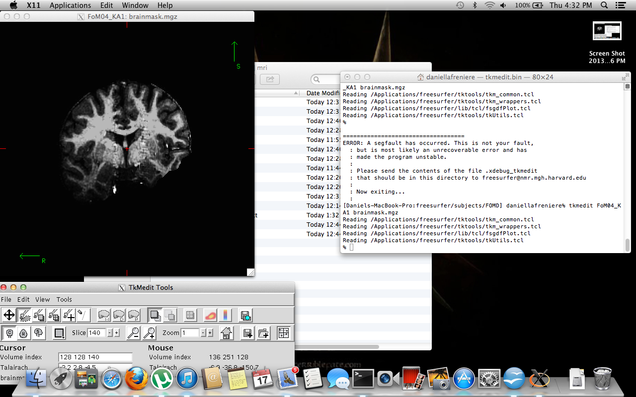
I'm very new to Freesurfer and am having some issues with the initial processing of our MPRAGE dicom files. So far, I'm able to get initial -autorecon1 steps to work but once this has finished and I open my brainmask.mgz files and the brain is incredibly dark in inferior regions. So much so that the skull strip appears to be removing large chunks of anterior temporal lobes. What remains of the temporal lobes is incredibly dark with grey matter almost indistinguishable from CSF.
I would really like to look into freesurfer for cortical thickness analysis of subcortical regions since it seems like such an awesome program but I have really been struggling to get a decent looking brain. It's probably noteworthy that these brains have always been quite dark but I'm usually able to correct them to a point where they're pretty good in other programs.
I have read about a few solutions to the problem but am having trouble implementing them. One suggestion was to use the -mprage flag; unfortunately, I'm not sure how to run this or when. Another suggestion was to use control points but I don't yet have the red and blue segmentation boundary lines - I'm using tkmedit FoM04_KA brainmask.mgz -aux T1.mgz to view the files. For reference, this is the line I use to run autorecon1: Recon-all -autorecon1 -subjid FoM04_KA
Does anyone have any suggestions as to what I could do? It would be greatly appreciated. I've attached a screenshot to illustrate. Also, we used a 4.7T Varian scanner with 0.375/0.375/1mm voxels. I believe we used a gradient echo multislice sequence. In addition, all subjects' data have been obtained so there is no chance of changing sequences…
Best wishes,
Dan

Wow, that's a huge bias field! Control points will help uniformity, but the snr may not be good enough for good results Bruce
On Jan 18, 2013, at 11:09 AM, Dan LaFreniere lafreniere.dj@gmail.com wrote:
Hey all,
I'm very new to Freesurfer and am having some issues with the initial processing of our MPRAGE dicom files. So far, I'm able to get initial -autorecon1 steps to work but once this has finished and I open my brainmask.mgz files and the brain is incredibly dark in inferior regions. So much so that the skull strip appears to be removing large chunks of anterior temporal lobes. What remains of the temporal lobes is incredibly dark with grey matter almost indistinguishable from CSF.
I would really like to look into freesurfer for cortical thickness analysis of subcortical regions since it seems like such an awesome program but I have really been struggling to get a decent looking brain. It's probably noteworthy that these brains have always been quite dark but I'm usually able to correct them to a point where they're pretty good in other programs.
I have read about a few solutions to the problem but am having trouble implementing them. One suggestion was to use the -mprage flag; unfortunately, I'm not sure how to run this or when. Another suggestion was to use control points but I don't yet have the red and blue segmentation boundary lines - I'm using tkmedit FoM04_KA brainmask.mgz -aux T1.mgz to view the files. For reference, this is the line I use to run autorecon1: Recon-all -autorecon1 -subjid FoM04_KA
Does anyone have any suggestions as to what I could do? It would be greatly appreciated. I've attached a screenshot to illustrate. Also, we used a 4.7T Varian scanner with 0.375/0.375/1mm voxels. I believe we used a gradient echo multislice sequence. In addition, all subjects' data have been obtained so there is no chance of changing sequences…
Best wishes,
Dan
<02 - Screen Shot.png> _______________________________________________ Freesurfer mailing list Freesurfer@nmr.mgh.harvard.edu https://mail.nmr.mgh.harvard.edu/mailman/listinfo/freesurfer
freesurfer@nmr.mgh.harvard.edu
-
 Bruce Fischl
 -
 Dan LaFreniere