
Dear Freesurfers!!
I've encountered two strange segmentation "errors", if they are errors at all.
Please see the attached fs.png:
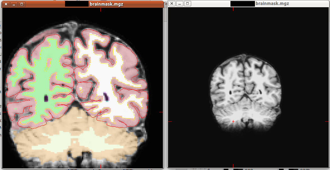
You can see that there are excess pial tissues on the superior part of the brain. The pial surface is defined correctly and the excess pial tissue is outside the red line yet it is coloured pink. My question, is :
Does this have any impact on the results?
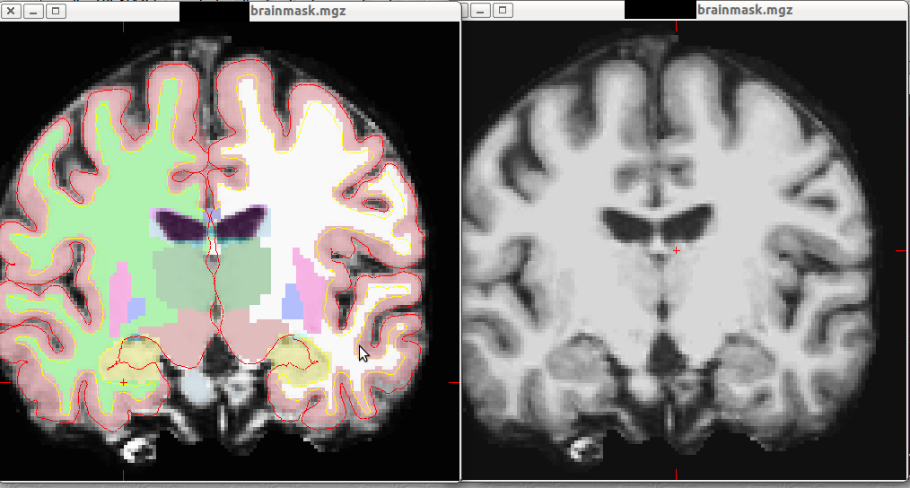
The second is related to the hippocampus.
Please see the attached fs2.png:
The pial surface around the hippocampus looks a bit weird. It actually discards 1/3 of the hippocampus (it is outside the red line).
Do you have any suggestions regarding the two problems.
I'm using FS 4.5 on several 64bit linux distributions.

The stuff outside the pial surface won't affect things, which is one of the reasons we prefer the surface-based measured of cortical morphometry.
  Similarly, we don't use the surface-based stuff in the hippocampus, so you should be all set (since many of the cortical assumptions aren't
  valid there)

  cheers
  Bruce
On Mon, 3 Oct 2011, Gergely Orsi wrote:
  Dear Freesurfers!!

  I've encountered two strange segmentation "errors", if they are errors at
  all.


  Please see the attached fs.png:

  You can see that there are excess pial tissues on the superior part of the
  brain. The pial surface is
  defined correctly and the excess pial tissue is outside the red line yet it
  is coloured pink. My question, is :

  Does this have any impact on the results?


  The second is related to the hippocampus.

  Please see the attached fs2.png:

  The pial surface around the hippocampus looks a bit weird.
  It actually discards 1/3 of the hippocampus (it is outside the red line).

  Do you have any suggestions regarding the two problems.

  I'm using FS 4.5 on several 64bit linux distributions.


  --
  Gergely Orsi
  biologist - research associate
  Diagnostic Center of Pécs
  H-7623 Pécs, Rét str. 2.
  Tel.: 0036-70-3174676
  E-mail: gergo.orsi@gmail.com
Thank you very much Bruce!
Have a nice day/night.
2011/10/3 Bruce Fischl fischl@nmr.mgh.harvard.edu
The stuff outside the pial surface won't affect things, which is one of the reasons we prefer the surface-based measured of cortical morphometry.
Similarly, we don't use the surface-based stuff in the hippocampus, so you should be all set (since many of the cortical assumptions aren't valid there)
 cheers
 Bruce
On Mon, 3 Oct 2011, Gergely Orsi wrote:
 Dear Freesurfers!!

 I've encountered two strange segmentation "errors", if they are
errors at all.
 Please see the attached fs.png:

 You can see that there are excess pial tissues on the superior part
of the brain. The pial surface is defined correctly and the excess pial tissue is outside the red line yet it is coloured pink. My question, is :
 Does this have any impact on the results?


 The second is related to the hippocampus.

 Please see the attached fs2.png:

 The pial surface around the hippocampus looks a bit weird.
 It actually discards 1/3 of the hippocampus (it is outside the red
line).
 Do you have any suggestions regarding the two problems.

 I'm using FS 4.5 on several 64bit linux distributions.


 --
 Gergely Orsi
 biologist - research associate
 Diagnostic Center of Pécs
 H-7623 Pécs, Rét str. 2.
 Tel.: 0036-70-3174676
 E-mail: gergo.orsi@gmail.com
The information in this e-mail is intended only for the person to whom it is addressed. If you believe this e-mail was sent to you in error and the e-mail contains patient information, please contact the Partners Compliance HelpLine at http://www.partners.org/**compliancelinehttp://www.partners.org/complianceline. If the e-mail was sent to you in error but does not contain patient information, please contact the sender and properly dispose of the e-mail.
Dear Bruce and others,
We have just ran into strange ICV volumes.
For all our 100+ subjets, the total icv is around 1500cm3 (around 1300-1700).
For the new group (only 7 subjects) the average ICV is around 1200 (ranging from 900-1500).
This is almost microcephalia, but they are normal.
If we take a look at the brainseg volume or brain mask volumes they are larger than the total ICV (for e.g. subject 5 has brainmaskvol: 1440; brainseg vol: 1021; ICV 1036)
This is not right. It's kind'a strange that someone has bigger brain than skull :)
I was using freesurfer 4.5 for both groups.
Do you have any idea what did I miss?
Thanks,
Gergo
2011/10/3 Bruce Fischl fischl@nmr.mgh.harvard.edu
The stuff outside the pial surface won't affect things, which is one of the reasons we prefer the surface-based measured of cortical morphometry.
Similarly, we don't use the surface-based stuff in the hippocampus, so you should be all set (since many of the cortical assumptions aren't valid there)
cheers Bruce
On Mon, 3 Oct 2011, Gergely Orsi wrote:
Dear Freesurfers!!
I've encountered two strange segmentation "errors", if they are errors at all.
Please see the attached fs.png:
You can see that there are excess pial tissues on the superior part of the brain. The pial surface is defined correctly and the excess pial tissue is outside the red line yet it is coloured pink. My question, is :
Does this have any impact on the results?
The second is related to the hippocampus.
Please see the attached fs2.png:
The pial surface around the hippocampus looks a bit weird. It actually discards 1/3 of the hippocampus (it is outside the red line).
Do you have any suggestions regarding the two problems.
I'm using FS 4.5 on several 64bit linux distributions.
-- Gergely Orsi biologist - research associate Diagnostic Center of Pécs H-7623 Pécs, Rét str. 2. Tel.: 0036-70-3174676 E-mail: gergo.orsi@gmail.com
The information in this e-mail is intended only for the person to whom it is addressed. If you believe this e-mail was sent to you in error and the e-mail contains patient information, please contact the Partners Compliance HelpLine at http://www.partners.org/**compliancelinehttp://www.partners.org/complianceline. If the e-mail was sent to you in error but does not contain patient information, please contact the sender and properly dispose of the e-mail.
can you send us an example of one that is way off? On Tue, 25 Oct 2011, Gergely Orsi wrote:
Dear Bruce and others,
We have just ran into strange ICV volumes.
For all our 100+ subjets, the total icv is around 1500cm3 (around 1300-1700).
For the new group (only 7 subjects) the average ICV is around 1200 (ranging from 900-1500).
This is almost microcephalia, but they are normal.
If we take a look at the brainseg volume or brain mask volumes they are larger than the total ICV (for e.g. subject 5 has brainmaskvol: 1440; brainseg vol: 1021; ICV 1036)
This is not right. It's kind'a strange that someone has bigger brain than skull :)
I was using freesurfer 4.5 for both groups.
Do you have any idea what did I miss?
Thanks,
Gergo
2011/10/3 Bruce Fischl fischl@nmr.mgh.harvard.edu The stuff outside the pial surface won't affect things, which is one of the reasons we prefer the surface-based measured of cortical morphometry.
Similarly, we don't use the surface-based stuff in the hippocampus, so you should be all set (since many of the cortical assumptions aren't valid there) cheers BruceOn Mon, 3 Oct 2011, Gergely Orsi wrote:
Dear Freesurfers!! I've encountered two strange segmentation "errors", if they are errors at all. Please see the attached fs.png: You can see that there are excess pial tissues on the superior part of the brain. The pial surface is defined correctly and the excess pial tissue is outside the red line yet it is coloured pink. My question, is : Does this have any impact on the results? The second is related to the hippocampus. Please see the attached fs2.png: The pial surface around the hippocampus looks a bit weird. It actually discards 1/3 of the hippocampus (it is outside the red line). Do you have any suggestions regarding the two problems. I'm using FS 4.5 on several 64bit linux distributions. -- Gergely Orsi biologist - research associate Diagnostic Center of Pécs H-7623 Pécs, Rét str. 2. Tel.: 0036-70-3174676 E-mail: gergo.orsi@gmail.comThe information in this e-mail is intended only for the person to whom it is addressed. If you believe this e-mail was sent to you in error and the e-mail contains patient information, please contact the Partners Compliance HelpLine at http://www.partners.org/complianceline . If the e-mail was sent to you in error but does not contain patient information, please contact the sender and properly dispose of the e-mail.
-- Gergely Orsi biologist - research associate Diagnostic Center of Pécs H-7623 Pécs, Rét str. 2. Tel.: 0036-70-3174676 E-mail: gergo.orsi@gmail.com
Here you are:
# Title Segmentation Statistics # # generating_program mri_segstats # cvs_version $Id: mri_segstats.c,v 1.33.2.5 2009/02/11 22:38:51 nicks Exp $ # cmdline mri_segstats --seg mri/aseg.mgz --sum stats/aseg.stats --pv mri/norm.mgz --excludeid 0 --brain-vol-from-seg --brainmask mri/brainmask.mgz --in mri/norm.mgz --in-intensity-name norm --in-intensity-units MR --etiv --subject SZEMAN2 --surf-wm-vol --ctab /home/kutato/freesurfer/ASegStatsLUT.txt # sysname Linux # hostname kutato16 # machine x86_64 # user kutato16 # anatomy_type volume # # SUBJECTS_DIR /home/kutato/freesurfer/subjects # subjectname SZEMAN2 # BrainMaskFile mri/brainmask.mgz # BrainMaskFileTimeStamp 2011/09/30 14:48:41 # Measure BrainMask, BrainMaskNVox, Number of Brain Mask Voxels, 1460346, unitless # Measure BrainMask, BrainMaskVol, Brain Mask Volume, 1460346.000000, mm^3 # Measure BrainSegNotVent, BrainSegVolNotVent, Brain Segmentation Volume Without Ventricles, 964476.000000, mm^3 # Measure BrainSeg, BrainSegNVox, Number of Brain Segmentation Voxels, 986721, unitless # Measure BrainSeg, BrainSegVol, Brain Segmentation Volume, 986721.000000, mm^3 # Measure IntraCranialVol, ICV, Intracranial Volume, 900902.902900, mm^3 # SegVolFile mri/aseg.mgz # SegVolFileTimeStamp 2011/09/30 21:17:55 # ColorTable /home/kutato/freesurfer/ASegStatsLUT.txt # ColorTableTimeStamp 2009/08/11 14:24:37 # InVolFile mri/norm.mgz # InVolFileTimeStamp 2011/09/30 15:23:28 # InVolFrame 0 # PVVolFile mri/norm.mgz # PVVolFileTimeStamp 2011/09/30 15:23:28 # surface-based-volume mm3 lh-cerebral-white-matter 199672.312500 # surface-based-volume mm3 rh-cerebral-white-matter 200666.875000 # surface-based-volume mm3 tot-cerebral-white-matter 400339.187500 # ExcludeSegId 0 # VoxelVolume_mm3 1 # TableCol 1 ColHeader Index # TableCol 1 FieldName Index # TableCol 1 Units NA # TableCol 2 ColHeader SegId # TableCol 2 FieldName Segmentation Id # TableCol 2 Units NA # TableCol 3 ColHeader NVoxels # TableCol 3 FieldName Number of Voxels # TableCol 3 Units unitless # TableCol 4 ColHeader Volume_mm3 # TableCol 4 FieldName Volume # TableCol 4 Units mm^3 # TableCol 5 ColHeader StructName # TableCol 5 FieldName Structure Name # TableCol 5 Units NA # TableCol 6 ColHeader normMean # TableCol 6 FieldName Intensity normMean # TableCol 6 Units MR # TableCol 7 ColHeader normStdDev # TableCol 7 FieldName Itensity normStdDev # TableCol 7 Units MR # TableCol 8 ColHeader normMin # TableCol 8 FieldName Intensity normMin # TableCol 8 Units MR # TableCol 9 ColHeader normMax # TableCol 9 FieldName Intensity normMax # TableCol 9 Units MR # TableCol 10 ColHeader normRange # TableCol 10 FieldName Intensity normRange # TableCol 10 Units MR # NRows 49 # NTableCols 10 # ColHeaders Index SegId NVoxels Volume_mm3 StructName normMean normStdDev normMin normMax normRange 1 2 181224 181224.0 Left-Cerebral-White-Matter 103.0824 7.8273 37.0000 124.0000 87.0000 2 3 220289 220289.0 Left-Cerebral-Cortex 73.9965 10.0547 25.0000 109.0000 84.0000 3 4 9827 9827.0 Left-Lateral-Ventricle 38.7152 10.8311 21.0000 89.0000 68.0000 4 5 472 472.0 Left-Inf-Lat-Vent 58.1272 10.0729 36.0000 97.0000 61.0000 5 7 11866 11866.0 Left-Cerebellum-White-Matter 85.2734 7.6396 41.0000 110.0000 69.0000 6 8 29669 29669.0 Left-Cerebellum-Cortex 62.3768 10.6794 17.0000 94.0000 77.0000 7 10 6422 6422.0 Left-Thalamus-Proper 97.5967 8.1825 54.0000 114.0000 60.0000 8 11 3645 3645.0 Left-Caudate 86.3108 8.2356 52.0000 110.0000 58.0000 9 12 5670 5670.0 Left-Putamen 93.1821 5.7430 62.0000 111.0000 49.0000 10 13 1860 1860.0 Left-Pallidum 105.5083 3.8920 85.0000 116.0000 31.0000 11 14 1023 1023.0 3rd-Ventricle 46.9565 12.7555 28.0000 78.0000 50.0000 12 15 1027 1027.0 4th-Ventricle 34.1996 12.3672 11.0000 73.0000 62.0000 13 16 12081 12081.0 Brain-Stem 91.9390 11.8567 44.0000 113.0000 69.0000 14 17 2538 2538.0 Left-Hippocampus 80.2219 8.1671 29.0000 109.0000 80.0000 15 18 952 952.0 Left-Amygdala 80.3787 7.1404 55.0000 105.0000 50.0000 16 24 1785 1785.0 CSF 57.1199 13.1850 29.0000 95.0000 66.0000 17 26 592 592.0 Left-Accumbens-area 84.8315 5.7364 60.0000 104.0000 44.0000 18 28 3806 3806.0 Left-VentralDC 101.0622 9.6848 51.0000 115.0000 64.0000 19 30 190 190.0 Left-vessel 71.0192 10.0774 43.0000 96.0000 53.0000 20 31 1895 1895.0 Left-choroid-plexus 54.4754 12.4270 24.0000 96.0000 72.0000 21 41 188053 188053.0 Right-Cerebral-White-Matter 101.6959 7.7683 38.0000 122.0000 84.0000 22 42 216751 216751.0 Right-Cerebral-Cortex 73.3417 9.8270 24.0000 106.0000 82.0000 23 43 9286 9286.0 Right-Lateral-Ventricle 39.9627 11.6894 21.0000 93.0000 72.0000 24 44 246 246.0 Right-Inf-Lat-Vent 50.3990 10.5551 27.0000 78.0000 51.0000 25 46 11000 11000.0 Right-Cerebellum-White-Matter 87.6621 6.5420 44.0000 108.0000 64.0000 26 47 36257 36257.0 Right-Cerebellum-Cortex 65.1387 10.4182 21.0000 94.0000 73.0000 27 49 6546 6546.0 Right-Thalamus-Proper 97.0832 8.3261 54.0000 114.0000 60.0000 28 50 3700 3700.0 Right-Caudate 84.7295 8.0259 48.0000 116.0000 68.0000 29 51 4879 4879.0 Right-Putamen 92.2331 5.6211 64.0000 111.0000 47.0000 30 52 1459 1459.0 Right-Pallidum 104.8717 3.4972 88.0000 114.0000 26.0000 31 53 3925 3925.0 Right-Hippocampus 78.0625 7.5244 49.0000 107.0000 58.0000 32 54 1406 1406.0 Right-Amygdala 79.3659 6.0069 55.0000 107.0000 52.0000 33 58 604 604.0 Right-Accumbens-area 88.0638 5.7372 60.0000 108.0000 48.0000 34 60 3824 3824.0 Right-VentralDC 100.9456 10.2128 47.0000 113.0000 66.0000 35 62 72 72.0 Right-vessel 69.6552 6.7526 55.0000 84.0000 29.0000 36 63 1781 1781.0 Right-choroid-plexus 56.6733 10.7892 27.0000 93.0000 66.0000 37 72 12 12.0 5th-Ventricle 63.1875 6.7352 52.0000 75.0000 23.0000 38 77 1148 1148.0 WM-hypointensities 78.2284 11.3100 44.0000 109.0000 65.0000 39 78 0 0.0 Left-WM-hypointensities 0.0000 0.0000 0.0000 0.0000 0.0000 40 79 0 0.0 Right-WM-hypointensities 0.0000 0.0000 0.0000 0.0000 0.0000 41 80 28 28.0 non-WM-hypointensities 65.2750 11.5603 45.0000 89.0000 44.0000 42 81 0 0.0 Left-non-WM-hypointensities 0.0000 0.0000 0.0000 0.0000 0.0000 43 82 0 0.0 Right-non-WM-hypointensities 0.0000 0.0000 0.0000 0.0000 0.0000 44 85 344 344.0 Optic-Chiasm 85.2781 19.9109 32.0000 116.0000 84.0000 45 251 700 700.0 CC_Posterior 111.7594 8.1882 77.0000 119.0000 42.0000 46 252 319 319.0 CC_Mid_Posterior 104.1261 12.7185 65.0000 119.0000 54.0000 47 253 325 325.0 CC_Central 107.0531 9.7545 73.0000 119.0000 46.0000 48 254 348 348.0 CC_Mid_Anterior 106.5294 9.0198 80.0000 119.0000 39.0000 49 255 752 752.0 CC_Anterior 107.1329 9.3344 65.0000 120.0000 55.0000
2011/10/25 Bruce Fischl fischl@nmr.mgh.harvard.edu
can you send us an example of one that is way off?
On Tue, 25 Oct 2011, Gergely Orsi wrote:
Dear Bruce and others,
We have just ran into strange ICV volumes.
For all our 100+ subjets, the total icv is around 1500cm3 (around 1300-1700).
For the new group (only 7 subjects) the average ICV is around 1200 (ranging from 900-1500).
This is almost microcephalia, but they are normal.
If we take a look at the brainseg volume or brain mask volumes they are larger than the total ICV (for e.g. subject 5 has brainmaskvol: 1440; brainseg vol: 1021; ICV 1036)
This is not right. It's kind'a strange that someone has bigger brain than skull :)
I was using freesurfer 4.5 for both groups.
Do you have any idea what did I miss?
Thanks,
Gergo
2011/10/3 Bruce Fischl fischl@nmr.mgh.harvard.edu The stuff outside the pial surface won't affect things, which is one of the reasons we prefer the surface-based measured of cortical morphometry.
Similarly, we don't use the surface-based stuff in the hippocampus,so you should be all set (since many of the cortical assumptions aren't valid there)
cheers BruceOn Mon, 3 Oct 2011, Gergely Orsi wrote:
Dear Freesurfers!! I've encountered two strange segmentation "errors", if they areerrors at all.
Please see the attached fs.png: You can see that there are excess pial tissues on the superior partof the brain. The pial surface is defined correctly and the excess pial tissue is outside the red line yet it is coloured pink. My question, is :
Does this have any impact on the results? The second is related to the hippocampus. Please see the attached fs2.png: The pial surface around the hippocampus looks a bit weird. It actually discards 1/3 of the hippocampus (it is outside the redline).
Do you have any suggestions regarding the two problems. I'm using FS 4.5 on several 64bit linux distributions. -- Gergely Orsi biologist - research associate Diagnostic Center of Pécs H-7623 Pécs, Rét str. 2. Tel.: 0036-70-3174676 E-mail: gergo.orsi@gmail.comThe information in this e-mail is intended only for the person to whom it is addressed. If you believe this e-mail was sent to you in error and the e-mail contains patient information, please contact the Partners Compliance HelpLine at http://www.partners.org/**compliancelinehttp://www.partners.org/complianceline. If the e-mail was sent to you in error but does not contain patient information, please contact the sender and properly dispose of the e-mail.
-- Gergely Orsi biologist - research associate Diagnostic Center of Pécs H-7623 Pécs, Rét str. 2. Tel.: 0036-70-3174676 E-mail: gergo.orsi@gmail.com
I meant canyou tar and gzip the subject and ftp it to us On Tue, 25 Oct 2011, Gergely Orsi wrote:
Here you are:
# Title Segmentation Statistics # # generating_program mri_segstats # cvs_version $Id: mri_segstats.c,v 1.33.2.5 2009/02/11 22:38:51 nicks Exp $ # cmdline mri_segstats --seg mri/aseg.mgz --sum stats/aseg.stats --pv mri/norm.mgz --excludeid 0 --brain-vol-from-seg --brainmask mri/brainmask.mgz --in mri/norm.mgz --in-intensity-name norm --in-intensity-units MR --etiv --subject SZEMAN2 --surf-wm-vol --ctab /home/kutato/freesurfer/ASegStatsLUT.txt # sysname Linux # hostname kutato16 # machine x86_64 # user kutato16 # anatomy_type volume # # SUBJECTS_DIR /home/kutato/freesurfer/subjects # subjectname SZEMAN2 # BrainMaskFile mri/brainmask.mgz # BrainMaskFileTimeStamp 2011/09/30 14:48:41 # Measure BrainMask, BrainMaskNVox, Number of Brain Mask Voxels, 1460346, unitless # Measure BrainMask, BrainMaskVol, Brain Mask Volume, 1460346.000000, mm^3 # Measure BrainSegNotVent, BrainSegVolNotVent, Brain Segmentation Volume Without Ventricles, 964476.000000, mm^3 # Measure BrainSeg, BrainSegNVox, Number of Brain Segmentation Voxels, 986721, unitless # Measure BrainSeg, BrainSegVol, Brain Segmentation Volume, 986721.000000, mm^3 # Measure IntraCranialVol, ICV, Intracranial Volume, 900902.902900, mm^3 # SegVolFile mri/aseg.mgz # SegVolFileTimeStamp 2011/09/30 21:17:55 # ColorTable /home/kutato/freesurfer/ASegStatsLUT.txt # ColorTableTimeStamp 2009/08/11 14:24:37 # InVolFile mri/norm.mgz # InVolFileTimeStamp 2011/09/30 15:23:28 # InVolFrame 0 # PVVolFile mri/norm.mgz # PVVolFileTimeStamp 2011/09/30 15:23:28 # surface-based-volume mm3 lh-cerebral-white-matter 199672.312500 # surface-based-volume mm3 rh-cerebral-white-matter 200666.875000 # surface-based-volume mm3 tot-cerebral-white-matter 400339.187500 # ExcludeSegId 0 # VoxelVolume_mm3 1 # TableCol 1 ColHeader Index # TableCol 1 FieldName Index # TableCol 1 Units NA # TableCol 2 ColHeader SegId # TableCol 2 FieldName Segmentation Id # TableCol 2 Units NA # TableCol 3 ColHeader NVoxels # TableCol 3 FieldName Number of Voxels # TableCol 3 Units unitless # TableCol 4 ColHeader Volume_mm3 # TableCol 4 FieldName Volume # TableCol 4 Units mm^3 # TableCol 5 ColHeader StructName # TableCol 5 FieldName Structure Name # TableCol 5 Units NA # TableCol 6 ColHeader normMean # TableCol 6 FieldName Intensity normMean # TableCol 6 Units MR # TableCol 7 ColHeader normStdDev # TableCol 7 FieldName Itensity normStdDev # TableCol 7 Units MR # TableCol 8 ColHeader normMin # TableCol 8 FieldName Intensity normMin # TableCol 8 Units MR # TableCol 9 ColHeader normMax # TableCol 9 FieldName Intensity normMax # TableCol 9 Units MR # TableCol 10 ColHeader normRange # TableCol 10 FieldName Intensity normRange # TableCol 10 Units MR # NRows 49 # NTableCols 10 # ColHeaders Index SegId NVoxels Volume_mm3 StructName normMean normStdDev normMin normMax normRange 1 2 181224 181224.0 Left-Cerebral-White-Matter 103.0824 7.8273 37.0000 124.0000 87.0000 2 3 220289 220289.0 Left-Cerebral-Cortex 73.9965 10.0547 25.0000 109.0000 84.0000 3 4 9827 9827.0 Left-Lateral-Ventricle 38.7152 10.8311 21.0000 89.0000 68.0000 4 5 472 472.0 Left-Inf-Lat-Vent 58.1272 10.0729 36.0000 97.0000 61.0000 5 7 11866 11866.0 Left-Cerebellum-White-Matter 85.2734 7.6396 41.0000 110.0000 69.0000 6 8 29669 29669.0 Left-Cerebellum-Cortex 62.3768 10.6794 17.0000 94.0000 77.0000 7 10 6422 6422.0 Left-Thalamus-Proper 97.5967 8.1825 54.0000 114.0000 60.0000 8 11 3645 3645.0 Left-Caudate 86.3108 8.2356 52.0000 110.0000 58.0000 9 12 5670 5670.0 Left-Putamen 93.1821 5.7430 62.0000 111.0000 49.0000 10 13 1860 1860.0 Left-Pallidum 105.5083 3.8920 85.0000 116.0000 31.0000 11 14 1023 1023.0 3rd-Ventricle 46.9565 12.7555 28.0000 78.0000 50.0000 12 15 1027 1027.0 4th-Ventricle 34.1996 12.3672 11.0000 73.0000 62.0000 13 16 12081 12081.0 Brain-Stem 91.9390 11.8567 44.0000 113.0000 69.0000 14 17 2538 2538.0 Left-Hippocampus 80.2219 8.1671 29.0000 109.0000 80.0000 15 18 952 952.0 Left-Amygdala 80.3787 7.1404 55.0000 105.0000 50.0000 16 24 1785 1785.0 CSF 57.1199 13.1850 29.0000 95.0000 66.0000 17 26 592 592.0 Left-Accumbens-area 84.8315 5.7364 60.0000 104.0000 44.0000 18 28 3806 3806.0 Left-VentralDC 101.0622 9.6848 51.0000 115.0000 64.0000 19 30 190 190.0 Left-vessel 71.0192 10.0774 43.0000 96.0000 53.0000 20 31 1895 1895.0 Left-choroid-plexus 54.4754 12.4270 24.0000 96.0000 72.0000 21 41 188053 188053.0 Right-Cerebral-White-Matter 101.6959 7.7683 38.0000 122.0000 84.0000 22 42 216751 216751.0 Right-Cerebral-Cortex 73.3417 9.8270 24.0000 106.0000 82.0000 23 43 9286 9286.0 Right-Lateral-Ventricle 39.9627 11.6894 21.0000 93.0000 72.0000 24 44 246 246.0 Right-Inf-Lat-Vent 50.3990 10.5551 27.0000 78.0000 51.0000 25 46 11000 11000.0 Right-Cerebellum-White-Matter 87.6621 6.5420 44.0000 108.0000 64.0000 26 47 36257 36257.0 Right-Cerebellum-Cortex 65.1387 10.4182 21.0000 94.0000 73.0000 27 49 6546 6546.0 Right-Thalamus-Proper 97.0832 8.3261 54.0000 114.0000 60.0000 28 50 3700 3700.0 Right-Caudate 84.7295 8.0259 48.0000 116.0000 68.0000 29 51 4879 4879.0 Right-Putamen 92.2331 5.6211 64.0000 111.0000 47.0000 30 52 1459 1459.0 Right-Pallidum 104.8717 3.4972 88.0000 114.0000 26.0000 31 53 3925 3925.0 Right-Hippocampus 78.0625 7.5244 49.0000 107.0000 58.0000 32 54 1406 1406.0 Right-Amygdala 79.3659 6.0069 55.0000 107.0000 52.0000 33 58 604 604.0 Right-Accumbens-area 88.0638 5.7372 60.0000 108.0000 48.0000 34 60 3824 3824.0 Right-VentralDC 100.9456 10.2128 47.0000 113.0000 66.0000 35 62 72 72.0 Right-vessel 69.6552 6.7526 55.0000 84.0000 29.0000 36 63 1781 1781.0 Right-choroid-plexus 56.6733 10.7892 27.0000 93.0000 66.0000 37 72 12 12.0 5th-Ventricle 63.1875 6.7352 52.0000 75.0000 23.0000 38 77 1148 1148.0 WM-hypointensities 78.2284 11.3100 44.0000 109.0000 65.0000 39 78 0 0.0 Left-WM-hypointensities 0.0000 0.0000 0.0000 0.0000 0.0000 40 79 0 0.0 Right-WM-hypointensities 0.0000 0.0000 0.0000 0.0000 0.0000 41 80 28 28.0 non-WM-hypointensities 65.2750 11.5603 45.0000 89.0000 44.0000 42 81 0 0.0 Left-non-WM-hypointensities 0.0000 0.0000 0.0000 0.0000 0.0000 43 82 0 0.0 Right-non-WM-hypointensities 0.0000 0.0000 0.0000 0.0000 0.0000 44 85 344 344.0 Optic-Chiasm 85.2781 19.9109 32.0000 116.0000 84.0000 45 251 700 700.0 CC_Posterior 111.7594 8.1882 77.0000 119.0000 42.0000 46 252 319 319.0 CC_Mid_Posterior 104.1261 12.7185 65.0000 119.0000 54.0000 47 253 325 325.0 CC_Central 107.0531 9.7545 73.0000 119.0000 46.0000 48 254 348 348.0 CC_Mid_Anterior 106.5294 9.0198 80.0000 119.0000 39.0000 49 255 752 752.0 CC_Anterior 107.1329 9.3344 65.0000 120.0000 55.0000
2011/10/25 Bruce Fischl fischl@nmr.mgh.harvard.edu can you send us an example of one that is way off?
On Tue, 25 Oct 2011, Gergely Orsi wrote:
  Dear Bruce and others,

  We have just ran into strange ICV volumes.

  For all our 100+ subjets, the total icv is around 1500cm3
  (around 1300-1700).

  For the new group (only 7 subjects) the average ICV is
  around 1200  (ranging from 900-1500).

  This is almost microcephalia, but they are normal.

  If we take a look at the brainseg volume or brain mask
  volumes they are larger than the total ICV (for e.g.
  subject 5 has brainmaskvol: 1440; brainseg
  vol: 1021; ICV 1036)

  This is not right. It's kind'a strange that someone has
  bigger brain than skull :)

  I was using freesurfer 4.5 for both groups.


  Do you have any idea what did I miss?

  Thanks,

  Gergo


  2011/10/3 Bruce Fischl <fischl@nmr.mgh.harvard.edu>
       The stuff outside the pial surface won't affect
  things, which is one of the reasons we prefer the
  surface-based measured of cortical
       morphometry.

       Similarly, we don't use the surface-based stuff in
  the hippocampus, so you should be all set (since many of
  the cortical assumptions aren't
       valid there)

       cheers
       Bruce


  On Mon, 3 Oct 2011, Gergely Orsi wrote:

       Dear Freesurfers!!

       I've encountered two strange segmentation "errors",
  if they are errors at
       all.


       Please see the attached fs.png:

       You can see that there are excess pial tissues on the
  superior part of the
       brain. The pial surface is
       defined correctly and the excess pial tissue is
  outside the red line yet it
       is coloured pink. My question, is :

       Does this have any impact on the results?


       The second is related to the hippocampus.

       Please see the attached fs2.png:

       The pial surface around the hippocampus looks a bit
  weird.
       It actually discards 1/3 of the hippocampus (it is
  outside the red line).

       Do you have any suggestions regarding the two
  problems.

       I'm using FS 4.5 on several 64bit linux
  distributions.


       --
       Gergely Orsi
       biologist - research associate
       Diagnostic Center of Pécs
       H-7623 Pécs, Rét str. 2.
       Tel.: 0036-70-3174676
       E-mail: gergo.orsi@gmail.com


  The information in this e-mail is intended only for the
  person to whom it is
  addressed. If you believe this e-mail was sent to you in
  error and the e-mail
  contains patient information, please contact the Partners
  Compliance HelpLine at
  http://www.partners.org/complianceline . If the e-mail was
  sent to you in error
  but does not contain patient information, please contact
  the sender and properly
  dispose of the e-mail.


  --
  Gergely Orsi
  biologist - research associate
  Diagnostic Center of Pécs
  H-7623 Pécs, Rét str. 2.
  Tel.: 0036-70-3174676
  E-mail: gergo.orsi@gmail.com
-- Gergely Orsi biologist - research associate Diagnostic Center of Pécs H-7623 Pécs, Rét str. 2. Tel.: 0036-70-3174676 E-mail: gergo.orsi@gmail.com
Yes of course, just a minute.
2011/10/25 Bruce Fischl fischl@nmr.mgh.harvard.edu
I meant canyou tar and gzip the subject and ftp it to us
On Tue, 25 Oct 2011, Gergely Orsi wrote:
Here you are:
# Title Segmentation Statistics # # generating_program mri_segstats # cvs_version $Id: mri_segstats.c,v 1.33.2.5 2009/02/11 22:38:51 nicks Exp $ # cmdline mri_segstats --seg mri/aseg.mgz --sum stats/aseg.stats --pv mri/norm.mgz --excludeid 0 --brain-vol-from-seg --brainmask mri/brainmask.mgz --in mri/norm.mgz --in-intensity-name norm --in-intensity-units MR --etiv --subject SZEMAN2 --surf-wm-vol --ctab /home/kutato/freesurfer/**ASegStatsLUT.txt # sysname Linux # hostname kutato16 # machine x86_64 # user kutato16 # anatomy_type volume # # SUBJECTS_DIR /home/kutato/freesurfer/**subjects # subjectname SZEMAN2 # BrainMaskFile mri/brainmask.mgz # BrainMaskFileTimeStamp 2011/09/30 14:48:41 # Measure BrainMask, BrainMaskNVox, Number of Brain Mask Voxels, 1460346, unitless # Measure BrainMask, BrainMaskVol, Brain Mask Volume, 1460346.000000, mm^3 # Measure BrainSegNotVent, BrainSegVolNotVent, Brain Segmentation Volume Without Ventricles, 964476.000000, mm^3 # Measure BrainSeg, BrainSegNVox, Number of Brain Segmentation Voxels, 986721, unitless # Measure BrainSeg, BrainSegVol, Brain Segmentation Volume, 986721.000000, mm^3 # Measure IntraCranialVol, ICV, Intracranial Volume, 900902.902900, mm^3 # SegVolFile mri/aseg.mgz # SegVolFileTimeStamp 2011/09/30 21:17:55 # ColorTable /home/kutato/freesurfer/**ASegStatsLUT.txt # ColorTableTimeStamp 2009/08/11 14:24:37 # InVolFile mri/norm.mgz # InVolFileTimeStamp 2011/09/30 15:23:28 # InVolFrame 0 # PVVolFile mri/norm.mgz # PVVolFileTimeStamp 2011/09/30 15:23:28 # surface-based-volume mm3 lh-cerebral-white-matter 199672.312500 # surface-based-volume mm3 rh-cerebral-white-matter 200666.875000 # surface-based-volume mm3 tot-cerebral-white-matter 400339.187500 # ExcludeSegId 0 # VoxelVolume_mm3 1 # TableCol 1 ColHeader Index # TableCol 1 FieldName Index # TableCol 1 Units NA # TableCol 2 ColHeader SegId # TableCol 2 FieldName Segmentation Id # TableCol 2 Units NA # TableCol 3 ColHeader NVoxels # TableCol 3 FieldName Number of Voxels # TableCol 3 Units unitless # TableCol 4 ColHeader Volume_mm3 # TableCol 4 FieldName Volume # TableCol 4 Units mm^3 # TableCol 5 ColHeader StructName # TableCol 5 FieldName Structure Name # TableCol 5 Units NA # TableCol 6 ColHeader normMean # TableCol 6 FieldName Intensity normMean # TableCol 6 Units MR # TableCol 7 ColHeader normStdDev # TableCol 7 FieldName Itensity normStdDev # TableCol 7 Units MR # TableCol 8 ColHeader normMin # TableCol 8 FieldName Intensity normMin # TableCol 8 Units MR # TableCol 9 ColHeader normMax # TableCol 9 FieldName Intensity normMax # TableCol 9 Units MR # TableCol 10 ColHeader normRange # TableCol 10 FieldName Intensity normRange # TableCol 10 Units MR # NRows 49 # NTableCols 10 # ColHeaders Index SegId NVoxels Volume_mm3 StructName normMean normStdDev normMin normMax normRange 1 2 181224 181224.0 Left-Cerebral-White-Matter ** 103.0824 7.8273 37.0000 124.0000 87.0000 2 3 220289 220289.0 Left-Cerebral-Cortex ** 73.9965 10.0547 25.0000 109.0000 84.0000 3 4 9827 9827.0 Left-Lateral-Ventricle ** 38.7152 10.8311 21.0000 89.0000 68.0000 4 5 472 472.0 Left-Inf-Lat-Vent ** 58.1272 10.0729 36.0000 97.0000 61.0000 5 7 11866 11866.0 Left-Cerebellum-White-Matter ** 85.2734 7.6396 41.0000 110.0000 69.0000 6 8 29669 29669.0 Left-Cerebellum-Cortex ** 62.3768 10.6794 17.0000 94.0000 77.0000 7 10 6422 6422.0 Left-Thalamus-Proper ** 97.5967 8.1825 54.0000 114.0000 60.0000 8 11 3645 3645.0 Left-Caudate ** 86.3108 8.2356 52.0000 110.0000 58.0000 9 12 5670 5670.0 Left-Putamen ** 93.1821 5.7430 62.0000 111.0000 49.0000 10 13 1860 1860.0 Left-Pallidum ** 105.5083 3.8920 85.0000 116.0000 31.0000 11 14 1023 1023.0 3rd-Ventricle ** 46.9565 12.7555 28.0000 78.0000 50.0000 12 15 1027 1027.0 4th-Ventricle ** 34.1996 12.3672 11.0000 73.0000 62.0000 13 16 12081 12081.0 Brain-Stem ** 91.9390 11.8567 44.0000 113.0000 69.0000 14 17 2538 2538.0 Left-Hippocampus ** 80.2219 8.1671 29.0000 109.0000 80.0000 15 18 952 952.0 Left-Amygdala ** 80.3787 7.1404 55.0000 105.0000 50.0000 16 24 1785 1785.0 CSF ** 57.1199 13.1850 29.0000 95.0000 66.0000 17 26 592 592.0 Left-Accumbens-area ** 84.8315 5.7364 60.0000 104.0000 44.0000 18 28 3806 3806.0 Left-VentralDC ** 101.0622 9.6848 51.0000 115.0000 64.0000 19 30 190 190.0 Left-vessel ** 71.0192 10.0774 43.0000 96.0000 53.0000 20 31 1895 1895.0 Left-choroid-plexus ** 54.4754 12.4270 24.0000 96.0000 72.0000 21 41 188053 188053.0 Right-Cerebral-White-Matter ** 101.6959 7.7683 38.0000 122.0000 84.0000 22 42 216751 216751.0 Right-Cerebral-Cortex ** 73.3417 9.8270 24.0000 106.0000 82.0000 23 43 9286 9286.0 Right-Lateral-Ventricle ** 39.9627 11.6894 21.0000 93.0000 72.0000 24 44 246 246.0 Right-Inf-Lat-Vent ** 50.3990 10.5551 27.0000 78.0000 51.0000 25 46 11000 11000.0 Right-Cerebellum-White-Matter ** 87.6621 6.5420 44.0000 108.0000 64.0000 26 47 36257 36257.0 Right-Cerebellum-Cortex ** 65.1387 10.4182 21.0000 94.0000 73.0000 27 49 6546 6546.0 Right-Thalamus-Proper ** 97.0832 8.3261 54.0000 114.0000 60.0000 28 50 3700 3700.0 Right-Caudate ** 84.7295 8.0259 48.0000 116.0000 68.0000 29 51 4879 4879.0 Right-Putamen ** 92.2331 5.6211 64.0000 111.0000 47.0000 30 52 1459 1459.0 Right-Pallidum ** 104.8717 3.4972 88.0000 114.0000 26.0000 31 53 3925 3925.0 Right-Hippocampus ** 78.0625 7.5244 49.0000 107.0000 58.0000 32 54 1406 1406.0 Right-Amygdala ** 79.3659 6.0069 55.0000 107.0000 52.0000 33 58 604 604.0 Right-Accumbens-area ** 88.0638 5.7372 60.0000 108.0000 48.0000 34 60 3824 3824.0 Right-VentralDC ** 100.9456 10.2128 47.0000 113.0000 66.0000 35 62 72 72.0 Right-vessel ** 69.6552 6.7526 55.0000 84.0000 29.0000 36 63 1781 1781.0 Right-choroid-plexus ** 56.6733 10.7892 27.0000 93.0000 66.0000 37 72 12 12.0 5th-Ventricle ** 63.1875 6.7352 52.0000 75.0000 23.0000 38 77 1148 1148.0 WM-hypointensities ** 78.2284 11.3100 44.0000 109.0000 65.0000 39 78 0 0.0 Left-WM-hypointensities ** 0.0000 0.0000 0.0000 0.0000 0.0000 40 79 0 0.0 Right-WM-hypointensities ** 0.0000 0.0000 0.0000 0.0000 0.0000 41 80 28 28.0 non-WM-hypointensities ** 65.2750 11.5603 45.0000 89.0000 44.0000 42 81 0 0.0 Left-non-WM-hypointensities ** 0.0000 0.0000 0.0000 0.0000 0.0000 43 82 0 0.0 Right-non-WM-hypointensities ** 0.0000 0.0000 0.0000 0.0000 0.0000 44 85 344 344.0 Optic-Chiasm ** 85.2781 19.9109 32.0000 116.0000 84.0000 45 251 700 700.0 CC_Posterior ** 111.7594 8.1882 77.0000 119.0000 42.0000 46 252 319 319.0 CC_Mid_Posterior ** 104.1261 12.7185 65.0000 119.0000 54.0000 47 253 325 325.0 CC_Central ** 107.0531 9.7545 73.0000 119.0000 46.0000 48 254 348 348.0 CC_Mid_Anterior ** 106.5294 9.0198 80.0000 119.0000 39.0000 49 255 752 752.0 CC_Anterior ** 107.1329 9.3344 65.0000 120.0000 55.0000
2011/10/25 Bruce Fischl fischl@nmr.mgh.harvard.edu can you send us an example of one that is way off?
On Tue, 25 Oct 2011, Gergely Orsi wrote:
 Dear Bruce and others,

 We have just ran into strange ICV volumes.

 For all our 100+ subjets, the total icv is around 1500cm3
 (around 1300-1700).

 For the new group (only 7 subjects) the average ICV is
 around 1200  (ranging from 900-1500).

 This is almost microcephalia, but they are normal.

 If we take a look at the brainseg volume or brain mask
 volumes they are larger than the total ICV (for e.g.
 subject 5 has brainmaskvol: 1440; brainseg
 vol: 1021; ICV 1036)

 This is not right. It's kind'a strange that someone has
 bigger brain than skull :)

 I was using freesurfer 4.5 for both groups.


 Do you have any idea what did I miss?

 Thanks,

 Gergo


 2011/10/3 Bruce Fischl <fischl@nmr.mgh.harvard.edu>
      The stuff outside the pial surface won't affect
 things, which is one of the reasons we prefer the
 surface-based measured of cortical
      morphometry.

      Similarly, we don't use the surface-based stuff in
 the hippocampus, so you should be all set (since many of
 the cortical assumptions aren't
      valid there)

      cheers
      Bruce


 On Mon, 3 Oct 2011, Gergely Orsi wrote:

      Dear Freesurfers!!

      I've encountered two strange segmentation "errors",
 if they are errors at
      all.


      Please see the attached fs.png:

      You can see that there are excess pial tissues on the
 superior part of the
      brain. The pial surface is
      defined correctly and the excess pial tissue is
 outside the red line yet it
      is coloured pink. My question, is :

      Does this have any impact on the results?


      The second is related to the hippocampus.

      Please see the attached fs2.png:

      The pial surface around the hippocampus looks a bit
 weird.
      It actually discards 1/3 of the hippocampus (it is
 outside the red line).

      Do you have any suggestions regarding the two
 problems.

      I'm using FS 4.5 on several 64bit linux
 distributions.


      --
      Gergely Orsi
      biologist - research associate
      Diagnostic Center of Pécs
      H-7623 Pécs, Rét str. 2.
      Tel.: 0036-70-3174676
      E-mail: gergo.orsi@gmail.com


 The information in this e-mail is intended only for the
 person to whom it is
 addressed. If you believe this e-mail was sent to you in
 error and the e-mail
 contains patient information, please contact the Partners
 Compliance HelpLine at
 http://www.partners.org/**complianceline<http://www.partners.org/complianceline>. If the e-mail was
 sent to you in error
 but does not contain patient information, please contact
 the sender and properly
 dispose of the e-mail.


 --
 Gergely Orsi
 biologist - research associate
 Diagnostic Center of Pécs
 H-7623 Pécs, Rét str. 2.
 Tel.: 0036-70-3174676
 E-mail: gergo.orsi@gmail.com
-- Gergely Orsi biologist - research associate Diagnostic Center of Pécs H-7623 Pécs, Rét str. 2. Tel.: 0036-70-3174676 E-mail: gergo.orsi@gmail.com
SZEMAN2.tar.gz have just been uploaded.
Thank you
Gergo
2011/10/25 Gergely Orsi gergo.orsi@gmail.com
Yes of course, just a minute.
2011/10/25 Bruce Fischl fischl@nmr.mgh.harvard.edu
I meant canyou tar and gzip the subject and ftp it to us
On Tue, 25 Oct 2011, Gergely Orsi wrote:
Here you are:
# Title Segmentation Statistics # # generating_program mri_segstats # cvs_version $Id: mri_segstats.c,v 1.33.2.5 2009/02/11 22:38:51 nicks Exp $ # cmdline mri_segstats --seg mri/aseg.mgz --sum stats/aseg.stats --pv mri/norm.mgz --excludeid 0 --brain-vol-from-seg --brainmask mri/brainmask.mgz --in mri/norm.mgz --in-intensity-name norm --in-intensity-units MR --etiv --subject SZEMAN2 --surf-wm-vol --ctab /home/kutato/freesurfer/**ASegStatsLUT.txt # sysname Linux # hostname kutato16 # machine x86_64 # user kutato16 # anatomy_type volume # # SUBJECTS_DIR /home/kutato/freesurfer/**subjects # subjectname SZEMAN2 # BrainMaskFile mri/brainmask.mgz # BrainMaskFileTimeStamp 2011/09/30 14:48:41 # Measure BrainMask, BrainMaskNVox, Number of Brain Mask Voxels, 1460346, unitless # Measure BrainMask, BrainMaskVol, Brain Mask Volume, 1460346.000000, mm^3 # Measure BrainSegNotVent, BrainSegVolNotVent, Brain Segmentation Volume Without Ventricles, 964476.000000, mm^3 # Measure BrainSeg, BrainSegNVox, Number of Brain Segmentation Voxels, 986721, unitless # Measure BrainSeg, BrainSegVol, Brain Segmentation Volume, 986721.000000, mm^3 # Measure IntraCranialVol, ICV, Intracranial Volume, 900902.902900, mm^3 # SegVolFile mri/aseg.mgz # SegVolFileTimeStamp 2011/09/30 21:17:55 # ColorTable /home/kutato/freesurfer/**ASegStatsLUT.txt # ColorTableTimeStamp 2009/08/11 14:24:37 # InVolFile mri/norm.mgz # InVolFileTimeStamp 2011/09/30 15:23:28 # InVolFrame 0 # PVVolFile mri/norm.mgz # PVVolFileTimeStamp 2011/09/30 15:23:28 # surface-based-volume mm3 lh-cerebral-white-matter 199672.312500 # surface-based-volume mm3 rh-cerebral-white-matter 200666.875000 # surface-based-volume mm3 tot-cerebral-white-matter 400339.187500 # ExcludeSegId 0 # VoxelVolume_mm3 1 # TableCol 1 ColHeader Index # TableCol 1 FieldName Index # TableCol 1 Units NA # TableCol 2 ColHeader SegId # TableCol 2 FieldName Segmentation Id # TableCol 2 Units NA # TableCol 3 ColHeader NVoxels # TableCol 3 FieldName Number of Voxels # TableCol 3 Units unitless # TableCol 4 ColHeader Volume_mm3 # TableCol 4 FieldName Volume # TableCol 4 Units mm^3 # TableCol 5 ColHeader StructName # TableCol 5 FieldName Structure Name # TableCol 5 Units NA # TableCol 6 ColHeader normMean # TableCol 6 FieldName Intensity normMean # TableCol 6 Units MR # TableCol 7 ColHeader normStdDev # TableCol 7 FieldName Itensity normStdDev # TableCol 7 Units MR # TableCol 8 ColHeader normMin # TableCol 8 FieldName Intensity normMin # TableCol 8 Units MR # TableCol 9 ColHeader normMax # TableCol 9 FieldName Intensity normMax # TableCol 9 Units MR # TableCol 10 ColHeader normRange # TableCol 10 FieldName Intensity normRange # TableCol 10 Units MR # NRows 49 # NTableCols 10 # ColHeaders Index SegId NVoxels Volume_mm3 StructName normMean normStdDev normMin normMax normRange 1 2 181224 181224.0 Left-Cerebral-White-Matter ** 103.0824 7.8273 37.0000 124.0000 87.0000 2 3 220289 220289.0 Left-Cerebral-Cortex ** 73.9965 10.0547 25.0000 109.0000 84.0000 3 4 9827 9827.0 Left-Lateral-Ventricle ** 38.7152 10.8311 21.0000 89.0000 68.0000 4 5 472 472.0 Left-Inf-Lat-Vent ** 58.1272 10.0729 36.0000 97.0000 61.0000 5 7 11866 11866.0 Left-Cerebellum-White-Matter ** 85.2734 7.6396 41.0000 110.0000 69.0000 6 8 29669 29669.0 Left-Cerebellum-Cortex ** 62.3768 10.6794 17.0000 94.0000 77.0000 7 10 6422 6422.0 Left-Thalamus-Proper ** 97.5967 8.1825 54.0000 114.0000 60.0000 8 11 3645 3645.0 Left-Caudate ** 86.3108 8.2356 52.0000 110.0000 58.0000 9 12 5670 5670.0 Left-Putamen ** 93.1821 5.7430 62.0000 111.0000 49.0000 10 13 1860 1860.0 Left-Pallidum ** 105.5083 3.8920 85.0000 116.0000 31.0000 11 14 1023 1023.0 3rd-Ventricle ** 46.9565 12.7555 28.0000 78.0000 50.0000 12 15 1027 1027.0 4th-Ventricle ** 34.1996 12.3672 11.0000 73.0000 62.0000 13 16 12081 12081.0 Brain-Stem ** 91.9390 11.8567 44.0000 113.0000 69.0000 14 17 2538 2538.0 Left-Hippocampus ** 80.2219 8.1671 29.0000 109.0000 80.0000 15 18 952 952.0 Left-Amygdala ** 80.3787 7.1404 55.0000 105.0000 50.0000 16 24 1785 1785.0 CSF ** 57.1199 13.1850 29.0000 95.0000 66.0000 17 26 592 592.0 Left-Accumbens-area ** 84.8315 5.7364 60.0000 104.0000 44.0000 18 28 3806 3806.0 Left-VentralDC ** 101.0622 9.6848 51.0000 115.0000 64.0000 19 30 190 190.0 Left-vessel ** 71.0192 10.0774 43.0000 96.0000 53.0000 20 31 1895 1895.0 Left-choroid-plexus ** 54.4754 12.4270 24.0000 96.0000 72.0000 21 41 188053 188053.0 Right-Cerebral-White-Matter ** 101.6959 7.7683 38.0000 122.0000 84.0000 22 42 216751 216751.0 Right-Cerebral-Cortex ** 73.3417 9.8270 24.0000 106.0000 82.0000 23 43 9286 9286.0 Right-Lateral-Ventricle ** 39.9627 11.6894 21.0000 93.0000 72.0000 24 44 246 246.0 Right-Inf-Lat-Vent ** 50.3990 10.5551 27.0000 78.0000 51.0000 25 46 11000 11000.0 Right-Cerebellum-White-Matter ** 87.6621 6.5420 44.0000 108.0000 64.0000 26 47 36257 36257.0 Right-Cerebellum-Cortex ** 65.1387 10.4182 21.0000 94.0000 73.0000 27 49 6546 6546.0 Right-Thalamus-Proper ** 97.0832 8.3261 54.0000 114.0000 60.0000 28 50 3700 3700.0 Right-Caudate ** 84.7295 8.0259 48.0000 116.0000 68.0000 29 51 4879 4879.0 Right-Putamen ** 92.2331 5.6211 64.0000 111.0000 47.0000 30 52 1459 1459.0 Right-Pallidum ** 104.8717 3.4972 88.0000 114.0000 26.0000 31 53 3925 3925.0 Right-Hippocampus ** 78.0625 7.5244 49.0000 107.0000 58.0000 32 54 1406 1406.0 Right-Amygdala ** 79.3659 6.0069 55.0000 107.0000 52.0000 33 58 604 604.0 Right-Accumbens-area ** 88.0638 5.7372 60.0000 108.0000 48.0000 34 60 3824 3824.0 Right-VentralDC ** 100.9456 10.2128 47.0000 113.0000 66.0000 35 62 72 72.0 Right-vessel ** 69.6552 6.7526 55.0000 84.0000 29.0000 36 63 1781 1781.0 Right-choroid-plexus ** 56.6733 10.7892 27.0000 93.0000 66.0000 37 72 12 12.0 5th-Ventricle ** 63.1875 6.7352 52.0000 75.0000 23.0000 38 77 1148 1148.0 WM-hypointensities ** 78.2284 11.3100 44.0000 109.0000 65.0000 39 78 0 0.0 Left-WM-hypointensities ** 0.0000 0.0000 0.0000 0.0000 0.0000 40 79 0 0.0 Right-WM-hypointensities ** 0.0000 0.0000 0.0000 0.0000 0.0000 41 80 28 28.0 non-WM-hypointensities ** 65.2750 11.5603 45.0000 89.0000 44.0000 42 81 0 0.0 Left-non-WM-hypointensities ** 0.0000 0.0000 0.0000 0.0000 0.0000 43 82 0 0.0 Right-non-WM-hypointensities ** 0.0000 0.0000 0.0000 0.0000 0.0000 44 85 344 344.0 Optic-Chiasm ** 85.2781 19.9109 32.0000 116.0000 84.0000 45 251 700 700.0 CC_Posterior ** 111.7594 8.1882 77.0000 119.0000 42.0000 46 252 319 319.0 CC_Mid_Posterior ** 104.1261 12.7185 65.0000 119.0000 54.0000 47 253 325 325.0 CC_Central ** 107.0531 9.7545 73.0000 119.0000 46.0000 48 254 348 348.0 CC_Mid_Anterior ** 106.5294 9.0198 80.0000 119.0000 39.0000 49 255 752 752.0 CC_Anterior ** 107.1329 9.3344 65.0000 120.0000 55.0000
2011/10/25 Bruce Fischl fischl@nmr.mgh.harvard.edu can you send us an example of one that is way off?
On Tue, 25 Oct 2011, Gergely Orsi wrote:
 Dear Bruce and others,

 We have just ran into strange ICV volumes.

 For all our 100+ subjets, the total icv is around 1500cm3
 (around 1300-1700).

 For the new group (only 7 subjects) the average ICV is
 around 1200  (ranging from 900-1500).

 This is almost microcephalia, but they are normal.

 If we take a look at the brainseg volume or brain mask
 volumes they are larger than the total ICV (for e.g.
 subject 5 has brainmaskvol: 1440; brainseg
 vol: 1021; ICV 1036)

 This is not right. It's kind'a strange that someone has
 bigger brain than skull :)

 I was using freesurfer 4.5 for both groups.


 Do you have any idea what did I miss?

 Thanks,

 Gergo


 2011/10/3 Bruce Fischl <fischl@nmr.mgh.harvard.edu>
      The stuff outside the pial surface won't affect
 things, which is one of the reasons we prefer the
 surface-based measured of cortical
      morphometry.

      Similarly, we don't use the surface-based stuff in
 the hippocampus, so you should be all set (since many of
 the cortical assumptions aren't
      valid there)

      cheers
      Bruce


 On Mon, 3 Oct 2011, Gergely Orsi wrote:

      Dear Freesurfers!!

      I've encountered two strange segmentation "errors",
 if they are errors at
      all.


      Please see the attached fs.png:

      You can see that there are excess pial tissues on the
 superior part of the
      brain. The pial surface is
      defined correctly and the excess pial tissue is
 outside the red line yet it
      is coloured pink. My question, is :

      Does this have any impact on the results?


      The second is related to the hippocampus.

      Please see the attached fs2.png:

      The pial surface around the hippocampus looks a bit
 weird.
      It actually discards 1/3 of the hippocampus (it is
 outside the red line).

      Do you have any suggestions regarding the two
 problems.

      I'm using FS 4.5 on several 64bit linux
 distributions.


      --
      Gergely Orsi
      biologist - research associate
      Diagnostic Center of Pécs
      H-7623 Pécs, Rét str. 2.
      Tel.: 0036-70-3174676
      E-mail: gergo.orsi@gmail.com


 The information in this e-mail is intended only for the
 person to whom it is
 addressed. If you believe this e-mail was sent to you in
 error and the e-mail
 contains patient information, please contact the Partners
 Compliance HelpLine at
 http://www.partners.org/**complianceline<http://www.partners.org/complianceline>. If the e-mail was
 sent to you in error
 but does not contain patient information, please contact
 the sender and properly
 dispose of the e-mail.


 --
 Gergely Orsi
 biologist - research associate
 Diagnostic Center of Pécs
 H-7623 Pécs, Rét str. 2.
 Tel.: 0036-70-3174676
 E-mail: gergo.orsi@gmail.com
-- Gergely Orsi biologist - research associate Diagnostic Center of Pécs H-7623 Pécs, Rét str. 2. Tel.: 0036-70-3174676 E-mail: gergo.orsi@gmail.com
-- Gergely Orsi biologist - research associate Diagnostic Center of Pécs H-7623 Pécs, Rét str. 2. Tel.: 0036-70-3174676 E-mail: gergo.orsi@gmail.com
I think your acquisition is cutting off some cerebellum, and in any case has a very steep intensity drop off in the cerebellum and brainstem. Is it slab selective in that direction? You might try increasing the size of the slab or lengthening your slab-select pulse, or including some oversampling in that direction.
On Tue, 25 Oct 2011, Gergely Orsi wrote:
SZEMAN2.tar.gz have just been uploaded.
Thank you
Gergo
2011/10/25 Gergely Orsi gergo.orsi@gmail.com Yes of course, just a minute.
2011/10/25 Bruce Fischl fischl@nmr.mgh.harvard.edu I meant canyou tar and gzip the subject and ftp it to us
On Tue, 25 Oct 2011, Gergely Orsi wrote:
  Here you are:

  # Title Segmentation Statistics
  #
  # generating_program mri_segstats
  # cvs_version $Id: mri_segstats.c,v 1.33.2.5 2009/02/11 22:38:51 nicks Exp $
  # cmdline mri_segstats --seg mri/aseg.mgz --sum stats/aseg.stats --pv
  mri/norm.mgz --excludeid 0 --brain-vol-from-seg --brainmask
  mri/brainmask.mgz --in mri/norm.mgz --in-intensity-name norm
  --in-intensity-units MR --etiv --subject SZEMAN2 --surf-wm-vol --ctab
  /home/kutato/freesurfer/ASegStatsLUT.txt
  # sysname  Linux
  # hostname kutato16
  # machine  x86_64
  # user     kutato16
  # anatomy_type volume
  #
  # SUBJECTS_DIR /home/kutato/freesurfer/subjects
  # subjectname SZEMAN2
  # BrainMaskFile  mri/brainmask.mgz
  # BrainMaskFileTimeStamp  2011/09/30 14:48:41
  # Measure BrainMask, BrainMaskNVox, Number of Brain Mask Voxels, 1460346,
  unitless
  # Measure BrainMask, BrainMaskVol, Brain Mask Volume, 1460346.000000, mm^3
  # Measure BrainSegNotVent, BrainSegVolNotVent, Brain Segmentation Volume
  Without Ventricles, 964476.000000, mm^3
  # Measure BrainSeg, BrainSegNVox, Number of Brain Segmentation Voxels,
  986721, unitless
  # Measure BrainSeg, BrainSegVol, Brain Segmentation Volume, 986721.000000,
  mm^3
  # Measure IntraCranialVol, ICV, Intracranial Volume, 900902.902900, mm^3
  # SegVolFile mri/aseg.mgz
  # SegVolFileTimeStamp  2011/09/30 21:17:55
  # ColorTable /home/kutato/freesurfer/ASegStatsLUT.txt
  # ColorTableTimeStamp 2009/08/11 14:24:37
  # InVolFile  mri/norm.mgz
  # InVolFileTimeStamp  2011/09/30 15:23:28
  # InVolFrame 0
  # PVVolFile  mri/norm.mgz
  # PVVolFileTimeStamp  2011/09/30 15:23:28
  # surface-based-volume mm3 lh-cerebral-white-matter 199672.312500
  # surface-based-volume mm3 rh-cerebral-white-matter 200666.875000
  # surface-based-volume mm3 tot-cerebral-white-matter 400339.187500
  # ExcludeSegId 0
  # VoxelVolume_mm3 1
  # TableCol  1 ColHeader Index
  # TableCol  1 FieldName Index
  # TableCol  1 Units     NA
  # TableCol  2 ColHeader SegId
  # TableCol  2 FieldName Segmentation Id
  # TableCol  2 Units     NA
  # TableCol  3 ColHeader NVoxels
  # TableCol  3 FieldName Number of Voxels
  # TableCol  3 Units     unitless
  # TableCol  4 ColHeader Volume_mm3
  # TableCol  4 FieldName Volume
  # TableCol  4 Units     mm^3
  # TableCol  5 ColHeader StructName
  # TableCol  5 FieldName Structure Name
  # TableCol  5 Units     NA
  # TableCol  6 ColHeader normMean
  # TableCol  6 FieldName Intensity normMean
  # TableCol  6 Units     MR
  # TableCol  7 ColHeader normStdDev
  # TableCol  7 FieldName Itensity normStdDev
  # TableCol  7 Units     MR
  # TableCol  8 ColHeader normMin
  # TableCol  8 FieldName Intensity normMin
  # TableCol  8 Units     MR
  # TableCol  9 ColHeader normMax
  # TableCol  9 FieldName Intensity normMax
  # TableCol  9 Units     MR
  # TableCol 10 ColHeader normRange
  # TableCol 10 FieldName Intensity normRange
  # TableCol 10 Units     MR
  # NRows 49
  # NTableCols 10
  # ColHeaders  Index SegId NVoxels Volume_mm3 StructName normMean normStdDev
  normMin normMax normRange
    1   2    181224   181224.0  Left-Cerebral-White-Matter       103.0824
  7.8273    37.0000   124.0000    87.0000
    2   3    220289   220289.0  Left-Cerebral-Cortex              73.9965
  10.0547    25.0000   109.0000    84.0000
    3   4      9827     9827.0  Left-Lateral-Ventricle            38.7152
  10.8311    21.0000    89.0000    68.0000
    4   5       472      472.0  Left-Inf-Lat-Vent                 58.1272
  10.0729    36.0000    97.0000    61.0000
    5   7     11866    11866.0  Left-Cerebellum-White-Matter      85.2734
  7.6396    41.0000   110.0000    69.0000
    6   8     29669    29669.0  Left-Cerebellum-Cortex            62.3768
  10.6794    17.0000    94.0000    77.0000
    7  10      6422     6422.0  Left-Thalamus-Proper              97.5967
  8.1825    54.0000   114.0000    60.0000
    8  11      3645     3645.0  Left-Caudate                      86.3108
  8.2356    52.0000   110.0000    58.0000
    9  12      5670     5670.0  Left-Putamen                      93.1821
  5.7430    62.0000   111.0000    49.0000
   10  13      1860     1860.0  Left-Pallidum                    105.5083
  3.8920    85.0000   116.0000    31.0000
   11  14      1023     1023.0  3rd-Ventricle                     46.9565
  12.7555    28.0000    78.0000    50.0000
   12  15      1027     1027.0  4th-Ventricle                     34.1996
  12.3672    11.0000    73.0000    62.0000
   13  16     12081    12081.0  Brain-Stem                        91.9390
  11.8567    44.0000   113.0000    69.0000
   14  17      2538     2538.0  Left-Hippocampus                  80.2219
  8.1671    29.0000   109.0000    80.0000
   15  18       952      952.0  Left-Amygdala                     80.3787
  7.1404    55.0000   105.0000    50.0000
   16  24      1785     1785.0  CSF                               57.1199
  13.1850    29.0000    95.0000    66.0000
   17  26       592      592.0  Left-Accumbens-area               84.8315
  5.7364    60.0000   104.0000    44.0000
   18  28      3806     3806.0  Left-VentralDC                   101.0622
  9.6848    51.0000   115.0000    64.0000
   19  30       190      190.0  Left-vessel                       71.0192
  10.0774    43.0000    96.0000    53.0000
   20  31      1895     1895.0  Left-choroid-plexus               54.4754
  12.4270    24.0000    96.0000    72.0000
   21  41    188053   188053.0  Right-Cerebral-White-Matter      101.6959
  7.7683    38.0000   122.0000    84.0000
   22  42    216751   216751.0  Right-Cerebral-Cortex             73.3417
  9.8270    24.0000   106.0000    82.0000
   23  43      9286     9286.0  Right-Lateral-Ventricle           39.9627
  11.6894    21.0000    93.0000    72.0000
   24  44       246      246.0  Right-Inf-Lat-Vent                50.3990
  10.5551    27.0000    78.0000    51.0000
   25  46     11000    11000.0  Right-Cerebellum-White-Matter     87.6621
  6.5420    44.0000   108.0000    64.0000
   26  47     36257    36257.0  Right-Cerebellum-Cortex           65.1387
  10.4182    21.0000    94.0000    73.0000
   27  49      6546     6546.0  Right-Thalamus-Proper             97.0832
  8.3261    54.0000   114.0000    60.0000
   28  50      3700     3700.0  Right-Caudate                     84.7295
  8.0259    48.0000   116.0000    68.0000
   29  51      4879     4879.0  Right-Putamen                     92.2331
  5.6211    64.0000   111.0000    47.0000
   30  52      1459     1459.0  Right-Pallidum                   104.8717
  3.4972    88.0000   114.0000    26.0000
   31  53      3925     3925.0  Right-Hippocampus                 78.0625
  7.5244    49.0000   107.0000    58.0000
   32  54      1406     1406.0  Right-Amygdala                    79.3659
  6.0069    55.0000   107.0000    52.0000
   33  58       604      604.0  Right-Accumbens-area              88.0638
  5.7372    60.0000   108.0000    48.0000
   34  60      3824     3824.0  Right-VentralDC                  100.9456
  10.2128    47.0000   113.0000    66.0000
   35  62        72       72.0  Right-vessel                      69.6552
  6.7526    55.0000    84.0000    29.0000
   36  63      1781     1781.0  Right-choroid-plexus              56.6733
  10.7892    27.0000    93.0000    66.0000
   37  72        12       12.0  5th-Ventricle                     63.1875
  6.7352    52.0000    75.0000    23.0000
   38  77      1148     1148.0  WM-hypointensities                78.2284
  11.3100    44.0000   109.0000    65.0000
   39  78         0        0.0  Left-WM-hypointensities            0.0000
  0.0000     0.0000     0.0000     0.0000
   40  79         0        0.0  Right-WM-hypointensities           0.0000
  0.0000     0.0000     0.0000     0.0000
   41  80        28       28.0  non-WM-hypointensities            65.2750
  11.5603    45.0000    89.0000    44.0000
   42  81         0        0.0  Left-non-WM-hypointensities        0.0000
  0.0000     0.0000     0.0000     0.0000
   43  82         0        0.0  Right-non-WM-hypointensities       0.0000
  0.0000     0.0000     0.0000     0.0000
   44  85       344      344.0  Optic-Chiasm                      85.2781
  19.9109    32.0000   116.0000    84.0000
   45 251       700      700.0  CC_Posterior                     111.7594
  8.1882    77.0000   119.0000    42.0000
   46 252       319      319.0  CC_Mid_Posterior                 104.1261
  12.7185    65.0000   119.0000    54.0000
   47 253       325      325.0  CC_Central                       107.0531
  9.7545    73.0000   119.0000    46.0000
   48 254       348      348.0  CC_Mid_Anterior                  106.5294
  9.0198    80.0000   119.0000    39.0000
   49 255       752      752.0  CC_Anterior                      107.1329
  9.3344    65.0000   120.0000    55.0000


  2011/10/25 Bruce Fischl <fischl@nmr.mgh.harvard.edu>
       can you send us an example of one that is way off?

  On Tue, 25 Oct 2011, Gergely Orsi wrote:

       Dear Bruce and others,

       We have just ran into strange ICV volumes.

       For all our 100+ subjets, the total icv is around 1500cm3
       (around 1300-1700).

       For the new group (only 7 subjects) the average ICV is
       around 1200  (ranging from 900-1500).

       This is almost microcephalia, but they are normal.

       If we take a look at the brainseg volume or brain mask
       volumes they are larger than the total ICV (for e.g.
       subject 5 has brainmaskvol: 1440; brainseg
       vol: 1021; ICV 1036)

       This is not right. It's kind'a strange that someone has
       bigger brain than skull :)

       I was using freesurfer 4.5 for both groups.


       Do you have any idea what did I miss?

       Thanks,

       Gergo


       2011/10/3 Bruce Fischl <fischl@nmr.mgh.harvard.edu>
            The stuff outside the pial surface won't affect
       things, which is one of the reasons we prefer the
       surface-based measured of cortical
            morphometry.

            Similarly, we don't use the surface-based stuff in
       the hippocampus, so you should be all set (since many of
       the cortical assumptions aren't
            valid there)

            cheers
            Bruce


       On Mon, 3 Oct 2011, Gergely Orsi wrote:

            Dear Freesurfers!!

            I've encountered two strange segmentation "errors",
       if they are errors at
            all.


            Please see the attached fs.png:

            You can see that there are excess pial tissues on the
       superior part of the
            brain. The pial surface is
            defined correctly and the excess pial tissue is
       outside the red line yet it
            is coloured pink. My question, is :

            Does this have any impact on the results?


            The second is related to the hippocampus.

            Please see the attached fs2.png:

            The pial surface around the hippocampus looks a bit
       weird.
            It actually discards 1/3 of the hippocampus (it is
       outside the red line).

            Do you have any suggestions regarding the two
       problems.

            I'm using FS 4.5 on several 64bit linux
       distributions.


            --
            Gergely Orsi
            biologist - research associate
            Diagnostic Center of Pécs
            H-7623 Pécs, Rét str. 2.
            Tel.: 0036-70-3174676
            E-mail: gergo.orsi@gmail.com


       The information in this e-mail is intended only for the
       person to whom it is
       addressed. If you believe this e-mail was sent to you in
       error and the e-mail
       contains patient information, please contact the Partners
       Compliance HelpLine at
       http://www.partners.org/complianceline . If the e-mail was
       sent to you in error
       but does not contain patient information, please contact
       the sender and properly
       dispose of the e-mail.


       --
       Gergely Orsi
       biologist - research associate
       Diagnostic Center of Pécs
       H-7623 Pécs, Rét str. 2.
       Tel.: 0036-70-3174676
       E-mail: gergo.orsi@gmail.com


  --
  Gergely Orsi
  biologist - research associate
  Diagnostic Center of Pécs
  H-7623 Pécs, Rét str. 2.
  Tel.: 0036-70-3174676
  E-mail: gergo.orsi@gmail.com
-- Gergely Orsi biologist - research associate Diagnostic Center of Pécs H-7623 Pécs, Rét str. 2. Tel.: 0036-70-3174676 E-mail: gergo.orsi@gmail.com
-- Gergely Orsi biologist - research associate Diagnostic Center of Pécs H-7623 Pécs, Rét str. 2. Tel.: 0036-70-3174676 E-mail: gergo.orsi@gmail.com
Yes.
This was our guess also, the problem is that these subject were scanned with an old MPRAGE sequence that has only 160 slices and 0.94mm slice thickness, thus it does not provide full brain coverage, part of the cerebellum is missing for the big headed subjects.
That is the main reason for the cerebellum cutoff.
Is it a good idea to normalize the brain volumes (total gray m., total white m., ventricles, CSF) with brain mask volume instead of ICV?
We just want to evaluate these 7 subjects, they have 2 time points (24 hours post injury and 3 months later) and want to compensate for different skull size. The volume values will be correlated with other parameters like MD, FA, LI, clinical scores, etc.
They were all measured with the same non-Freesurfer optimized MPRAGE sequence (TR:1900ms, TE:3,41ms, FOV: 240mm, FOV Phase:87,5%, slice thickness: 0,94mm, number of slices :160, phase encoding direction: R->L, TI:900ms, flip angle:9° matrix:256×256, bandwith:180Hz/pixel, resolution: 0,94×0,94×0,94mm.)
Can you suggest any other measured parameter that we can use to normalize the volumes?
One more question, how can we assess the total left and right hemisphere' volume?
Thank you very much.
Gergo
2011/10/25 Bruce Fischl fischl@nmr.mgh.harvard.edu
I think your acquisition is cutting off some cerebellum, and in any case has a very steep intensity drop off in the cerebellum and brainstem. Is it slab selective in that direction? You might try increasing the size of the slab or lengthening your slab-select pulse, or including some oversampling in that direction.
On Tue, 25 Oct 2011, Gergely Orsi wrote:
SZEMAN2.tar.gz have just been uploaded.
Thank you
Gergo
2011/10/25 Gergely Orsi gergo.orsi@gmail.com Yes of course, just a minute.
2011/10/25 Bruce Fischl fischl@nmr.mgh.harvard.edu I meant canyou tar and gzip the subject and ftp it to us
On Tue, 25 Oct 2011, Gergely Orsi wrote:
 Here you are:

 # Title Segmentation Statistics
 #
 # generating_program mri_segstats
 # cvs_version $Id: mri_segstats.c,v 1.33.2.5 2009/02/11 22:38:51
nicks Exp $ # cmdline mri_segstats --seg mri/aseg.mgz --sum stats/aseg.stats --pv mri/norm.mgz --excludeid 0 --brain-vol-from-seg --brainmask mri/brainmask.mgz --in mri/norm.mgz --in-intensity-name norm --in-intensity-units MR --etiv --subject SZEMAN2 --surf-wm-vol --ctab /home/kutato/freesurfer/**ASegStatsLUT.txt # sysname Linux # hostname kutato16 # machine x86_64 # user kutato16 # anatomy_type volume # # SUBJECTS_DIR /home/kutato/freesurfer/**subjects # subjectname SZEMAN2 # BrainMaskFile mri/brainmask.mgz # BrainMaskFileTimeStamp 2011/09/30 14:48:41 # Measure BrainMask, BrainMaskNVox, Number of Brain Mask Voxels, 1460346, unitless # Measure BrainMask, BrainMaskVol, Brain Mask Volume, 1460346.000000, mm^3 # Measure BrainSegNotVent, BrainSegVolNotVent, Brain Segmentation Volume Without Ventricles, 964476.000000, mm^3 # Measure BrainSeg, BrainSegNVox, Number of Brain Segmentation Voxels, 986721, unitless # Measure BrainSeg, BrainSegVol, Brain Segmentation Volume, 986721.000000, mm^3 # Measure IntraCranialVol, ICV, Intracranial Volume, 900902.902900, mm^3 # SegVolFile mri/aseg.mgz # SegVolFileTimeStamp 2011/09/30 21:17:55 # ColorTable /home/kutato/freesurfer/**ASegStatsLUT.txt # ColorTableTimeStamp 2009/08/11 14:24:37 # InVolFile mri/norm.mgz # InVolFileTimeStamp 2011/09/30 15:23:28 # InVolFrame 0 # PVVolFile mri/norm.mgz # PVVolFileTimeStamp 2011/09/30 15:23:28 # surface-based-volume mm3 lh-cerebral-white-matter 199672.312500 # surface-based-volume mm3 rh-cerebral-white-matter 200666.875000 # surface-based-volume mm3 tot-cerebral-white-matter 400339.187500 # ExcludeSegId 0 # VoxelVolume_mm3 1 # TableCol 1 ColHeader Index # TableCol 1 FieldName Index # TableCol 1 Units NA # TableCol 2 ColHeader SegId # TableCol 2 FieldName Segmentation Id # TableCol 2 Units NA # TableCol 3 ColHeader NVoxels # TableCol 3 FieldName Number of Voxels # TableCol 3 Units unitless # TableCol 4 ColHeader Volume_mm3 # TableCol 4 FieldName Volume # TableCol 4 Units mm^3 # TableCol 5 ColHeader StructName # TableCol 5 FieldName Structure Name # TableCol 5 Units NA # TableCol 6 ColHeader normMean # TableCol 6 FieldName Intensity normMean # TableCol 6 Units MR # TableCol 7 ColHeader normStdDev # TableCol 7 FieldName Itensity normStdDev # TableCol 7 Units MR # TableCol 8 ColHeader normMin # TableCol 8 FieldName Intensity normMin # TableCol 8 Units MR # TableCol 9 ColHeader normMax # TableCol 9 FieldName Intensity normMax # TableCol 9 Units MR # TableCol 10 ColHeader normRange # TableCol 10 FieldName Intensity normRange # TableCol 10 Units MR # NRows 49 # NTableCols 10 # ColHeaders Index SegId NVoxels Volume_mm3 StructName normMean normStdDev normMin normMax normRange 1 2 181224 181224.0 Left-Cerebral-White-Matter ** 103.0824 7.8273 37.0000 124.0000 87.0000 2 3 220289 220289.0 Left-Cerebral-Cortex ** 73.9965 10.0547 25.0000 109.0000 84.0000 3 4 9827 9827.0 Left-Lateral-Ventricle ** 38.7152 10.8311 21.0000 89.0000 68.0000 4 5 472 472.0 Left-Inf-Lat-Vent ** 58.1272 10.0729 36.0000 97.0000 61.0000 5 7 11866 11866.0 Left-Cerebellum-White-Matter ** 85.2734 7.6396 41.0000 110.0000 69.0000 6 8 29669 29669.0 Left-Cerebellum-Cortex ** 62.3768 10.6794 17.0000 94.0000 77.0000 7 10 6422 6422.0 Left-Thalamus-Proper ** 97.5967 8.1825 54.0000 114.0000 60.0000 8 11 3645 3645.0 Left-Caudate ** 86.3108 8.2356 52.0000 110.0000 58.0000 9 12 5670 5670.0 Left-Putamen ** 93.1821 5.7430 62.0000 111.0000 49.0000 10 13 1860 1860.0 Left-Pallidum ** 105.5083 3.8920 85.0000 116.0000 31.0000 11 14 1023 1023.0 3rd-Ventricle ** 46.9565 12.7555 28.0000 78.0000 50.0000 12 15 1027 1027.0 4th-Ventricle ** 34.1996 12.3672 11.0000 73.0000 62.0000 13 16 12081 12081.0 Brain-Stem ** 91.9390 11.8567 44.0000 113.0000 69.0000 14 17 2538 2538.0 Left-Hippocampus ** 80.2219 8.1671 29.0000 109.0000 80.0000 15 18 952 952.0 Left-Amygdala ** 80.3787 7.1404 55.0000 105.0000 50.0000 16 24 1785 1785.0 CSF ** 57.1199 13.1850 29.0000 95.0000 66.0000 17 26 592 592.0 Left-Accumbens-area ** 84.8315 5.7364 60.0000 104.0000 44.0000 18 28 3806 3806.0 Left-VentralDC ** 101.0622 9.6848 51.0000 115.0000 64.0000 19 30 190 190.0 Left-vessel ** 71.0192 10.0774 43.0000 96.0000 53.0000 20 31 1895 1895.0 Left-choroid-plexus ** 54.4754 12.4270 24.0000 96.0000 72.0000 21 41 188053 188053.0 Right-Cerebral-White-Matter ** 101.6959 7.7683 38.0000 122.0000 84.0000 22 42 216751 216751.0 Right-Cerebral-Cortex ** 73.3417 9.8270 24.0000 106.0000 82.0000 23 43 9286 9286.0 Right-Lateral-Ventricle ** 39.9627 11.6894 21.0000 93.0000 72.0000 24 44 246 246.0 Right-Inf-Lat-Vent ** 50.3990 10.5551 27.0000 78.0000 51.0000 25 46 11000 11000.0 Right-Cerebellum-White-Matter ** 87.6621 6.5420 44.0000 108.0000 64.0000 26 47 36257 36257.0 Right-Cerebellum-Cortex ** 65.1387 10.4182 21.0000 94.0000 73.0000 27 49 6546 6546.0 Right-Thalamus-Proper ** 97.0832 8.3261 54.0000 114.0000 60.0000 28 50 3700 3700.0 Right-Caudate ** 84.7295 8.0259 48.0000 116.0000 68.0000 29 51 4879 4879.0 Right-Putamen ** 92.2331 5.6211 64.0000 111.0000 47.0000 30 52 1459 1459.0 Right-Pallidum ** 104.8717 3.4972 88.0000 114.0000 26.0000 31 53 3925 3925.0 Right-Hippocampus ** 78.0625 7.5244 49.0000 107.0000 58.0000 32 54 1406 1406.0 Right-Amygdala ** 79.3659 6.0069 55.0000 107.0000 52.0000 33 58 604 604.0 Right-Accumbens-area ** 88.0638 5.7372 60.0000 108.0000 48.0000 34 60 3824 3824.0 Right-VentralDC ** 100.9456 10.2128 47.0000 113.0000 66.0000 35 62 72 72.0 Right-vessel ** 69.6552 6.7526 55.0000 84.0000 29.0000 36 63 1781 1781.0 Right-choroid-plexus ** 56.6733 10.7892 27.0000 93.0000 66.0000 37 72 12 12.0 5th-Ventricle ** 63.1875 6.7352 52.0000 75.0000 23.0000 38 77 1148 1148.0 WM-hypointensities ** 78.2284 11.3100 44.0000 109.0000 65.0000 39 78 0 0.0 Left-WM-hypointensities ** 0.0000 0.0000 0.0000 0.0000 0.0000 40 79 0 0.0 Right-WM-hypointensities ** 0.0000 0.0000 0.0000 0.0000 0.0000 41 80 28 28.0 non-WM-hypointensities ** 65.2750 11.5603 45.0000 89.0000 44.0000 42 81 0 0.0 Left-non-WM-hypointensities ** 0.0000 0.0000 0.0000 0.0000 0.0000 43 82 0 0.0 Right-non-WM-hypointensities ** 0.0000 0.0000 0.0000 0.0000 0.0000 44 85 344 344.0 Optic-Chiasm ** 85.2781 19.9109 32.0000 116.0000 84.0000 45 251 700 700.0 CC_Posterior ** 111.7594 8.1882 77.0000 119.0000 42.0000 46 252 319 319.0 CC_Mid_Posterior ** 104.1261 12.7185 65.0000 119.0000 54.0000 47 253 325 325.0 CC_Central ** 107.0531 9.7545 73.0000 119.0000 46.0000 48 254 348 348.0 CC_Mid_Anterior ** 106.5294 9.0198 80.0000 119.0000 39.0000 49 255 752 752.0 CC_Anterior ** 107.1329 9.3344 65.0000 120.0000 55.0000
 2011/10/25 Bruce Fischl <fischl@nmr.mgh.harvard.edu>
      can you send us an example of one that is way off?

 On Tue, 25 Oct 2011, Gergely Orsi wrote:

      Dear Bruce and others,

      We have just ran into strange ICV volumes.

      For all our 100+ subjets, the total icv is around 1500cm3
      (around 1300-1700).

      For the new group (only 7 subjects) the average ICV is
      around 1200  (ranging from 900-1500).

      This is almost microcephalia, but they are normal.

      If we take a look at the brainseg volume or brain mask
      volumes they are larger than the total ICV (for e.g.
      subject 5 has brainmaskvol: 1440; brainseg
      vol: 1021; ICV 1036)

      This is not right. It's kind'a strange that someone has
      bigger brain than skull :)

      I was using freesurfer 4.5 for both groups.


      Do you have any idea what did I miss?

      Thanks,

      Gergo


      2011/10/3 Bruce Fischl <fischl@nmr.mgh.harvard.edu>
           The stuff outside the pial surface won't affect
      things, which is one of the reasons we prefer the
      surface-based measured of cortical
           morphometry.

           Similarly, we don't use the surface-based stuff in
      the hippocampus, so you should be all set (since many of
      the cortical assumptions aren't
           valid there)

           cheers
           Bruce


      On Mon, 3 Oct 2011, Gergely Orsi wrote:

           Dear Freesurfers!!

           I've encountered two strange segmentation "errors",
      if they are errors at
           all.


           Please see the attached fs.png:

           You can see that there are excess pial tissues on the
      superior part of the
           brain. The pial surface is
           defined correctly and the excess pial tissue is
      outside the red line yet it
           is coloured pink. My question, is :

           Does this have any impact on the results?


           The second is related to the hippocampus.

           Please see the attached fs2.png:

           The pial surface around the hippocampus looks a bit
      weird.
           It actually discards 1/3 of the hippocampus (it is
      outside the red line).

           Do you have any suggestions regarding the two
      problems.

           I'm using FS 4.5 on several 64bit linux
      distributions.


           --
           Gergely Orsi
           biologist - research associate
           Diagnostic Center of Pécs
           H-7623 Pécs, Rét str. 2.
           Tel.: 0036-70-3174676
           E-mail: gergo.orsi@gmail.com


      The information in this e-mail is intended only for the
      person to whom it is
      addressed. If you believe this e-mail was sent to you in
      error and the e-mail
      contains patient information, please contact the Partners
      Compliance HelpLine at
      http://www.partners.org/**complianceline<http://www.partners.org/complianceline>. If the e-mail was
      sent to you in error
      but does not contain patient information, please contact
      the sender and properly
      dispose of the e-mail.


      --
      Gergely Orsi
      biologist - research associate
      Diagnostic Center of Pécs
      H-7623 Pécs, Rét str. 2.
      Tel.: 0036-70-3174676
      E-mail: gergo.orsi@gmail.com


 --
 Gergely Orsi
 biologist - research associate
 Diagnostic Center of Pécs
 H-7623 Pécs, Rét str. 2.
 Tel.: 0036-70-3174676
 E-mail: gergo.orsi@gmail.com
-- Gergely Orsi biologist - research associate Diagnostic Center of Pécs H-7623 Pécs, Rét str. 2. Tel.: 0036-70-3174676 E-mail: gergo.orsi@gmail.com
-- Gergely Orsi biologist - research associate Diagnostic Center of Pécs H-7623 Pécs, Rét str. 2. Tel.: 0036-70-3174676 E-mail: gergo.orsi@gmail.com
total brain volume would be a more conservative normalization - it would test the hypothesis that some brain regions are atrophying faster than average.
cheers Bruce On Thu, 27 Oct 2011, Gergely Orsi wrote:
Yes.
This was our guess also, the problem is that these subject were scanned with an old MPRAGE sequence that has only 160 slices and 0.94mm slice thickness, thus it does not provide full brain coverage, part of the cerebellum is missing for the big headed subjects.
That is the main reason for the cerebellum cutoff.
Is it a good idea to normalize the brain volumes (total gray m., total white m., ventricles, CSF) with brain mask volume instead of ICV?
We just want to evaluate these 7 subjects, they have 2 time points (24 hours post injury and 3 months later) and want to compensate for different skull size. The volume values will be correlated with other parameters like MD, FA, LI, clinical scores, etc.
They were all measured with the same non-Freesurfer optimized MPRAGE sequence (TR:1900ms, TE:3,41ms, FOV: 240mm, FOV Phase:87,5%, slice thickness: 0,94mm, number of slices :160, phase encoding direction: R->L, TI:900ms, flip angle:9° matrix:256×256, bandwith:180Hz/pixel, resolution: 0,94×0,94×0,94mm.)
Can you suggest any other measured parameter that we can use to normalize the volumes?
One more question, how can we assess the total left and right hemisphere' volume?
Thank you very much.
Gergo
2011/10/25 Bruce Fischl fischl@nmr.mgh.harvard.edu I think your acquisition is cutting off some cerebellum, and in any case has a very steep intensity drop off in the cerebellum and brainstem. Is it slab selective in that direction? You might try increasing the size of the slab or lengthening your slab-select pulse, or including some oversampling in that direction.
On Tue, 25 Oct 2011, Gergely Orsi wrote:
  SZEMAN2.tar.gz have just been uploaded.

  Thank you

  Gergo

  2011/10/25 Gergely Orsi <gergo.orsi@gmail.com>
       Yes of course, just a minute.


  2011/10/25 Bruce Fischl <fischl@nmr.mgh.harvard.edu>
       I meant canyou tar and gzip the subject and ftp it to
  us

  On Tue, 25 Oct 2011, Gergely Orsi wrote:

       Here you are:

       # Title Segmentation Statistics
       #
       # generating_program mri_segstats
       # cvs_version $Id: mri_segstats.c,v 1.33.2.5
  2009/02/11 22:38:51 nicks Exp $
       # cmdline mri_segstats --seg mri/aseg.mgz --sum
  stats/aseg.stats --pv
       mri/norm.mgz --excludeid 0 --brain-vol-from-seg
  --brainmask
       mri/brainmask.mgz --in mri/norm.mgz
  --in-intensity-name norm
       --in-intensity-units MR --etiv --subject SZEMAN2
  --surf-wm-vol --ctab
       /home/kutato/freesurfer/ASegStatsLUT.txt
       # sysname  Linux
       # hostname kutato16
       # machine  x86_64
       # user     kutato16
       # anatomy_type volume
       #
       # SUBJECTS_DIR /home/kutato/freesurfer/subjects
       # subjectname SZEMAN2
       # BrainMaskFile  mri/brainmask.mgz
       # BrainMaskFileTimeStamp  2011/09/30 14:48:41
       # Measure BrainMask, BrainMaskNVox, Number of Brain
  Mask Voxels, 1460346,
       unitless
       # Measure BrainMask, BrainMaskVol, Brain Mask Volume,
  1460346.000000, mm^3
       # Measure BrainSegNotVent, BrainSegVolNotVent, Brain
  Segmentation Volume
       Without Ventricles, 964476.000000, mm^3
       # Measure BrainSeg, BrainSegNVox, Number of Brain
  Segmentation Voxels,
       986721, unitless
       # Measure BrainSeg, BrainSegVol, Brain Segmentation
  Volume, 986721.000000,
       mm^3
       # Measure IntraCranialVol, ICV, Intracranial Volume,
  900902.902900, mm^3
       # SegVolFile mri/aseg.mgz
       # SegVolFileTimeStamp  2011/09/30 21:17:55
       # ColorTable /home/kutato/freesurfer/ASegStatsLUT.txt
       # ColorTableTimeStamp 2009/08/11 14:24:37
       # InVolFile  mri/norm.mgz
       # InVolFileTimeStamp  2011/09/30 15:23:28
       # InVolFrame 0
       # PVVolFile  mri/norm.mgz
       # PVVolFileTimeStamp  2011/09/30 15:23:28
       # surface-based-volume mm3 lh-cerebral-white-matter
  199672.312500
       # surface-based-volume mm3 rh-cerebral-white-matter
  200666.875000
       # surface-based-volume mm3 tot-cerebral-white-matter
  400339.187500
       # ExcludeSegId 0
       # VoxelVolume_mm3 1
       # TableCol  1 ColHeader Index
       # TableCol  1 FieldName Index
       # TableCol  1 Units     NA
       # TableCol  2 ColHeader SegId
       # TableCol  2 FieldName Segmentation Id
       # TableCol  2 Units     NA
       # TableCol  3 ColHeader NVoxels
       # TableCol  3 FieldName Number of Voxels
       # TableCol  3 Units     unitless
       # TableCol  4 ColHeader Volume_mm3
       # TableCol  4 FieldName Volume
       # TableCol  4 Units     mm^3
       # TableCol  5 ColHeader StructName
       # TableCol  5 FieldName Structure Name
       # TableCol  5 Units     NA
       # TableCol  6 ColHeader normMean
       # TableCol  6 FieldName Intensity normMean
       # TableCol  6 Units     MR
       # TableCol  7 ColHeader normStdDev
       # TableCol  7 FieldName Itensity normStdDev
       # TableCol  7 Units     MR
       # TableCol  8 ColHeader normMin
       # TableCol  8 FieldName Intensity normMin
       # TableCol  8 Units     MR
       # TableCol  9 ColHeader normMax
       # TableCol  9 FieldName Intensity normMax
       # TableCol  9 Units     MR
       # TableCol 10 ColHeader normRange
       # TableCol 10 FieldName Intensity normRange
       # TableCol 10 Units     MR
       # NRows 49
       # NTableCols 10
       # ColHeaders  Index SegId NVoxels Volume_mm3
  StructName normMean normStdDev
       normMin normMax normRange
         1   2    181224   181224.0
  Left-Cerebral-White-Matter       103.0824
       7.8273    37.0000   124.0000    87.0000
         2   3    220289   220289.0
  Left-Cerebral-Cortex              73.9965
       10.0547    25.0000   109.0000    84.0000
         3   4      9827     9827.0
  Left-Lateral-Ventricle            38.7152
       10.8311    21.0000    89.0000    68.0000
         4   5       472      472.0
  Left-Inf-Lat-Vent                 58.1272
       10.0729    36.0000    97.0000    61.0000
         5   7     11866    11866.0
  Left-Cerebellum-White-Matter      85.2734
       7.6396    41.0000   110.0000    69.0000
         6   8     29669    29669.0
  Left-Cerebellum-Cortex            62.3768
       10.6794    17.0000    94.0000    77.0000
         7  10      6422     6422.0
  Left-Thalamus-Proper              97.5967
       8.1825    54.0000   114.0000    60.0000
         8  11      3645     3645.0
  Left-Caudate                      86.3108
       8.2356    52.0000   110.0000    58.0000
         9  12      5670     5670.0
  Left-Putamen                      93.1821
       5.7430    62.0000   111.0000    49.0000
        10  13      1860     1860.0
  Left-Pallidum                    105.5083
       3.8920    85.0000   116.0000    31.0000
        11  14      1023     1023.0
  3rd-Ventricle                     46.9565
       12.7555    28.0000    78.0000    50.0000
        12  15      1027     1027.0
  4th-Ventricle                     34.1996
       12.3672    11.0000    73.0000    62.0000
        13  16     12081    12081.0
  Brain-Stem                        91.9390
       11.8567    44.0000   113.0000    69.0000
        14  17      2538     2538.0
  Left-Hippocampus                  80.2219
       8.1671    29.0000   109.0000    80.0000
        15  18       952      952.0
  Left-Amygdala                     80.3787
       7.1404    55.0000   105.0000    50.0000
        16  24      1785     1785.0
  CSF                               57.1199
       13.1850    29.0000    95.0000    66.0000
        17  26       592      592.0
  Left-Accumbens-area               84.8315
       5.7364    60.0000   104.0000    44.0000
        18  28      3806     3806.0
  Left-VentralDC                   101.0622
       9.6848    51.0000   115.0000    64.0000
        19  30       190      190.0
  Left-vessel                       71.0192
       10.0774    43.0000    96.0000    53.0000
        20  31      1895     1895.0
  Left-choroid-plexus               54.4754
       12.4270    24.0000    96.0000    72.0000
        21  41    188053   188053.0
  Right-Cerebral-White-Matter      101.6959
       7.7683    38.0000   122.0000    84.0000
        22  42    216751   216751.0
  Right-Cerebral-Cortex             73.3417
       9.8270    24.0000   106.0000    82.0000
        23  43      9286     9286.0
  Right-Lateral-Ventricle           39.9627
       11.6894    21.0000    93.0000    72.0000
        24  44       246      246.0
  Right-Inf-Lat-Vent                50.3990
       10.5551    27.0000    78.0000    51.0000
        25  46     11000    11000.0
  Right-Cerebellum-White-Matter     87.6621
       6.5420    44.0000   108.0000    64.0000
        26  47     36257    36257.0
  Right-Cerebellum-Cortex           65.1387
       10.4182    21.0000    94.0000    73.0000
        27  49      6546     6546.0
  Right-Thalamus-Proper             97.0832
       8.3261    54.0000   114.0000    60.0000
        28  50      3700     3700.0
  Right-Caudate                     84.7295
       8.0259    48.0000   116.0000    68.0000
        29  51      4879     4879.0
  Right-Putamen                     92.2331
       5.6211    64.0000   111.0000    47.0000
        30  52      1459     1459.0
  Right-Pallidum                   104.8717
       3.4972    88.0000   114.0000    26.0000
        31  53      3925     3925.0
  Right-Hippocampus                 78.0625
       7.5244    49.0000   107.0000    58.0000
        32  54      1406     1406.0
  Right-Amygdala                    79.3659
       6.0069    55.0000   107.0000    52.0000
        33  58       604      604.0
  Right-Accumbens-area              88.0638
       5.7372    60.0000   108.0000    48.0000
        34  60      3824     3824.0
  Right-VentralDC                  100.9456
       10.2128    47.0000   113.0000    66.0000
        35  62        72       72.0
  Right-vessel                      69.6552
       6.7526    55.0000    84.0000    29.0000
        36  63      1781     1781.0
  Right-choroid-plexus              56.6733
       10.7892    27.0000    93.0000    66.0000
        37  72        12       12.0
  5th-Ventricle                     63.1875
       6.7352    52.0000    75.0000    23.0000
        38  77      1148     1148.0
  WM-hypointensities                78.2284
       11.3100    44.0000   109.0000    65.0000
        39  78         0        0.0
  Left-WM-hypointensities            0.0000
       0.0000     0.0000     0.0000     0.0000
        40  79         0        0.0
  Right-WM-hypointensities           0.0000
       0.0000     0.0000     0.0000     0.0000
        41  80        28       28.0
  non-WM-hypointensities            65.2750
       11.5603    45.0000    89.0000    44.0000
        42  81         0        0.0
  Left-non-WM-hypointensities        0.0000
       0.0000     0.0000     0.0000     0.0000
        43  82         0        0.0
  Right-non-WM-hypointensities       0.0000
       0.0000     0.0000     0.0000     0.0000
        44  85       344      344.0
  Optic-Chiasm                      85.2781
       19.9109    32.0000   116.0000    84.0000
        45 251       700      700.0
  CC_Posterior                     111.7594
       8.1882    77.0000   119.0000    42.0000
        46 252       319      319.0
  CC_Mid_Posterior                 104.1261
       12.7185    65.0000   119.0000    54.0000
        47 253       325      325.0
  CC_Central                       107.0531
       9.7545    73.0000   119.0000    46.0000
        48 254       348      348.0
  CC_Mid_Anterior                  106.5294
       9.0198    80.0000   119.0000    39.0000
        49 255       752      752.0
  CC_Anterior                      107.1329
       9.3344    65.0000   120.0000    55.0000


       2011/10/25 Bruce Fischl <fischl@nmr.mgh.harvard.edu>
            can you send us an example of one that is way
  off?

       On Tue, 25 Oct 2011, Gergely Orsi wrote:

            Dear Bruce and others,

            We have just ran into strange ICV volumes.

            For all our 100+ subjets, the total icv is
  around 1500cm3
            (around 1300-1700).

            For the new group (only 7 subjects) the average
  ICV is
            around 1200  (ranging from 900-1500).

            This is almost microcephalia, but they are
  normal.

            If we take a look at the brainseg volume or
  brain mask
            volumes they are larger than the total ICV (for
  e.g.
            subject 5 has brainmaskvol: 1440; brainseg
            vol: 1021; ICV 1036)

            This is not right. It's kind'a strange that
  someone has
            bigger brain than skull :)

            I was using freesurfer 4.5 for both groups.


            Do you have any idea what did I miss?

            Thanks,

            Gergo


            2011/10/3 Bruce Fischl
  <fischl@nmr.mgh.harvard.edu>
                 The stuff outside the pial surface won't
  affect
            things, which is one of the reasons we prefer
  the
            surface-based measured of cortical
                 morphometry.

                 Similarly, we don't use the surface-based
  stuff in
            the hippocampus, so you should be all set (since
  many of
            the cortical assumptions aren't
                 valid there)

                 cheers
                 Bruce


            On Mon, 3 Oct 2011, Gergely Orsi wrote:

                 Dear Freesurfers!!

                 I've encountered two strange segmentation
  "errors",
            if they are errors at
                 all.


                 Please see the attached fs.png:

                 You can see that there are excess pial
  tissues on the
            superior part of the
                 brain. The pial surface is
                 defined correctly and the excess pial
  tissue is
            outside the red line yet it
                 is coloured pink. My question, is :

                 Does this have any impact on the results?


                 The second is related to the hippocampus.

                 Please see the attached fs2.png:

                 The pial surface around the hippocampus
  looks a bit
            weird.
                 It actually discards 1/3 of the hippocampus
  (it is
            outside the red line).

                 Do you have any suggestions regarding the
  two
            problems.

                 I'm using FS 4.5 on several 64bit linux
            distributions.


                 --
                 Gergely Orsi
                 biologist - research associate
                 Diagnostic Center of Pécs
                 H-7623 Pécs, Rét str. 2.
                 Tel.: 0036-70-3174676
                 E-mail: gergo.orsi@gmail.com


            The information in this e-mail is intended only
  for the
            person to whom it is
            addressed. If you believe this e-mail was sent
  to you in
            error and the e-mail
            contains patient information, please contact the
  Partners
            Compliance HelpLine at
            http://www.partners.org/complianceline . If the
  e-mail was
            sent to you in error
            but does not contain patient information, please
  contact
            the sender and properly
            dispose of the e-mail.


            --
            Gergely Orsi
            biologist - research associate
            Diagnostic Center of Pécs
            H-7623 Pécs, Rét str. 2.
            Tel.: 0036-70-3174676
            E-mail: gergo.orsi@gmail.com


       --
       Gergely Orsi
       biologist - research associate
       Diagnostic Center of Pécs
       H-7623 Pécs, Rét str. 2.
       Tel.: 0036-70-3174676
       E-mail: gergo.orsi@gmail.com


  --
  Gergely Orsi
  biologist - research associate
  Diagnostic Center of Pécs
  H-7623 Pécs, Rét str. 2.
  Tel.: 0036-70-3174676
  E-mail: gergo.orsi@gmail.com


  --
  Gergely Orsi
  biologist - research associate
  Diagnostic Center of Pécs
  H-7623 Pécs, Rét str. 2.
  Tel.: 0036-70-3174676
  E-mail: gergo.orsi@gmail.com
-- Gergely Orsi biologist - research associate Diagnostic Center of Pécs H-7623 Pécs, Rét str. 2. Tel.: 0036-70-3174676 E-mail: gergo.orsi@gmail.com

you can also check the accuracy of the registration used to compute the eTIV with:
cd $SUBJECTS_DIR/$subject/mri/transforms
tkregister2 --lta talairach_with_skull.lta \ --targ $FREESURFER_HOME/average/RB_all_withskull_2008-03-26.gca \ --mov ../nu_noneck.mgz --reg junk.reg.dat
doug
Gergely Orsi wrote:
Dear Bruce and others,
We have just ran into strange ICV volumes.
For all our 100+ subjets, the total icv is around 1500cm3 (around 1300-1700).
For the new group (only 7 subjects) the average ICV is around 1200 (ranging from 900-1500).
This is almost microcephalia, but they are normal.
If we take a look at the brainseg volume or brain mask volumes they are larger than the total ICV (for e.g. subject 5 has brainmaskvol: 1440; brainseg vol: 1021; ICV 1036)
This is not right. It's kind'a strange that someone has bigger brain than skull :)
I was using freesurfer 4.5 for both groups.
Do you have any idea what did I miss?
Thanks,
Gergo
2011/10/3 Bruce Fischl <fischl@nmr.mgh.harvard.edu mailto:fischl@nmr.mgh.harvard.edu>
The stuff outside the pial surface won't affect things, which is
one of the reasons we prefer the surface-based measured of
cortical morphometry.

Similarly, we don't use the surface-based stuff in the
hippocampus, so you should be all set (since many of the cortical
assumptions aren't valid there)

cheers
Bruce


On Mon, 3 Oct 2011, Gergely Orsi wrote:

    Dear Freesurfers!!

    I've encountered two strange segmentation "errors", if they
    are errors at
    all.


    Please see the attached fs.png:

    You can see that there are excess pial tissues on the superior
    part of the
    brain. The pial surface is
    defined correctly and the excess pial tissue is outside the
    red line yet it
    is coloured pink. My question, is :

    Does this have any impact on the results?


    The second is related to the hippocampus.

    Please see the attached fs2.png:

    The pial surface around the hippocampus looks a bit weird.
    It actually discards 1/3 of the hippocampus (it is outside the
    red line).

    Do you have any suggestions regarding the two problems.

    I'm using FS 4.5 on several 64bit linux distributions.


    --
    Gergely Orsi
    biologist - research associate
    Diagnostic Center of Pécs
    H-7623 Pécs, Rét str. 2.
    Tel.: 0036-70-3174676
    E-mail: gergo.orsi@gmail.com <mailto:gergo.orsi@gmail.com>


The information in this e-mail is intended only for the person to
whom it is
addressed. If you believe this e-mail was sent to you in error and
the e-mail
contains patient information, please contact the Partners
Compliance HelpLine at
http://www.partners.org/complianceline . If the e-mail was sent to
you in error
but does not contain patient information, please contact the
sender and properly
dispose of the e-mail.
-- Gergely Orsi biologist - research associate Diagnostic Center of Pécs H-7623 Pécs, Rét str. 2. Tel.: 0036-70-3174676 E-mail: gergo.orsi@gmail.com mailto:gergo.orsi@gmail.com
Freesurfer mailing list Freesurfer@nmr.mgh.harvard.edu https://mail.nmr.mgh.harvard.edu/mailman/listinfo/freesurfer
freesurfer@nmr.mgh.harvard.edu
-
 Bruce Fischl
 -
 Douglas N Greve
 -
 Gergely Orsi