
Hi Liz, I cannot say for sure without seeing the posteriors, but that totally looks like it is part of the alveus – which we leave out of the hippo subfield segmentation. Cheers, /Eugenio
Juan Eugenio Iglesias Senior research fellow CMIC (UCL), MGH (HMS) and CSAIL (MIT) http://www.jeiglesias.com
From: freesurfer-bounces@nmr.mgh.harvard.edu on behalf of Lizbeth Ayoub lizbeth.ayoub@utoronto.ca Reply-To: Freesurfer support list freesurfer@nmr.mgh.harvard.edu Date: Wednesday, September 16, 2020 at 23:41 To: "freesurfer@nmr.mgh.harvard.edu" freesurfer@nmr.mgh.harvard.edu Subject: [Freesurfer] Hippocampal segmentation -quality check
External Email - Use Caution Hi everyone,
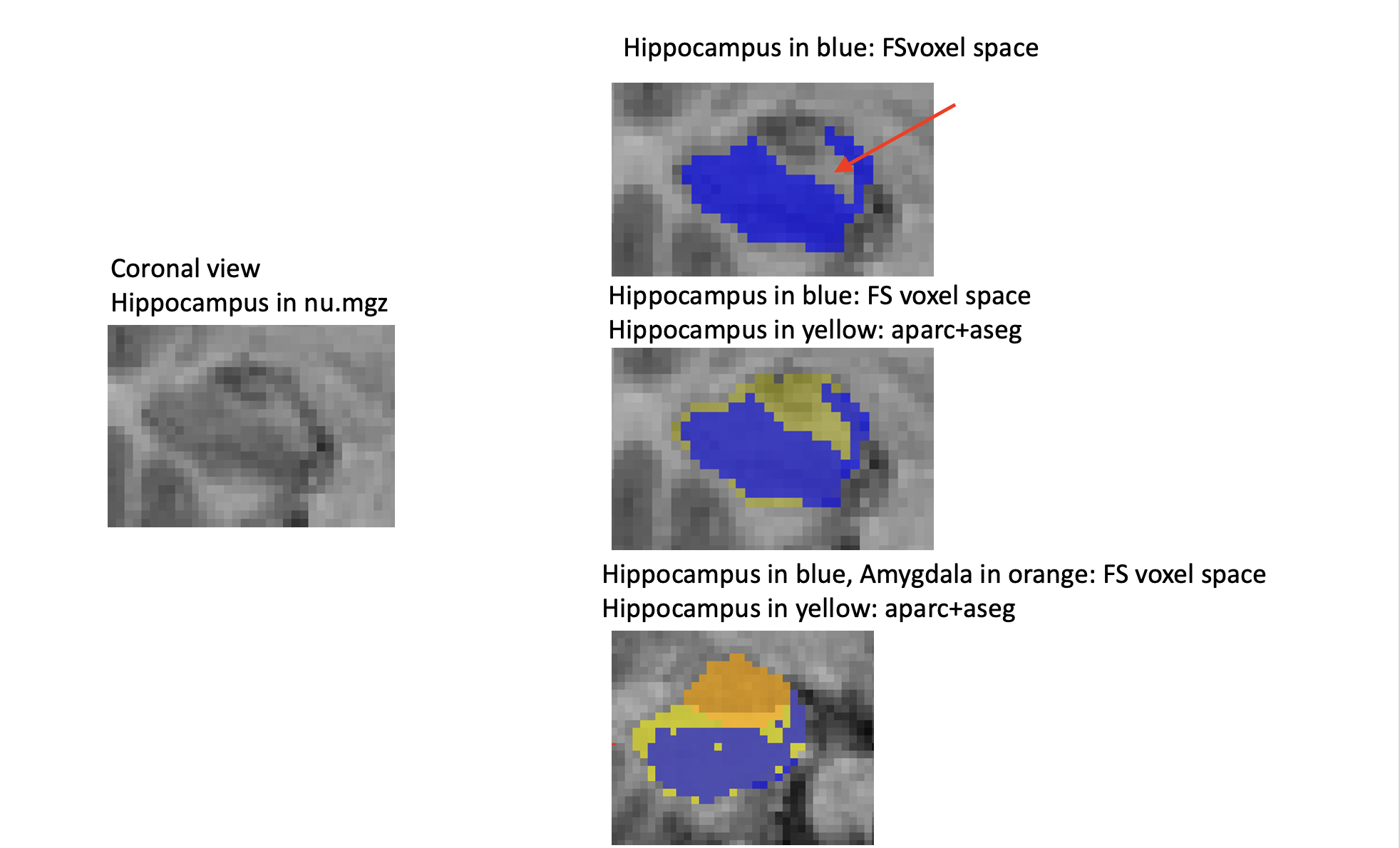
I am in the process of checking my hippocampal segmentations. I am finding that the hippocampal mask is often underestimating certain grey matter areas perhaps pertaining to the hippocampus? For example, the area pointed by the red arrow, here below, was initially covered by the apart + aseg hippocampal mask. Would the FS voxel space mask of the hippocampus pass quality check or would you say that a part of the hippocampal grey matter would be missing ?
Thank you for your helpful insight,
Liz
Liz[cid:image001.png@01D68C86.C1F12140]
freesurfer@nmr.mgh.harvard.edu
-
 Iglesias Gonzalez, Juan E.