
External Email - Use Caution
Dear freesurfer experts,
I re-send this question because i didn't received (or missed) the answer from the first time i sent it.
I am performing a longitudinal study (2 time points, 1 year apart) in patients with Multiple Sclerosis using freesurfer 6.0.
After following the tutorial for the CROSS, the BASE (where I only performed few manual edits of the surfaces) and the LONG steps, I am facing a problem with the results of the longitudinal pipeline:
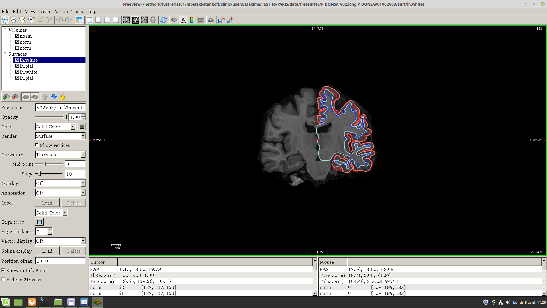
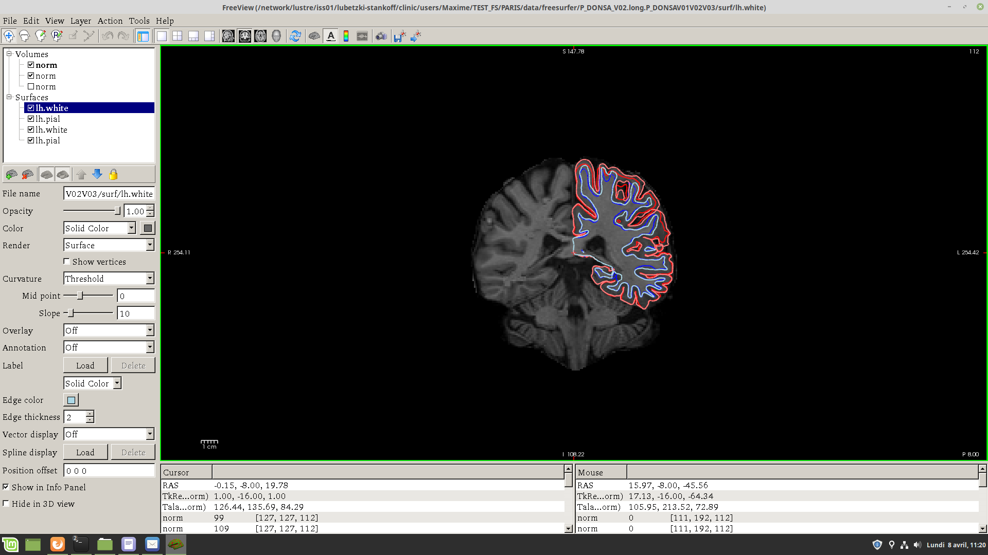
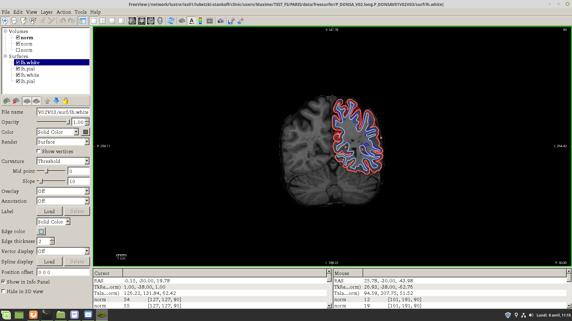
If I overlay the final surfaces of the different time points (the white and pial surfaces of the LONG step) on the BASE template using the command given in the fs tutorial, there are important differences between the two time points (see figure attached). This happened only on a subset of my subjects.
One of our collaborators in this study suggested that this might be because the patient was positioned differently in the two time points, in particular with respect to the z axis (and indeed the major differences between the two surfaces are in the topmost area of the brain).
Does anybody have experienced the same problem? And how can I correct for it?
Thank you in advance for your help.
(*in the captures surfaces of the first visit are in red and blue and in lighter tones for the second visit)

Hi Maxime,
also how do the surfaces look on their respective time point images from long? Do they look accurate there? Could be that there are differences across the time points that cause the surfaces to differ this much. Was the same hardware (scanner, coil) and same protocol used for both time points?
Best, Martin
On Tue, 2019-04-09 at 11:04 +0000, maxime GUILLAUME wrote:
External Email - Use CautionDear freesurfer experts,
I re-send this question because i didn't received (or missed) the answer from the first time i sent it.
I am performing a longitudinal study (2 time points, 1 year apart) in patients with Multiple Sclerosis using freesurfer 6.0.
After following the tutorial for the CROSS, the BASE (where I only performed few manual edits of the surfaces) and the LONG steps, I am facing a problem with the results of the longitudinal pipeline:
If I overlay the final surfaces of the different time points (the white and pial surfaces of the LONG step) on the BASE template using the command given in the fs tutorial, there are important differences between the two time points (see figure attached). This happened only on a subset of my subjects. One of our collaborators in this study suggested that this might be because the patient was positioned differently in the two time points, in particular with respect to the z axis (and indeed the major differences between the two surfaces are in the topmost area of the brain). Does anybody have experienced the same problem? And how can I correct for it?
Thank you in advance for your help.
(*in the captures surfaces of the first visit are in red and blue and in lighter tones for the second visit) _______________________________________________ Freesurfer mailing list Freesurfer@nmr.mgh.harvard.edu https://mail.nmr.mgh.harvard.edu/mailman/listinfo/freesurfer
freesurfer@nmr.mgh.harvard.edu
-
 Martin Reuter
 -
 maxime GUILLAUME