
External Email - Use Caution
Hello FreeSurfer Developers,
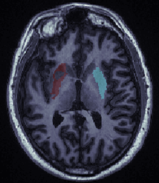
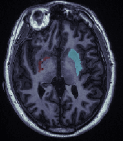
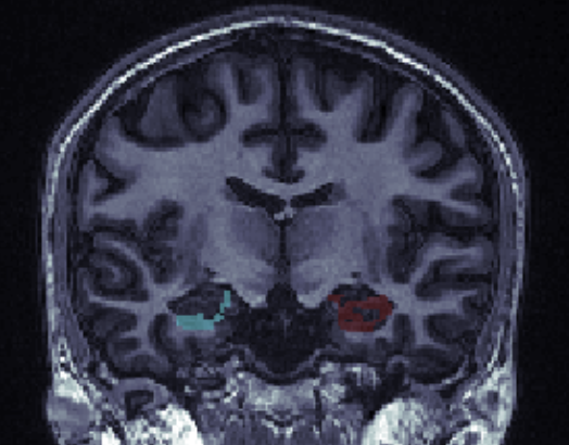
I am attempting to run recon-all on a group of nifti T1’s (parameters listed below). All the T1’s look good (no noticeable artifacts) and the cortical parcellations work well, and log files say completed without error. However, when looking at the subcortical segmentations, the thalamus, caudate and hippocampus appear consistently under segmented (screenshots below).
T1 MRI: 1.5 Tesla GE Signa HDxt scanner with an 8-channel head coil, using a three-dimensional fast spoiled gradient echo (3D-FSPGR) imaging sequence, with 188 sagittal slices scanned using the following parameters: repetition time (TR) = 11.70 ms, echo time (TE) = 5.12 ms, inversion time = 500 ms, flip angle = 8°, matrix size = 256×256, slice thickness = 1 mm, in-plane resolution = 1 mm^2.
We have tried a number of preprocessing steps on the T1 images but none of them appear to significantly improve the subcortical segmentations:
1. Normalizing the intensity of T1 voxels 2. Running FreeSurfer versions 5.3, 6, and 7 3. FastSurfer 4. DeNoising using ANTS 5. Bias field correction using ANTS
We did not find anyone with similar issues in the archive so any insights or guidance would be much appreciated.
Sincerely, Melody

Hippcampus actually looks like it might be ok; are those holes actually ventricle? I don't see a caudate label on there. Thalamus can often be underlabeled as there is so much WM in it. You can try running the thalamic nuclei segmentation, eg, segment_subregions thalamus --cross subject This will segment the nuclei, but you can just merge them all together to get the whole thalamus seg
On 1/30/2025 3:18 PM, Jeong Yeon Kang wrote:
External Email - Use Caution
Hello FreeSurfer Developers,
I am attempting to run recon-all on a group of nifti T1’s (parameters listed below). All the T1’s look good (no noticeable artifacts) and the cortical parcellations work well, and log files say completed without error. However, when looking at the subcortical segmentations, the thalamus, caudate and hippocampus appear consistently under segmented (screenshots below).
T1 MRI: 1.5 Tesla GE Signa HDxt scanner with an 8-channel head coil, using a three-dimensional fast spoiled gradient echo (3D-FSPGR) imaging sequence, with 188 sagittal slices scanned using the following parameters: repetition time (TR) = 11.70 ms, echo time (TE) = 5.12 ms, inversion time = 500 ms, flip angle = 8°, matrix size = 256×256, slice thickness = 1 mm, in-plane resolution = 1 mm^2.
We have tried a number of preprocessing steps on the T1 images but none of them appear to significantly improve the subcortical segmentations:
- Normalizing the intensity of T1 voxels
- Running FreeSurfer versions 5.3, 6, and 7
- FastSurfer
- DeNoising using ANTS
- Bias field correction using ANTS
We did not find anyone with similar issues in the archive so any insights or guidance would be much appreciated.
Sincerely, Melody
-- *Melody Jeong Yeon Kang* Ph.D. Student Imaging Genetics Center *USC* Stevens Neuroimaging and Informatics Institute Keck School of Medicine of *USC* University of Southern California Email: jkang395@usc.edu
Freesurfer mailing list Freesurfer@nmr.mgh.harvard.edu https://mail.nmr.mgh.harvard.edu/mailman/listinfo/freesurfer
External Email - Use Caution
Hi Dr. Greve,
Thank you very much for your reply, and we will definitely consider running the thalamic nuclei segmentation.
To provide additional information, for the thalamus, caudate (attached photos below), and hippocampus, undersegmented regions are labelled as "CSF" and not white matter in freeview. Please let me know if regions being labelled as CSF provides any clues as to why multiple subcortical ROIs in most of the subjects may be failing.
Sincerely, Melody
[image: Screen Shot 2025-02-12 at 11.48.27 AM.png]
[image: Screen Shot 2025-02-12 at 11.46.18 AM.png][image: Screen Shot 2025-02-12 at 11.46.30 AM.png]
On Mon, Feb 3, 2025 at 6:26 AM Douglas N. Greve dgreve@mgh.harvard.edu wrote:
Hippcampus actually looks like it might be ok; are those holes actually ventricle? I don't see a caudate label on there. Thalamus can often be underlabeled as there is so much WM in it. You can try running the thalamic nuclei segmentation, eg,
Hippcampus actually looks like it might be ok; are those holes actually ventricle? I don't see a caudate label on there. Thalamus can often be underlabeled as there is so much WM in it. You can try running the thalamic nuclei segmentation, eg, segment_subregions thalamus --cross subject This will segment the nuclei, but you can just merge them all together to get the whole thalamus seg
On 1/30/2025 3:18 PM, Jeong Yeon Kang wrote:
External Email - Use CautionHello FreeSurfer Developers,
I am attempting to run recon-all on a group of nifti T1’s (parameters listed below). All the T1’s look good (no noticeable artifacts) and the cortical parcellations work well, and log files say completed without error. However, when looking at the subcortical segmentations, the thalamus, caudate and hippocampus appear consistently under segmented (screenshots below).
T1 MRI: 1.5 Tesla GE Signa HDxt scanner with an 8-channel head coil, using a three-dimensional fast spoiled gradient echo (3D-FSPGR) imaging sequence, with 188 sagittal slices scanned using the following parameters: repetition time (TR) = 11.70 ms, echo time (TE) = 5.12 ms, inversion time = 500 ms, flip angle = 8°, matrix size = 256×256, slice thickness = 1 mm, in-plane resolution = 1 mm^2.
We have tried a number of preprocessing steps on the T1 images but none of them appear to significantly improve the subcortical segmentations:
- Normalizing the intensity of T1 voxels
- Running FreeSurfer versions 5.3, 6, and 7
- FastSurfer
- DeNoising using ANTS
- Bias field correction using ANTS
We did not find anyone with similar issues in the archive so any insights or guidance would be much appreciated.
Sincerely, Melody
-- *Melody Jeong Yeon Kang* Ph.D. Student Imaging Genetics Center *USC* Stevens Neuroimaging and Informatics Institute Keck School of Medicine of *USC* University of Southern California Email: jkang395@usc.edu
Freesurfer mailing listFreesurfer@nmr.mgh.harvard.eduhttps://mail.nmr.mgh.harvard.edu/mailman/listinfo/freesurfer https://secure-web.cisco.com/1GR2dbBk9-kh4KNfizPbpDmnJ-Q0OnAcCCzBWQSU8i3BUUDNCjwh1pFko5xv0H9Woa--TmUXIist308fQiV6A5kntBjiCw28iBY3RJB1L8yPcp38dOcufHvDtdccaDfZDei7N4fhXgLliO4svOynOZvf_ZvYVEzahuiQKd6HLiUZb18nVNTf3PwVCLMA28ZbZMFHfMeY4jh9RuioiqxzJFE4dFA3O1I33FivY-ARFfTTtxeaU2JT1F2GfOTJcSQ82oMhRjzIxD-EpKmHQMnZJtljUwb7Q2QLprvjctqt_z62R6xru-70RZnNqKGZBQt6ni0863l7B6VwdOQyR-sSDbg/https%3A%2F%2Furldefense.com%2Fv3%2F__https%3A%2F%2Fmail.nmr.mgh.harvard.edu%2Fmailman%2Flistinfo%2Ffreesurfer__;!!LIr3w8kk_Xxm!vE0TQ-mqnt5R_AgwuKxI5EIcA5O4i0kp6zqz3UmVUuSQNoP454tHw-N2ZJAVLMLeZ8C8K3Zy3wc3WdpW40rA3z8Z$
Freesurfer mailing list Freesurfer@nmr.mgh.harvard.edu
https://secure-web.cisco.com/1GR2dbBk9-kh4KNfizPbpDmnJ-Q0OnAcCCzBWQSU8i3BUUD... The information in this e-mail is intended only for the person to whom it is addressed. If you believe this e-mail was sent to you in error and the e-mail contains patient information, please contact the Mass General Brigham Compliance HelpLine at https://secure-web.cisco.com/1dA9F7iHI2XjFlNQ8JRKLFu-EJx0FzI2Bd1i71VY1FzrKJh... < https://secure-web.cisco.com/1dA9F7iHI2XjFlNQ8JRKLFu-EJx0FzI2Bd1i71VY1FzrKJh...
.
freesurfer@nmr.mgh.harvard.edu
-
 Douglas N. Greve
 -
 Jeong Yeon Kang