
Hello Freesurfers,
I'm putting *.IMG files into the program and after correcting talaraich transformations for them, I notice that when the reconstructions have completed, that there are entire lobes of white matter absent from the surfaces detected and from the wm.mgz file. I went through my hand at first to manually fill in the missing white matter for the first case, but after seeing how many were affected, I figured there was some setting I may be missing or some contrast level that may be off.
I had tried using control points, but after following the WIKI instructions on regenerating surfaces after placing them, I found no significant difference in white matter detection.
How do I ensure all of the white matter in my converted IMGs is identified?
Thank you kindly, Christopher Luna MIT Brain and Cognitive Sciences, Class of 2012 Desimone Lab, McGovern Institute for Brain Research MIT Archery Club Alpha Kappa Psi Colony at MIT

Hi Christopher,
  are you sure that the orientations are correct? That is, when you bring the orig.mgz up in tkmedit does the coronal view actually show the brain coronally? Img
  is a dangerous format to use as it doesn't contain orientation info. Do you have access to the data in some other format before it was convered to img?

  Bruce
On Mon, 1 Aug 2011, Christopher Luna wrote:
Hello Freesurfers, I'm putting *.IMG files into the program and after correcting talaraich transformations for them, I notice that when the reconstructions have completed, that there are entire lobes of white matter absent from the surfaces detected and from the wm.mgz file. I went through my hand at first to manually fill in the missing white matter for the first case, but after seeing how many were affected, I figured there was some setting I may be missing or some contrast level that may be off.
I had tried using control points, but after following the WIKI instructions on regenerating surfaces after placing them, I found no significant difference in white matter detection.
How do I ensure all of the white matter in my converted IMGs is identified?
Thank you kindly, Christopher Luna MIT Brain and Cognitive Sciences, Class of 2012Desimone Lab, McGovern Institute for Brain Research MIT Archery Club Alpha Kappa Psi Colony at MIT
Hi Bruce,
The orientations are correct - they weren't at first so I had to do a lot of work in tkregister2 to get the orientations correct. Unfortunately I only have access to the *.IMG and sometimes the *.HDR file for each subject.
Christopher Luna MIT Brain and Cognitive Sciences, Class of 2012 Desimone Lab, McGovern Institute for Brain Research MIT Archery Club Alpha Kappa Psi Colony at MIT
On Mon, Aug 1, 2011 at 12:16 PM, Bruce Fischl fischl@nmr.mgh.harvard.eduwrote:
Hi Christopher,
are you sure that the orientations are correct? That is, when you bring the orig.mgz up in tkmedit does the coronal view actually show the brain coronally? Img is a dangerous format to use as it doesn't contain orientation info. Do you have access to the data in some other format before it was convered to img?
Bruce
On Mon, 1 Aug 2011, Christopher Luna wrote:
Hello Freesurfers,
I'm putting *.IMG files into the program and after correcting talaraich transformations for them, I notice that when the reconstructions have completed, that there are entire lobes of white matter absent from the surfaces detected and from the wm.mgz file. I went through my hand at first to manually fill in the missing white matter for the first case, but after seeing how many were affected, I figured there was some setting I may be missing or some contrast level that may be off.
I had tried using control points, but after following the WIKI instructions on regenerating surfaces after placing them, I found no significant difference in white matter detection.
How do I ensure all of the white matter in my converted IMGs is identified?
Thank you kindly, Christopher Luna MIT Brain and Cognitive Sciences, Class of 2012Desimone Lab, McGovern Institute for Brain Research
MIT Archery Club Alpha Kappa Psi Colony at MIT
The information in this e-mail is intended only for the person to whom it is addressed. If you believe this e-mail was sent to you in error and the e-mail contains patient information, please contact the Partners Compliance HelpLine at http://www.partners.org/**compliancelinehttp://www.partners.org/complianceline. If the e-mail was sent to you in error but does not contain patient information, please contact the sender and properly dispose of the e-mail.
how do you know left/right is correct? In any case, if you upload the subject we'll take a look. Things very rarely fail that badly
  On Mon, 1 Aug 2011, Christopher Luna wrote:

  Hi Bruce,
  The orientations are correct - they weren't at first so I
  had to do a lot of work in tkregister2 to get the
  orientations correct. Unfortunately I only have access to
  the
  *.IMG and sometimes the *.HDR file for each subject.

  Christopher Luna
MIT Brain and Cognitive Sciences, Class of 2012Desimone Lab, McGovern Institute for Brain Research MIT Archery Club Alpha Kappa Psi Colony at MIT
On Mon, Aug 1, 2011 at 12:16 PM, Bruce Fischl fischl@nmr.mgh.harvard.edu wrote: Hi Christopher,
are you sure that the orientations are correct? That is, when you bring the orig.mgz up in tkmedit does the coronal view actually show the brain coronally? Img is a dangerous format to use as it doesn't contain orientation info. Do you have access to the data in some other format before it was convered to img? BruceOn Mon, 1 Aug 2011, Christopher Luna wrote:
Hello Freesurfers, I'm putting *.IMG files into the program and after correcting talaraich transformations for them, I notice that when the reconstructions have completed, that there are entire lobes of white matter absent from the surfaces detected and from the wm.mgz file. I went through my hand at first to manually fill in the missing white matter for the first case, but after seeing how many were affected, I figured there was some setting I may be missing or some contrast level that may be off.
I had tried using control points, but after following the WIKI instructions on regenerating surfaces after placing them, I found no significant difference in white matter detection.
How do I ensure all of the white matter in my converted IMGs is identified?
Thank you kindly, Christopher Luna MIT Brain and Cognitive Sciences, Class of 2012Desimone Lab, McGovern Institute for Brain Research MIT Archery Club Alpha Kappa Psi Colony at MIT
The information in this e-mail is intended only for the person to whom it is addressed. If you believe this e-mail was sent to you in error and the e-mail contains patient information, please contact the Partners Compliance HelpLine at http://www.partners.org/complianceline . If the e-mail was sent to you in error but does not contain patient information, please contact the sender and properly dispose of the e-mail.
Ah, I forgot about the left/right orientation. Where should I upload these files?
Thanks, Christopher Luna MIT Brain and Cognitive Sciences, Class of 2012 Desimone Lab, McGovern Institute for Brain Research MIT Archery Club Alpha Kappa Psi Colony at MIT
On Mon, Aug 1, 2011 at 12:23 PM, Bruce Fischl fischl@nmr.mgh.harvard.eduwrote:
how do you know left/right is correct? In any case, if you upload the subject we'll take a look. Things very rarely fail that badly
 On Mon, 1 Aug 2011, Christopher Luna wrote:

 Hi Bruce,
 The orientations are correct - they weren't at first so I
 had to do a lot of work in tkregister2 to get the
 orientations correct. Unfortunately I only have access to
 the
 *.IMG and sometimes the *.HDR file for each subject.

 Christopher Luna
MIT Brain and Cognitive Sciences, Class of 2012Desimone Lab, McGovern Institute for Brain Research MIT Archery Club Alpha Kappa Psi Colony at MIT
On Mon, Aug 1, 2011 at 12:16 PM, Bruce Fischl fischl@nmr.mgh.harvard.edu wrote: Hi Christopher,
are you sure that the orientations are correct? That is, when youbring the orig.mgz up in tkmedit does the coronal view actually show the brain coronally? Img is a dangerous format to use as it doesn't contain orientation info. Do you have access to the data in some other format before it was convered to img?
BruceOn Mon, 1 Aug 2011, Christopher Luna wrote:
Hello Freesurfers, I'm putting *.IMG files into the program and after correcting talaraich transformations for them, I notice that when the reconstructions have completed, that there are entire lobes of white matter absent from the surfaces detected and from the wm.mgz file. I went through my hand at first to manually fill in the missing white matter for the first case, but after seeing how many were affected, I figured there was some setting I may be missing or some contrast level that may be off.
I had tried using control points, but after following the WIKI instructions on regenerating surfaces after placing them, I found no significant difference in white matter detection.
How do I ensure all of the white matter in my converted IMGs is identified?
Thank you kindly, Christopher Luna MIT Brain and Cognitive Sciences, Class of 2012Desimone Lab, McGovern Institute for Brain Research MIT Archery Club Alpha Kappa Psi Colony at MIT
The information in this e-mail is intended only for the person to whom it is addressed. If you believe this e-mail was sent to you in error and the e-mail contains patient information, please contact the Partners Compliance HelpLine at http://www.partners.org/**compliancelinehttp://www.partners.org/complianceline. If the e-mail was sent to you in error but does not contain patient information, please contact the sender and properly dispose of the e-mail.
https://surfer.nmr.mgh.harvard.edu/fswiki/FtpFileExchange?highlight=%28ftp%2... On Mon, 1 Aug 2011, Christopher Luna wrote:
  Ah, I forgot about the left/right orientation. Where should I upload these
  files?
  Thanks,
  Christopher Luna
MIT Brain and Cognitive Sciences, Class of 2012Desimone Lab, McGovern Institute for Brain Research MIT Archery Club Alpha Kappa Psi Colony at MIT
On Mon, Aug 1, 2011 at 12:23 PM, Bruce Fischl fischl@nmr.mgh.harvard.edu wrote: how do you know left/right is correct? In any case, if you upload the subject we'll take a look. Things very rarely fail that badly
 On Mon, 1 Aug 2011, Christopher Luna wrote:

 Hi Bruce,
 The orientations are correct - they weren't at first so I
 had to do a lot of work in tkregister2 to get the
 orientations correct. Unfortunately I only have access to
 the
 *.IMG and sometimes the *.HDR file for each subject.

 Christopher Luna
MIT Brain and Cognitive Sciences, Class of 2012Desimone Lab, McGovern Institute for Brain Research MIT Archery Club Alpha Kappa Psi Colony at MIT
On Mon, Aug 1, 2011 at 12:16 PM, Bruce Fischl fischl@nmr.mgh.harvard.edu wrote: Hi Christopher,
are you sure that the orientations are correct? That is, when you bring the orig.mgz up in tkmedit does the coronal view actually show the brain coronally? Img is a dangerous format to use as it doesn't contain orientation info. Do you have access to the data in some other format before it was convered to img?
Bruce
On Mon, 1 Aug 2011, Christopher Luna wrote:
Hello Freesurfers, I'm putting *.IMG files into the program and after correcting talaraich transformations for them, I notice that when the reconstructions have completed, that there are entire lobes of white matter absent from the surfaces detected and from the wm.mgz file. I went through my hand at first to manually fill in the missing white matter for the first case, but after seeing how many were affected, I figured there was some setting I may be missing or some contrast level that may be off.
I had tried using control points, but after following the WIKI instructions on regenerating surfaces after placing them, I found no significant difference in white matter detection.
How do I ensure all of the white matter in my converted IMGs is identified?
Thank you kindly, Christopher Luna MIT Brain and Cognitive Sciences, Class of 2012Desimone Lab, McGovern Institute for Brain Research MIT Archery Club Alpha Kappa Psi Colony at MIT
The information in this e-mail is intended only for the person to whom it is addressed. If you believe this e-mail was sent to you in error and the e-mail contains patient information, please contact the Partners Compliance HelpLine at http://www.partners.org/complianceline . If the e-mail was sent to you in error but does not contain patient information, please contact the sender and properly dispose of the e-mail.
I've uploaded the IMG and HDR files, they are named after my email address.
Thank you kindly, Christopher Luna MIT Brain and Cognitive Sciences, Class of 2012 Desimone Lab, McGovern Institute for Brain Research MIT Archery Club Alpha Kappa Psi Colony at MIT
On Mon, Aug 1, 2011 at 12:40 PM, Bruce Fischl fischl@nmr.mgh.harvard.eduwrote:
https://surfer.nmr.mgh.**harvard.edu/fswiki/**FtpFileExchange?highlight=%* *28ftp%29https://surfer.nmr.mgh.harvard.edu/fswiki/FtpFileExchange?highlight=%28ftp%29
On Mon, 1 Aug 2011, Christopher Luna wrote:
Ah, I forgot about the left/right orientation. Where should I upload these
files? Thanks, Christopher Luna MIT Brain and Cognitive Sciences, Class of 2012Desimone Lab, McGovern Institute for Brain Research MIT Archery Club Alpha Kappa Psi Colony at MIT
On Mon, Aug 1, 2011 at 12:23 PM, Bruce Fischl <fischl@nmr.mgh.harvard.edu
wrote: how do you know left/right is correct? In any case, if you upload the subject we'll take a look. Things very rarely fail that badly
 On Mon, 1 Aug 2011, Christopher Luna wrote:

 Hi Bruce,
 The orientations are correct - they weren't at first so I
 had to do a lot of work in tkregister2 to get the
 orientations correct. Unfortunately I only have access to
 the
 *.IMG and sometimes the *.HDR file for each subject.

 Christopher Luna
MIT Brain and Cognitive Sciences, Class of 2012Desimone Lab, McGovern Institute for Brain Research MIT Archery Club Alpha Kappa Psi Colony at MIT
On Mon, Aug 1, 2011 at 12:16 PM, Bruce Fischl fischl@nmr.mgh.harvard.edu wrote: Hi Christopher,
are you sure that the orientations are correct? That is,when you bring the orig.mgz up in tkmedit does the coronal view actually show the brain coronally? Img is a dangerous format to use as it doesn't contain orientation info. Do you have access to the data in some other format before it was convered to img?
BruceOn Mon, 1 Aug 2011, Christopher Luna wrote:
Hello Freesurfers, I'm putting *.IMG files into the program and after correcting talaraich transformations for them, I notice that when the reconstructions have completed, that there are entire lobes of white matter absent from the surfaces detected and from the wm.mgz file. I went through my hand at first to manually fill in the missing white matter for the first case, but after seeing how many were affected, I figured there was some setting I may be missing or some contrast level that may be off.
I had tried using control points, but after following the WIKI instructions on regenerating surfaces after placing them, I found no significant difference in white matter detection.
How do I ensure all of the white matter in my converted IMGs is identified?
Thank you kindly, Christopher Luna MIT Brain and Cognitive Sciences, Class of 2012Desimone Lab, McGovern Institute for Brain Research MIT Archery Club Alpha Kappa Psi Colony at MIT
The information in this e-mail is intended only for the person to whom it is addressed. If you believe this e-mail was sent to you in error and the e-mail contains patient information, please contact the Partners Compliance HelpLine at http://www.partners.org/**compliancelinehttp://www.partners.org/complianceline. If the e-mail was sent to you in error but does not contain patient information, please contact the sender and properly dispose of the e-mail.
Hi Chris,
they are front/back reversed (a/p), which is why everything fails. I don't know how you are ever going to know left from right though. You should talk to whoever got you this data and see if they have some way to tell.
cheers Bruce
On Mon, 1 Aug 2011, Christopher Luna wrote:
I've uploaded the IMG and HDR files, they are named after my email address. Thank you kindly, Christopher Luna MIT Brain and Cognitive Sciences, Class of 2012Desimone Lab, McGovern Institute for Brain Research MIT Archery Club Alpha Kappa Psi Colony at MIT
On Mon, Aug 1, 2011 at 12:40 PM, Bruce Fischl fischl@nmr.mgh.harvard.edu wrote: https://surfer.nmr.mgh.harvard.edu/fswiki/FtpFileExchange?highlight=%28ftp%2... On Mon, 1 Aug 2011, Christopher Luna wrote:
 Ah, I forgot about the left/right orientation. Where should I upload these
 files?
 Thanks,
 Christopher Luna
MIT Brain and Cognitive Sciences, Class of 2012Desimone Lab, McGovern Institute for Brain Research MIT Archery Club Alpha Kappa Psi Colony at MIT
On Mon, Aug 1, 2011 at 12:23 PM, Bruce Fischl fischl@nmr.mgh.harvard.edu wrote: how do you know left/right is correct? In any case, if you upload the subject we'll take a look. Things very rarely fail that badly
 On Mon, 1 Aug 2011, Christopher Luna wrote:

 Hi Bruce,
 The orientations are correct - they weren't at first so I
 had to do a lot of work in tkregister2 to get the
 orientations correct. Unfortunately I only have access to
 the
 *.IMG and sometimes the *.HDR file for each subject.

 Christopher Luna
MIT Brain and Cognitive Sciences, Class of 2012Desimone Lab, McGovern Institute for Brain Research MIT Archery Club Alpha Kappa Psi Colony at MIT
On Mon, Aug 1, 2011 at 12:16 PM, Bruce Fischl fischl@nmr.mgh.harvard.edu wrote: Hi Christopher,
are you sure that the orientations are correct? That is, when you bring the orig.mgz up in tkmedit does the coronal view actually show the brain coronally? Img is a dangerous format to use as it doesn't contain orientation info. Do you have access to the data in some other format before it was convered to img?
Bruce
On Mon, 1 Aug 2011, Christopher Luna wrote:
Hello Freesurfers, I'm putting *.IMG files into the program and after correcting talaraich transformations for them, I notice that when the reconstructions have completed, that there are entire lobes of white matter absent from the surfaces detected and from the wm.mgz file. I went through my hand at first to manually fill in the missing white matter for the first case, but after seeing how many were affected, I figured there was some setting I may be missing or some contrast level that may be off.
I had tried using control points, but after following the WIKI instructions on regenerating surfaces after placing them, I found no significant difference in white matter detection.
How do I ensure all of the white matter in my converted IMGs is identified?
Thank you kindly, Christopher Luna MIT Brain and Cognitive Sciences, Class of 2012Desimone Lab, McGovern Institute for Brain Research MIT Archery Club Alpha Kappa Psi Colony at MIT
The information in this e-mail is intended only for the person to whom it is addressed. If you believe this e-mail was sent to you in error and the e-mail contains patient information, please contact the Partners Compliance HelpLine at http://www.partners.org/complianceline . If the e-mail was sent to you in error but does not contain patient information, please contact the sender and properly dispose of the e-mail.
Hi Bruce,
Thank you very much! We'll get in contact with them.
Best, Christopher Luna MIT Brain and Cognitive Sciences, Class of 2012 Desimone Lab, McGovern Institute for Brain Research MIT Archery Club Alpha Kappa Psi Colony at MIT
On Mon, Aug 1, 2011 at 2:52 PM, Bruce Fischl fischl@nmr.mgh.harvard.eduwrote:
Hi Chris,
they are front/back reversed (a/p), which is why everything fails. I don't know how you are ever going to know left from right though. You should talk to whoever got you this data and see if they have some way to tell.
cheers
Bruce
On Mon, 1 Aug 2011, Christopher Luna wrote:
I've uploaded the IMG and HDR files, they are named after my email
address. Thank you kindly, Christopher Luna MIT Brain and Cognitive Sciences, Class of 2012Desimone Lab, McGovern Institute for Brain Research MIT Archery Club Alpha Kappa Psi Colony at MIT
On Mon, Aug 1, 2011 at 12:40 PM, Bruce Fischl fischl@nmr.mgh.harvard.edu wrote: https://surfer.nmr.mgh.**harvard.edu/fswiki/** FtpFileExchange?highlight=%**28ftp%29https://surfer.nmr.mgh.harvard.edu/fswiki/FtpFileExchange?highlight=%28ftp%29 On Mon, 1 Aug 2011, Christopher Luna wrote:
Ah, I forgot about the left/right orientation. Where should I uploadthese files? Thanks, Christopher Luna MIT Brain and Cognitive Sciences, Class of 2012Desimone Lab, McGovern Institute for Brain Research MIT Archery Club Alpha Kappa Psi Colony at MIT
On Mon, Aug 1, 2011 at 12:23 PM, Bruce Fischl <fischl@nmr.mgh.harvard.edu
wrote: how do you know left/right is correct? In any case, if you upload the subject we'll take a look. Things very rarely fail that badly
 On Mon, 1 Aug 2011, Christopher Luna wrote:

 Hi Bruce,
 The orientations are correct - they weren't at first so I
 had to do a lot of work in tkregister2 to get the
 orientations correct. Unfortunately I only have access to
 the
 *.IMG and sometimes the *.HDR file for each subject.

 Christopher Luna
MIT Brain and Cognitive Sciences, Class of 2012Desimone Lab, McGovern Institute for Brain Research MIT Archery Club Alpha Kappa Psi Colony at MIT
On Mon, Aug 1, 2011 at 12:16 PM, Bruce Fischl fischl@nmr.mgh.harvard.edu wrote: Hi Christopher,
are you sure that the orientations are correct? That is,when you bring the orig.mgz up in tkmedit does the coronal view actually show the brain coronally? Img is a dangerous format to use as it doesn't contain orientation info. Do you have access to the data in some other format before it was convered to img?
BruceOn Mon, 1 Aug 2011, Christopher Luna wrote:
Hello Freesurfers, I'm putting *.IMG files into the program and after correcting talaraich transformations for them, I notice that when the reconstructions have completed, that there are entire lobes of white matter absent from the surfaces detected and from the wm.mgz file. I went through my hand at first to manually fill in the missing white matter for the first case, but after seeing how many were affected, I figured there was some setting I may be missing or some contrast level that may be off.
I had tried using control points, but after following the WIKI instructions on regenerating surfaces after placing them, I found no significant difference in white matter detection.
How do I ensure all of the white matter in my converted IMGs is identified?
Thank you kindly, Christopher Luna MIT Brain and Cognitive Sciences, Class of 2012Desimone Lab, McGovern Institute for Brain Research MIT Archery Club Alpha Kappa Psi Colony at MIT
The information in this e-mail is intended only for the person to whom it is addressed. If you believe this e-mail was sent to you in error and the e-mail contains patient information, please contact the Partners Compliance HelpLine at http://www.partners.org/**compliancelinehttp://www.partners.org/complianceline. If the e-mail was sent to you in error but does not contain patient information, please contact the sender and properly dispose of the e-mail.
Hello,
On this topic again, we tried fixing the coordinate system (we flipped A/P) as much as we could until we heard back from our colleagues.
And we also altered the white matter thresholds from mri_segment in case the missing white matter was due to some strange qualities in the original IMG files.
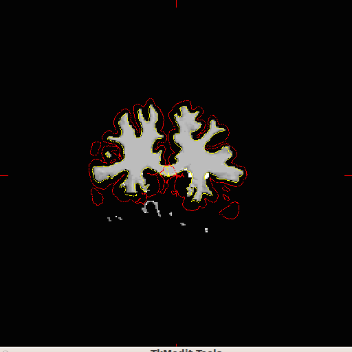
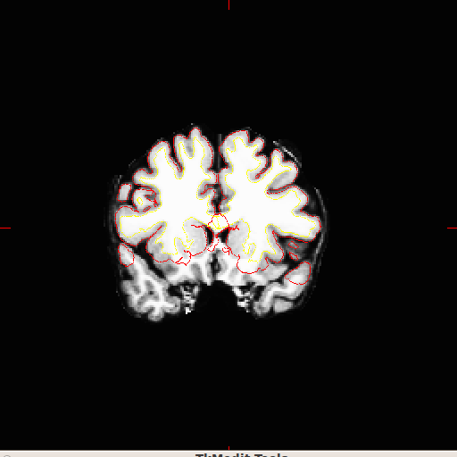
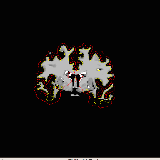
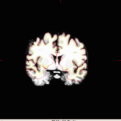
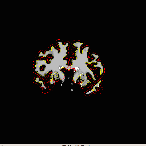
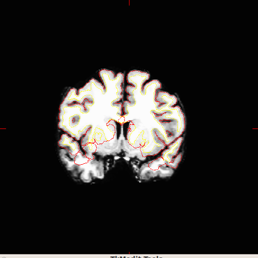
Unfortunately, this didn't work. I've attached some example pictures of what we end up with even after tinkering with the white threshold.
Are we going to have to add in this missing white matter by hand?
Christopher Luna MIT Brain and Cognitive Sciences, Class of 2012 Desimone Lab, McGovern Institute for Brain Research MIT Archery Club Alpha Kappa Psi Colony at MIT
On Mon, Aug 1, 2011 at 3:04 PM, Christopher Luna cdluna@mit.edu wrote:
Hi Bruce,
Thank you very much! We'll get in contact with them.
Best, Christopher Luna MIT Brain and Cognitive Sciences, Class of 2012 Desimone Lab, McGovern Institute for Brain Research MIT Archery Club Alpha Kappa Psi Colony at MIT
On Mon, Aug 1, 2011 at 2:52 PM, Bruce Fischl fischl@nmr.mgh.harvard.eduwrote:
Hi Chris,
  they are front/back reversed (a/p), which is why everything fails. I don't know how you are ever going to know left
  from right though. You should talk to whoever got you this data and see if they have some way to tell.

  cheers
  Bruce


  On Mon, 1 Aug 2011, Christopher Luna wrote:

  I've uploaded the IMG and HDR files, they are named after my email address.
  Thank you kindly,
  Christopher Luna
MIT Brain and Cognitive Sciences, Class of 2012Desimone Lab, McGovern Institute for Brain Research MIT Archery Club Alpha Kappa Psi Colony at MIT
On Mon, Aug 1, 2011 at 12:40 PM, Bruce Fischl < fischl@nmr.mgh.harvard.edu> wrote: https://surfer.nmr.mgh.**harvard.edu/fswiki/** FtpFileExchange?highlight=%**28ftp%29https://surfer.nmr.mgh.harvard.edu/fswiki/FtpFileExchange?highlight=%28ftp%29 On Mon, 1 Aug 2011, Christopher Luna wrote:
Ah, I forgot about the left/right orientation. Where should I uploadthese files? Thanks, Christopher Luna MIT Brain and Cognitive Sciences, Class of 2012Desimone Lab, McGovern Institute for Brain Research MIT Archery Club Alpha Kappa Psi Colony at MIT
On Mon, Aug 1, 2011 at 12:23 PM, Bruce Fischl < fischl@nmr.mgh.harvard.edu> wrote: how do you know left/right is correct? In any case, if you upload the subject we'll take a look. Things very rarely fail that badly
On Mon, 1 Aug 2011, Christopher Luna wrote: Hi Bruce, The orientations are correct - they weren't at first so I had to do a lot of work in tkregister2 to get the orientations correct. Unfortunately I only have access to the *.IMG and sometimes the *.HDR file for each subject. Christopher LunaMIT Brain and Cognitive Sciences, Class of 2012Desimone Lab, McGovern Institute for Brain Research MIT Archery Club Alpha Kappa Psi Colony at MIT
On Mon, Aug 1, 2011 at 12:16 PM, Bruce Fischl fischl@nmr.mgh.harvard.edu wrote: Hi Christopher,
are you sure that the orientations are correct? That is,when you bring the orig.mgz up in tkmedit does the coronal view actually show the brain coronally? Img is a dangerous format to use as it doesn't contain orientation info. Do you have access to the data in some other format before it was convered to img?
BruceOn Mon, 1 Aug 2011, Christopher Luna wrote:
Hello Freesurfers, I'm putting *.IMG files into the program and after correcting talaraich transformations for them, I notice that when the reconstructions have completed, that there are entire lobes of white matter absent from the surfaces detected and from the wm.mgz file. I went through my hand at first to manually fill in the missing white matter for the first case, but after seeing how many were affected, I figured there was some setting I may be missing or some contrast level that may be off.
I had tried using control points, but after following the WIKI instructions on regenerating surfaces after placing them, I found no significant difference in white matter detection.
How do I ensure all of the white matter in my converted IMGs is identified?
Thank you kindly, Christopher Luna MIT Brain and Cognitive Sciences, Class of 2012Desimone Lab, McGovern Institute for Brain Research MIT Archery Club Alpha Kappa Psi Colony at MIT
The information in this e-mail is intended only for the person to whom it is addressed. If you believe this e-mail was sent to you in error and the e-mail contains patient information, please contact the Partners Compliance HelpLine at http://www.partners.org/**compliancelinehttp://www.partners.org/complianceline. If the e-mail was sent to you in error but does not contain patient information, please contact the sender and properly dispose of the e-mail.
Eek, that looks awful! Can you upload this subject so we can figure out what is going on?
On Aug 9, 2011, at 4:09 PM, Christopher Luna cdluna@mit.edu wrote:
Hello,
On this topic again, we tried fixing the coordinate system (we flipped A/P) as much as we could until we heard back from our colleagues.
And we also altered the white matter thresholds from mri_segment in case the missing white matter was due to some strange qualities in the original IMG files.
Unfortunately, this didn't work. I've attached some example pictures of what we end up with even after tinkering with the white threshold.
Are we going to have to add in this missing white matter by hand?
Christopher Luna MIT Brain and Cognitive Sciences, Class of 2012 Desimone Lab, McGovern Institute for Brain Research MIT Archery Club Alpha Kappa Psi Colony at MIT
On Mon, Aug 1, 2011 at 3:04 PM, Christopher Luna cdluna@mit.edu wrote: Hi Bruce,
Thank you very much! We'll get in contact with them.
Best, Christopher Luna MIT Brain and Cognitive Sciences, Class of 2012 Desimone Lab, McGovern Institute for Brain Research MIT Archery Club Alpha Kappa Psi Colony at MIT
On Mon, Aug 1, 2011 at 2:52 PM, Bruce Fischl fischl@nmr.mgh.harvard.edu wrote: Hi Chris,
they are front/back reversed (a/p), which is why everything fails. I don't know how you are ever going to know left from right though. You should talk to whoever got you this data and see if they have some way to tell.
cheers
Bruce
On Mon, 1 Aug 2011, Christopher Luna wrote:
I've uploaded the IMG and HDR files, they are named after my email address. Thank you kindly, Christopher Luna MIT Brain and Cognitive Sciences, Class of 2012Desimone Lab, McGovern Institute for Brain Research MIT Archery Club Alpha Kappa Psi Colony at MIT
On Mon, Aug 1, 2011 at 12:40 PM, Bruce Fischl fischl@nmr.mgh.harvard.edu wrote: https://surfer.nmr.mgh.harvard.edu/fswiki/FtpFileExchange?highlight=%28ftp%2... On Mon, 1 Aug 2011, Christopher Luna wrote:
Ah, I forgot about the left/right orientation. Where should I upload these files? Thanks, Christopher LunaMIT Brain and Cognitive Sciences, Class of 2012Desimone Lab, McGovern Institute for Brain Research MIT Archery Club Alpha Kappa Psi Colony at MIT
On Mon, Aug 1, 2011 at 12:23 PM, Bruce Fischl fischl@nmr.mgh.harvard.edu wrote: how do you know left/right is correct? In any case, if you upload the subject we'll take a look. Things very rarely fail that badly
On Mon, 1 Aug 2011, Christopher Luna wrote: Hi Bruce, The orientations are correct - they weren't at first so I had to do a lot of work in tkregister2 to get the orientations correct. Unfortunately I only have access to the *.IMG and sometimes the *.HDR file for each subject. Christopher LunaMIT Brain and Cognitive Sciences, Class of 2012Desimone Lab, McGovern Institute for Brain Research MIT Archery Club Alpha Kappa Psi Colony at MIT
On Mon, Aug 1, 2011 at 12:16 PM, Bruce Fischl fischl@nmr.mgh.harvard.edu wrote: Hi Christopher,
are you sure that the orientations are correct? That is,when you bring the orig.mgz up in tkmedit does the coronal view actually show the brain coronally? Img is a dangerous format to use as it doesn't contain orientation info. Do you have access to the data in some other format before it was convered to img?
BruceOn Mon, 1 Aug 2011, Christopher Luna wrote:
Hello Freesurfers, I'm putting *.IMG files into the program and after correcting talaraich transformations for them, I notice that when the reconstructions have completed, that there are entire lobes of white matter absent from the surfaces detected and from the wm.mgz file. I went through my hand at first to manually fill in the missing white matter for the first case, but after seeing how many were affected, I figured there was some setting I may be missing or some contrast level that may be off.
I had tried using control points, but after following the WIKI instructions on regenerating surfaces after placing them, I found no significant difference in white matter detection.
How do I ensure all of the white matter in my converted IMGs is identified?
Thank you kindly, Christopher Luna MIT Brain and Cognitive Sciences, Class of 2012Desimone Lab, McGovern Institute for Brain Research MIT Archery Club Alpha Kappa Psi Colony at MIT
The information in this e-mail is intended only for the person to whom it is addressed. If you believe this e-mail was sent to you in error and the e-mail contains patient information, please contact the Partners Compliance HelpLine at http://www.partners.org/complianceline . If the e-mail was sent to you in error but does not contain patient information, please contact the sender and properly dispose of the e-mail.
  <example1a.png>

  <example1b.png>

  <example2a.png>

  <example2b.png>

  <example3a.png>

  <example3b.png>
Okay, I've uploaded them as cdluna_whitematter_002.img and *.hdr.
Thanks, Christopher Luna MIT Brain and Cognitive Sciences, Class of 2012 Desimone Lab, McGovern Institute for Brain Research MIT Archery Club Alpha Kappa Psi Colony at MIT
On Tue, Aug 9, 2011 at 11:02 PM, Bruce Fischl fischl@nmr.mgh.harvard.eduwrote:
Eek, that looks awful! Can you upload this subject so we can figure out what is going on?
On Aug 9, 2011, at 4:09 PM, Christopher Luna cdluna@mit.edu wrote:
Hello,
On this topic again, we tried fixing the coordinate system (we flipped A/P) as much as we could until we heard back from our colleagues.
And we also altered the white matter thresholds from mri_segment in case the missing white matter was due to some strange qualities in the original IMG files.
Unfortunately, this didn't work. I've attached some example pictures of what we end up with even after tinkering with the white threshold.
Are we going to have to add in this missing white matter by hand?
Christopher Luna MIT Brain and Cognitive Sciences, Class of 2012 Desimone Lab, McGovern Institute for Brain Research MIT Archery Club Alpha Kappa Psi Colony at MIT
On Mon, Aug 1, 2011 at 3:04 PM, Christopher Luna < cdluna@mit.edu cdluna@mit.edu> wrote:
Hi Bruce,
Thank you very much! We'll get in contact with them.
Best, Christopher Luna MIT Brain and Cognitive Sciences, Class of 2012 Desimone Lab, McGovern Institute for Brain Research MIT Archery Club Alpha Kappa Psi Colony at MIT
On Mon, Aug 1, 2011 at 2:52 PM, Bruce Fischl <fischl@nmr.mgh.harvard.edu fischl@nmr.mgh.harvard.edu> wrote:
Hi Chris,
they are front/back reversed (a/p), which is why everything fails. I don't know how you are ever going to know left from right though. You should talk to whoever got you this data and see if they have some way to tell.
 cheers
 Bruce


 On Mon, 1 Aug 2011, Christopher Luna wrote:

 I've uploaded the IMG and HDR files, they are named after my email
address. Thank you kindly, Christopher Luna MIT Brain and Cognitive Sciences, Class of 2012Desimone Lab, McGovern Institute for Brain Research MIT Archery Club Alpha Kappa Psi Colony at MIT
On Mon, Aug 1, 2011 at 12:40 PM, Bruce Fischl <fischl@nmr.mgh.harvard.edu fischl@nmr.mgh.harvard.edu> wrote: https://surfer.nmr.mgh.harvard.edu/fswiki/FtpFileExchange?highlight=%28ftp%29 https://surfer.nmr.mgh.**harvard.edu/fswiki/** FtpFileExchange?highlight=%**28ftp%29 On Mon, 1 Aug 2011, Christopher Luna wrote:
Ah, I forgot about the left/right orientation. Where should Iupload these files? Thanks, Christopher Luna MIT Brain and Cognitive Sciences, Class of 2012Desimone Lab, McGovern Institute for Brain Research MIT Archery Club Alpha Kappa Psi Colony at MIT
On Mon, Aug 1, 2011 at 12:23 PM, Bruce Fischl <fischl@nmr.mgh.harvard.edu fischl@nmr.mgh.harvard.edu> wrote: how do you know left/right is correct? In any case, if you upload the subject we'll take a look. Things very rarely fail that badly
 On Mon, 1 Aug 2011, Christopher Luna wrote:

 Hi Bruce,
 The orientations are correct - they weren't at first so I
 had to do a lot of work in tkregister2 to get the
 orientations correct. Unfortunately I only have access to
 the
 *.IMG and sometimes the *.HDR file for each subject.

 Christopher Luna
MIT Brain and Cognitive Sciences, Class of 2012Desimone Lab, McGovern Institute for Brain Research MIT Archery Club Alpha Kappa Psi Colony at MIT
On Mon, Aug 1, 2011 at 12:16 PM, Bruce Fischl < fischl@nmr.mgh.harvard.edufischl@nmr.mgh.harvard.edu> wrote: Hi Christopher,
are you sure that the orientations are correct? That is,when you bring the orig.mgz up in tkmedit does the coronal view actually show the brain coronally? Img is a dangerous format to use as it doesn't contain orientation info. Do you have access to the data in some other format before it was convered to img?
BruceOn Mon, 1 Aug 2011, Christopher Luna wrote:
Hello Freesurfers, I'm putting *.IMG files into the program and after correcting talaraich transformations for them, I notice that when the reconstructions have completed, that there are entire lobes of white matter absent from the surfaces detected and from the wm.mgz file. I went through my hand at first to manually fill in the missing white matter for the first case, but after seeing how many were affected, I figured there was some setting I may be missing or some contrast level that may be off.
I had tried using control points, but after following the WIKI instructions on regenerating surfaces after placing them, I found no significant difference in white matter detection.
How do I ensure all of the white matter in my converted IMGs is identified?
Thank you kindly, Christopher Luna MIT Brain and Cognitive Sciences, Class of 2012Desimone Lab, McGovern Institute for Brain Research MIT Archery Club Alpha Kappa Psi Colony at MIT
The information in this e-mail is intended only for the person to whom it is addressed. If you believe this e-mail was sent to you in error and the e-mail contains patient information, please contact the Partners Compliance HelpLine at http://www.partners.org/compliancelinehttp://www.partners.org/**complianceline . If the e-mail was sent to you in error but does not contain patient information, please contact the sender and properly dispose of the e-mail.
 <example1a.png>

 <example1b.png>

 <example2a.png>

 <example2b.png>

 <example3a.png>

 <example3b.png>
Hi Chris,
  when I bring that one up in freeview it looks like the a/p axis is still reversed (i.e. 'P' is at the front of the head)

  Bruce
  On Wed, 10 Aug 2011, Christopher Luna wrote:

  Okay, I've uploaded them as cdluna_whitematter_002.img and *.hdr.
  Thanks,
  Christopher Luna
MIT Brain and Cognitive Sciences, Class of 2012Desimone Lab, McGovern Institute for Brain Research MIT Archery Club Alpha Kappa Psi Colony at MIT
On Tue, Aug 9, 2011 at 11:02 PM, Bruce Fischl fischl@nmr.mgh.harvard.edu wrote: Eek, that looks awful! Can you upload this subject so we can figure out what is going on?
On Aug 9, 2011, at 4:09 PM, Christopher Luna cdluna@mit.edu wrote:
Hello,On this topic again, we tried fixing the coordinate system (we flipped A/P) as much as we could until we heard back from our colleagues.
And we also altered the white matter thresholds from mri_segment in case the missing white matter was due to some strange qualities in the original IMG files.
Unfortunately, this didn't work. I've attached some example pictures of what we end up with even after tinkering with the white threshold.
Are we going to have to add in this missing white matter by hand?
Christopher Luna MIT Brain and Cognitive Sciences, Class of 2012Desimone Lab, McGovern Institute for Brain Research MIT Archery Club Alpha Kappa Psi Colony at MIT
On Mon, Aug 1, 2011 at 3:04 PM, Christopher Luna cdluna@mit.edu wrote: Hi Bruce, Thank you very much! We'll get in contact with them.
Best, Christopher Luna MIT Brain and Cognitive Sciences, Class of 2012Desimone Lab, McGovern Institute for Brain Research MIT Archery Club Alpha Kappa Psi Colony at MIT
On Mon, Aug 1, 2011 at 2:52 PM, Bruce Fischl fischl@nmr.mgh.harvard.edu wrote: Hi Chris,
they are front/back reversed (a/p), which is why everything fails. I don't know how you are ever going to know left from right though. You should talk to whoever got you this data and see if they have some way to tell. cheers Bruce On Mon, 1 Aug 2011, Christopher Luna wrote: I've uploaded the IMG and HDR files, they are named after my email address. Thank you kindly, Christopher LunaMIT Brain and Cognitive Sciences, Class of 2012Desimone Lab, McGovern Institute for Brain Research MIT Archery Club Alpha Kappa Psi Colony at MIT
On Mon, Aug 1, 2011 at 12:40 PM, Bruce Fischl fischl@nmr.mgh.harvard.edu wrote: https://surfer.nmr.mgh.harvard.edu/fswiki/FtpFileExchange?highlight=%28ftp%2... On Mon, 1 Aug 2011, Christopher Luna wrote:
Ah, I forgot about the left/right orientation. Where should I upload these files? Thanks, Christopher Luna MIT Brain and Cognitive Sciences, Class of 2012Desimone Lab, McGovern Institute for Brain Research MIT Archery Club Alpha Kappa Psi Colony at MIT
On Mon, Aug 1, 2011 at 12:23 PM, Bruce Fischl fischl@nmr.mgh.harvard.edu wrote: how do you know left/right is correct? In any case, if you upload the subject we'll take a look. Things very rarely fail that badly
On Mon, 1 Aug 2011, Christopher Luna wrote:
Hi Bruce, The orientations are correct - they weren't at first so I had to do a lot of work in tkregister2 to get the orientations correct. Unfortunately I only have access to the *.IMG and sometimes the *.HDR file for each subject.
Christopher Luna MIT Brain and Cognitive Sciences, Class of 2012Desimone Lab, McGovern Institute for Brain Research MIT Archery Club Alpha Kappa Psi Colony at MIT
On Mon, Aug 1, 2011 at 12:16 PM, Bruce Fischl fischl@nmr.mgh.harvard.edu wrote: Hi Christopher,
are you sure that the orientations are correct? That is, when you bring the orig.mgz up in tkmedit does the coronal view actually show the brain coronally? Img is a dangerous format to use as it doesn't contain orientation info. Do you have access to the data in some other format before it was convered to img?
Bruce
On Mon, 1 Aug 2011, Christopher Luna wrote:
Hello Freesurfers, I'm putting *.IMG files into the program and after correcting talaraich transformations for them, I notice that when the reconstructions have completed, that there are entire lobes of white matter absent from the surfaces detected and from the wm.mgz file. I went through my hand at first to manually fill in the missing white matter for the first case, but after seeing how many were affected, I figured there was some setting I may be missing or some contrast level that may be off.
I had tried using control points, but after following the WIKI instructions on regenerating surfaces after placing them, I found no significant difference in white matter detection.
How do I ensure all of the white matter in my converted IMGs is identified?
Thank you kindly, Christopher Luna MIT Brain and Cognitive Sciences, Class of 2012Desimone Lab, McGovern Institute for Brain Research MIT Archery Club Alpha Kappa Psi Colony at MIT
The information in this e-mail is intended only for the person to whom it is addressed. If you believe this e-mail was sent to you in error and the e-mail contains patient information, please contact the Partners Compliance HelpLine at http://www.partners.org/complianceline . If the e-mail was sent to you in error but does not contain patient information, please contact the sender and properly dispose of the e-mail.
<example1a.png> <example1b.png> <example2a.png> <example2b.png> <example3a.png> <example3b.png>
Hi Bruce,
We tried to reverse A/P by reversing it the Talairach transform since recon-all wasn't able to do it automatically. I'm actually not sure how to reverse it in the IMG and HDR files.
Christopher Luna MIT Brain and Cognitive Sciences, Class of 2012 Desimone Lab, McGovern Institute for Brain Research MIT Archery Club Alpha Kappa Psi Colony at MIT
On Wed, Aug 10, 2011 at 2:19 PM, Bruce Fischl fischl@nmr.mgh.harvard.eduwrote:
Hi Chris,
when I bring that one up in freeview it looks like the a/p axis is still reversed (i.e. 'P' is at the front of the head)
  Bruce
  On Wed, 10 Aug 2011, Christopher Luna wrote:

  Okay, I've uploaded them as cdluna_whitematter_002.img and *.hdr.
  Thanks,
  Christopher Luna
MIT Brain and Cognitive Sciences, Class of 2012Desimone Lab, McGovern Institute for Brain Research MIT Archery Club Alpha Kappa Psi Colony at MIT
On Tue, Aug 9, 2011 at 11:02 PM, Bruce Fischl fischl@nmr.mgh.harvard.edu wrote: Eek, that looks awful! Can you upload this subject so we can figure out what is going on?
On Aug 9, 2011, at 4:09 PM, Christopher Luna cdluna@mit.edu wrote:
Hello,On this topic again, we tried fixing the coordinate system (we flipped A/P) as much as we could until we heard back from our colleagues.
And we also altered the white matter thresholds from mri_segment in case the missing white matter was due to some strange qualities in the original IMG files.
Unfortunately, this didn't work. I've attached some example pictures of what we end up with even after tinkering with the white threshold.
Are we going to have to add in this missing white matter by hand?
Christopher Luna MIT Brain and Cognitive Sciences, Class of 2012Desimone Lab, McGovern Institute for Brain Research MIT Archery Club Alpha Kappa Psi Colony at MIT
On Mon, Aug 1, 2011 at 3:04 PM, Christopher Luna cdluna@mit.edu wrote: Hi Bruce, Thank you very much! We'll get in contact with them.
Best, Christopher Luna MIT Brain and Cognitive Sciences, Class of 2012Desimone Lab, McGovern Institute for Brain Research MIT Archery Club Alpha Kappa Psi Colony at MIT
On Mon, Aug 1, 2011 at 2:52 PM, Bruce Fischl fischl@nmr.mgh.harvard.edu wrote: Hi Chris,
they are front/back reversed (a/p), which is why everything fails. Idon't know how you are ever going to know left from right though. You should talk to whoever got you this data and see if they have some way to tell.
cheers Bruce On Mon, 1 Aug 2011, Christopher Luna wrote: I've uploaded the IMG and HDR files, they are named after my emailaddress. Thank you kindly, Christopher Luna MIT Brain and Cognitive Sciences, Class of 2012Desimone Lab, McGovern Institute for Brain Research MIT Archery Club Alpha Kappa Psi Colony at MIT
On Mon, Aug 1, 2011 at 12:40 PM, Bruce Fischl fischl@nmr.mgh.harvard.edu wrote: https://surfer.nmr.mgh.**harvard.edu/fswiki/** FtpFileExchange?highlight=%**28ftp%29https://surfer.nmr.mgh.harvard.edu/fswiki/FtpFileExchange?highlight=%28ftp%29 On Mon, 1 Aug 2011, Christopher Luna wrote:
Ah, I forgot about the left/right orientation. Where should I uploadthese files? Thanks, Christopher Luna MIT Brain and Cognitive Sciences, Class of 2012Desimone Lab, McGovern Institute for Brain Research MIT Archery Club Alpha Kappa Psi Colony at MIT
On Mon, Aug 1, 2011 at 12:23 PM, Bruce Fischl <fischl@nmr.mgh.harvard.edu
wrote: how do you know left/right is correct? In any case, if you upload the subject we'll take a look. Things very rarely fail that badly
On Mon, 1 Aug 2011, Christopher Luna wrote: Hi Bruce, The orientations are correct - they weren't at first so I had to do a lot of work in tkregister2 to get the orientations correct. Unfortunately I only have access to the *.IMG and sometimes the *.HDR file for each subject. Christopher LunaMIT Brain and Cognitive Sciences, Class of 2012Desimone Lab, McGovern Institute for Brain Research MIT Archery Club Alpha Kappa Psi Colony at MIT
On Mon, Aug 1, 2011 at 12:16 PM, Bruce Fischl fischl@nmr.mgh.harvard.edu wrote: Hi Christopher,
are you sure that the orientations are correct? That is,when you bring the orig.mgz up in tkmedit does the coronal view actually show the brain coronally? Img is a dangerous format to use as it doesn't contain orientation info. Do you have access to the data in some other format before it was convered to img?
BruceOn Mon, 1 Aug 2011, Christopher Luna wrote:
Hello Freesurfers, I'm putting *.IMG files into the program and after correcting talaraich transformations for them, I notice that when the reconstructions have completed, that there are entire lobes of white matter absent from the surfaces detected and from the wm.mgz file. I went through my hand at first to manually fill in the missing white matter for the first case, but after seeing how many were affected, I figured there was some setting I may be missing or some contrast level that may be off.
I had tried using control points, but after following the WIKI instructions on regenerating surfaces after placing them, I found no significant difference in white matter detection.
How do I ensure all of the white matter in my converted IMGs is identified?
Thank you kindly, Christopher Luna MIT Brain and Cognitive Sciences, Class of 2012Desimone Lab, McGovern Institute for Brain Research MIT Archery Club Alpha Kappa Psi Colony at MIT
The information in this e-mail is intended only for the person to whom it is addressed. If you believe this e-mail was sent to you in error and the e-mail contains patient information, please contact the Partners Compliance HelpLine at http://www.partners.org/**compliancelinehttp://www.partners.org/complianceline. If the e-mail was sent to you in error but does not contain patient information, please contact the sender and properly dispose of the e-mail.
<example1a.png> <example1b.png> <example2a.png> <example2b.png> <example3a.png> <example3b.png>
Hi Chris,
that won't work for us. We assume it's in an ras coordinate system. I think you can do it with mri_convert, Doug probably knows how, but if you can't tell left from right it's going to be a problem forever. It's not worth running it through recon-all until the .img files comes up with the appropriate directions in freeview or tkmedit. Try looking at the mri_convert help
cheers Bruce
On Wed, 10 Aug 2011, Christopher Luna wrote:
Hi Bruce, We tried to reverse A/P by reversing it the Talairach transform since recon-all wasn't able to do it automatically. I'm actually not sure how to reverse it in the IMG and HDR files.
Christopher Luna MIT Brain and Cognitive Sciences, Class of 2012Desimone Lab, McGovern Institute for Brain Research MIT Archery Club Alpha Kappa Psi Colony at MIT
On Wed, Aug 10, 2011 at 2:19 PM, Bruce Fischl fischl@nmr.mgh.harvard.edu wrote: Hi Chris,
when I bring that one up in freeview it looks like the a/p axis is still reversed (i.e. 'P' is at the front of the head) Bruce On Wed, 10 Aug 2011, Christopher Luna wrote: Okay, I've uploaded them as cdluna_whitematter_002.img and *.hdr. Thanks, Christopher LunaMIT Brain and Cognitive Sciences, Class of 2012Desimone Lab, McGovern Institute for Brain Research MIT Archery Club Alpha Kappa Psi Colony at MIT
On Tue, Aug 9, 2011 at 11:02 PM, Bruce Fischl fischl@nmr.mgh.harvard.edu wrote: Eek, that looks awful! Can you upload this subject so we can figure out what is going on?
On Aug 9, 2011, at 4:09 PM, Christopher Luna cdluna@mit.edu wrote:
Hello, On this topic again, we tried fixing the coordinate system (we flipped A/P) as much as we could until we heard back from our colleagues.
And we also altered the white matter thresholds from mri_segment in case the missing white matter was due to some strange qualities in the original IMG files.
Unfortunately, this didn't work. I've attached some example pictures of what we end up with even after tinkering with the white threshold.
Are we going to have to add in this missing white matter by hand?
Christopher Luna MIT Brain and Cognitive Sciences, Class of 2012Desimone Lab, McGovern Institute for Brain Research MIT Archery Club Alpha Kappa Psi Colony at MIT
On Mon, Aug 1, 2011 at 3:04 PM, Christopher Luna cdluna@mit.edu wrote: Hi Bruce, Thank you very much! We'll get in contact with them.
Best, Christopher Luna MIT Brain and Cognitive Sciences, Class of 2012Desimone Lab, McGovern Institute for Brain Research MIT Archery Club Alpha Kappa Psi Colony at MIT
On Mon, Aug 1, 2011 at 2:52 PM, Bruce Fischl fischl@nmr.mgh.harvard.edu wrote: Hi Chris,
they are front/back reversed (a/p), which is why everything fails. I don't know how you are ever going to know left from right though. You should talk to whoever got you this data and see if they have some way to tell.
cheers Bruce
On Mon, 1 Aug 2011, Christopher Luna wrote:
I've uploaded the IMG and HDR files, they are named after my email address. Thank you kindly, Christopher Luna MIT Brain and Cognitive Sciences, Class of 2012Desimone Lab, McGovern Institute for Brain Research MIT Archery Club Alpha Kappa Psi Colony at MIT
On Mon, Aug 1, 2011 at 12:40 PM, Bruce Fischl fischl@nmr.mgh.harvard.edu wrote: https://surfer.nmr.mgh.harvard.edu/fswiki/FtpFileExchange?highlight=%28ftp%2... On Mon, 1 Aug 2011, Christopher Luna wrote:
Ah, I forgot about the left/right orientation. Where should I upload these files? Thanks, Christopher Luna MIT Brain and Cognitive Sciences, Class of 2012Desimone Lab, McGovern Institute for Brain Research MIT Archery Club Alpha Kappa Psi Colony at MIT
On Mon, Aug 1, 2011 at 12:23 PM, Bruce Fischl fischl@nmr.mgh.harvard.edu wrote: how do you know left/right is correct? In any case, if you upload the subject we'll take a look. Things very rarely fail that badly
On Mon, 1 Aug 2011, Christopher Luna wrote:
Hi Bruce, The orientations are correct - they weren't at first so I had to do a lot of work in tkregister2 to get the orientations correct. Unfortunately I only have access to the *.IMG and sometimes the *.HDR file for each subject.
Christopher Luna MIT Brain and Cognitive Sciences, Class of 2012Desimone Lab, McGovern Institute for Brain Research MIT Archery Club Alpha Kappa Psi Colony at MIT
On Mon, Aug 1, 2011 at 12:16 PM, Bruce Fischl fischl@nmr.mgh.harvard.edu wrote: Hi Christopher,
are you sure that the orientations are correct? That is, when you bring the orig.mgz up in tkmedit does the coronal view actually show the brain coronally? Img is a dangerous format to use as it doesn't contain orientation info. Do you have access to the data in some other format before it was convered to img?
Bruce
On Mon, 1 Aug 2011, Christopher Luna wrote:
Hello Freesurfers, I'm putting *.IMG files into the program and after correcting talaraich transformations for them, I notice that when the reconstructions have completed, that there are entire lobes of white matter absent from the surfaces detected and from the wm.mgz file. I went through my hand at first to manually fill in the missing white matter for the first case, but after seeing how many were affected, I figured there was some setting I may be missing or some contrast level that may be off.
I had tried using control points, but after following the WIKI instructions on regenerating surfaces after placing them, I found no significant difference in white matter detection.
How do I ensure all of the white matter in my converted IMGs is identified?
Thank you kindly, Christopher Luna MIT Brain and Cognitive Sciences, Class of 2012Desimone Lab, McGovern Institute for Brain Research MIT Archery Club Alpha Kappa Psi Colony at MIT
The information in this e-mail is intended only for the person to whom it is addressed. If you believe this e-mail was sent to you in error and the e-mail contains patient information, please contact the Partners Compliance HelpLine at http://www.partners.org/complianceline . If the e-mail was sent to you in error but does not contain patient information, please contact the sender and properly dispose of the e-mail.
<example1a.png>
<example1b.png>
<example2a.png>
<example2b.png>
<example3a.png>
<example3b.png>

One thing you can try is to look at the current orientation with mri_info --orientation f.img This will print out a 3-letter string like LPS or LSA. Create a new string by replacing the A with a P or P with an A. then mri_convert f.img --in_orientation NewString f.new.nii where NewString is the 3-letter string you created. Try loading this into tkmedit or freeview to make sure that it is oriented correctly. But, like Bruce says, you won't be able to tell left from right (unless you have fiducials). doug
Bruce Fischl wrote:
Hi Chris,
that won't work for us. We assume it's in an ras coordinate system. I think you can do it with mri_convert, Doug probably knows how, but if you can't tell left from right it's going to be a problem forever. It's not worth running it through recon-all until the .img files comes up with the appropriate directions in freeview or tkmedit. Try looking at the mri_convert help
cheers Bruce
On Wed, 10 Aug 2011, Christopher Luna wrote:
Hi Bruce, We tried to reverse A/P by reversing it the Talairach transform since recon-all wasn't able to do it automatically. I'm actually not sure how to reverse it in the IMG and HDR files.
Christopher Luna MIT Brain and Cognitive Sciences, Class of 2012Desimone Lab, McGovern Institute for Brain Research MIT Archery Club Alpha Kappa Psi Colony at MIT
On Wed, Aug 10, 2011 at 2:19 PM, Bruce Fischl fischl@nmr.mgh.harvard.edu wrote: Hi Chris,
when I bring that one up in freeview it looks like the a/p axisis still reversed (i.e. 'P' is at the front of the head)
Bruce On Wed, 10 Aug 2011, Christopher Luna wrote: Okay, I've uploaded them as cdluna_whitematter_002.img and *.hdr. Thanks, Christopher LunaMIT Brain and Cognitive Sciences, Class of 2012Desimone Lab, McGovern Institute for Brain Research MIT Archery Club Alpha Kappa Psi Colony at MIT
On Tue, Aug 9, 2011 at 11:02 PM, Bruce Fischl fischl@nmr.mgh.harvard.edu wrote: Eek, that looks awful! Can you upload this subject so we can figure out what is going on?
On Aug 9, 2011, at 4:09 PM, Christopher Luna cdluna@mit.edu wrote:
Hello,On this topic again, we tried fixing the coordinate system (we flipped A/P) as much as we could until we heard back from our colleagues.
And we also altered the white matter thresholds from mri_segment in case the missing white matter was due to some strange qualities in the original IMG files.
Unfortunately, this didn't work. I've attached some example pictures of what we end up with even after tinkering with the white threshold.
Are we going to have to add in this missing white matter by hand?
Christopher Luna MIT Brain and Cognitive Sciences, Class of 2012Desimone Lab, McGovern Institute for Brain Research MIT Archery Club Alpha Kappa Psi Colony at MIT
On Mon, Aug 1, 2011 at 3:04 PM, Christopher Luna cdluna@mit.edu wrote: Hi Bruce, Thank you very much! We'll get in contact with them.
Best, Christopher Luna MIT Brain and Cognitive Sciences, Class of 2012Desimone Lab, McGovern Institute for Brain Research MIT Archery Club Alpha Kappa Psi Colony at MIT
On Mon, Aug 1, 2011 at 2:52 PM, Bruce Fischl fischl@nmr.mgh.harvard.edu wrote: Hi Chris,
they are front/back reversed (a/p), which is why everythingfails. I don't know how you are ever going to know left from right though. You should talk to whoever got you this data and see if they have some way to tell.
cheers Bruce On Mon, 1 Aug 2011, Christopher Luna wrote: I've uploaded the IMG and HDR files, they are named after myemail address. Thank you kindly, Christopher Luna MIT Brain and Cognitive Sciences, Class of 2012Desimone Lab, McGovern Institute for Brain Research MIT Archery Club Alpha Kappa Psi Colony at MIT
On Mon, Aug 1, 2011 at 12:40 PM, Bruce Fischl fischl@nmr.mgh.harvard.edu wrote:
https://surfer.nmr.mgh.harvard.edu/fswiki/FtpFileExchange?highlight=%28ftp%2...
 On Mon, 1 Aug 2011, Christopher Luna wrote:

 Ah, I forgot about the left/right orientation. Where should I
upload these files? Thanks, Christopher Luna MIT Brain and Cognitive Sciences, Class of 2012Desimone Lab, McGovern Institute for Brain Research MIT Archery Club Alpha Kappa Psi Colony at MIT
On Mon, Aug 1, 2011 at 12:23 PM, Bruce Fischl fischl@nmr.mgh.harvard.edu wrote: how do you know left/right is correct? In any case, if you upload the subject we'll take a look. Things very rarely fail that badly
 On Mon, 1 Aug 2011, Christopher Luna wrote:

 Hi Bruce,
 The orientations are correct - they weren't at first so I
 had to do a lot of work in tkregister2 to get the
 orientations correct. Unfortunately I only have access to
 the
 *.IMG and sometimes the *.HDR file for each subject.

 Christopher Luna
MIT Brain and Cognitive Sciences, Class of 2012Desimone Lab, McGovern Institute for Brain Research MIT Archery Club Alpha Kappa Psi Colony at MIT
On Mon, Aug 1, 2011 at 12:16 PM, Bruce Fischl fischl@nmr.mgh.harvard.edu wrote: Hi Christopher,
are you sure that the orientations are correct? That is,when you bring the orig.mgz up in tkmedit does the coronal view actually show the brain coronally? Img is a dangerous format to use as it doesn't contain orientation info. Do you have access to the data in some other format before it was convered to img?
BruceOn Mon, 1 Aug 2011, Christopher Luna wrote:
Hello Freesurfers, I'm putting *.IMG files into the program and after correcting talaraich transformations for them, I notice that when the reconstructions have completed, that there are entire lobes of white matter absent from the surfaces detected and from the wm.mgz file. I went through my hand at first to manually fill in the missing white matter for the first case, but after seeing how many were affected, I figured there was some setting I may be missing or some contrast level that may be off.
I had tried using control points, but after following the WIKI instructions on regenerating surfaces after placing them, I found no significant difference in white matter detection.
How do I ensure all of the white matter in my converted IMGs is identified?
Thank you kindly, Christopher Luna MIT Brain and Cognitive Sciences, Class of 2012Desimone Lab, McGovern Institute for Brain Research MIT Archery Club Alpha Kappa Psi Colony at MIT
The information in this e-mail is intended only for the person to whom it is addressed. If you believe this e-mail was sent to you in error and the e-mail contains patient information, please contact the Partners Compliance HelpLine at http://www.partners.org/complianceline . If the e-mail was sent to you in error but does not contain patient information, please contact the sender and properly dispose of the e-mail.
 <example1a.png>

 <example1b.png>

 <example2a.png>

 <example2b.png>

 <example3a.png>

 <example3b.png>
Freesurfer mailing list Freesurfer@nmr.mgh.harvard.edu https://mail.nmr.mgh.harvard.edu/mailman/listinfo/freesurfer
This fixed everything! Thank you!
Christopher Luna MIT Brain and Cognitive Sciences, Class of 2012 Desimone Lab, McGovern Institute for Brain Research MIT Archery Club Alpha Kappa Psi Colony at MIT
On Wed, Aug 10, 2011 at 2:43 PM, Douglas N Greve greve@nmr.mgh.harvard.eduwrote:
One thing you can try is to look at the current orientation with mri_info --orientation f.img This will print out a 3-letter string like LPS or LSA. Create a new string by replacing the A with a P or P with an A. then mri_convert f.img --in_orientation NewString f.new.nii where NewString is the 3-letter string you created. Try loading this into tkmedit or freeview to make sure that it is oriented correctly. But, like Bruce says, you won't be able to tell left from right (unless you have fiducials). doug
Bruce Fischl wrote:
Hi Chris,
that won't work for us. We assume it's in an ras coordinate system. I think you can do it with mri_convert, Doug probably knows how, but if you can't tell left from right it's going to be a problem forever. It's not worth running it through recon-all until the .img files comes up with the appropriate directions in freeview or tkmedit. Try looking at the mri_convert help
cheers Bruce
On Wed, 10 Aug 2011, Christopher Luna wrote:
Hi Bruce,
We tried to reverse A/P by reversing it the Talairach transform since recon-all wasn't able to do it automatically. I'm actually not sure how to reverse it in the IMG and HDR files.
Christopher Luna MIT Brain and Cognitive Sciences, Class of 2012Desimone Lab, McGovern Institute for Brain Research MIT Archery Club Alpha Kappa Psi Colony at MIT
On Wed, Aug 10, 2011 at 2:19 PM, Bruce Fischl < fischl@nmr.mgh.harvard.edu> wrote: Hi Chris,
when I bring that one up in freeview it looks like the a/p axis isstill reversed (i.e. 'P' is at the front of the head)
 Bruce
 On Wed, 10 Aug 2011, Christopher Luna wrote:

 Okay, I've uploaded them as cdluna_whitematter_002.img and *.hdr.
 Thanks,
 Christopher Luna
MIT Brain and Cognitive Sciences, Class of 2012Desimone Lab, McGovern Institute for Brain Research MIT Archery Club Alpha Kappa Psi Colony at MIT
On Tue, Aug 9, 2011 at 11:02 PM, Bruce Fischl < fischl@nmr.mgh.harvard.edu> wrote: Eek, that looks awful! Can you upload this subject so we can figure out what is going on?
On Aug 9, 2011, at 4:09 PM, Christopher Luna cdluna@mit.edu wrote:
Hello,On this topic again, we tried fixing the coordinate system (we flipped A/P) as much as we could until we heard back from our colleagues.
And we also altered the white matter thresholds from mri_segment in case the missing white matter was due to some strange qualities in the original IMG files.
Unfortunately, this didn't work. I've attached some example pictures of what we end up with even after tinkering with the white threshold.
Are we going to have to add in this missing white matter by hand?
Christopher Luna MIT Brain and Cognitive Sciences, Class of 2012Desimone Lab, McGovern Institute for Brain Research MIT Archery Club Alpha Kappa Psi Colony at MIT
On Mon, Aug 1, 2011 at 3:04 PM, Christopher Luna cdluna@mit.edu wrote: Hi Bruce, Thank you very much! We'll get in contact with them.
Best, Christopher Luna MIT Brain and Cognitive Sciences, Class of 2012Desimone Lab, McGovern Institute for Brain Research MIT Archery Club Alpha Kappa Psi Colony at MIT
On Mon, Aug 1, 2011 at 2:52 PM, Bruce Fischl fischl@nmr.mgh.harvard.edu wrote: Hi Chris,
they are front/back reversed (a/p), which is why everything fails. Idon't know how you are ever going to know left from right though. You should talk to whoever got you this data and see if they have some way to tell.
cheers
Bruce


On Mon, 1 Aug 2011, Christopher Luna wrote:

I've uploaded the IMG and HDR files, they are named after my email
address. Thank you kindly, Christopher Luna MIT Brain and Cognitive Sciences, Class of 2012Desimone Lab, McGovern Institute for Brain Research MIT Archery Club Alpha Kappa Psi Colony at MIT
On Mon, Aug 1, 2011 at 12:40 PM, Bruce Fischl < fischl@nmr.mgh.harvard.edu> wrote: https://surfer.nmr.mgh.**harvard.edu/fswiki/** FtpFileExchange?highlight=%**28ftp%29https://surfer.nmr.mgh.harvard.edu/fswiki/FtpFileExchange?highlight=%28ftp%29 On Mon, 1 Aug 2011, Christopher Luna wrote:
Ah, I forgot about the left/right orientation. Where should I uploadthese files? Thanks, Christopher Luna MIT Brain and Cognitive Sciences, Class of 2012Desimone Lab, McGovern Institute for Brain Research MIT Archery Club Alpha Kappa Psi Colony at MIT
On Mon, Aug 1, 2011 at 12:23 PM, Bruce Fischl < fischl@nmr.mgh.harvard.edu> wrote: how do you know left/right is correct? In any case, if you upload the subject we'll take a look. Things very rarely fail that badly
On Mon, 1 Aug 2011, Christopher Luna wrote:

Hi Bruce,
The orientations are correct - they weren't at first so I
had to do a lot of work in tkregister2 to get the
orientations correct. Unfortunately I only have access to
the
*.IMG and sometimes the *.HDR file for each subject.

Christopher Luna
MIT Brain and Cognitive Sciences, Class of 2012Desimone Lab, McGovern Institute for Brain Research MIT Archery Club Alpha Kappa Psi Colony at MIT
On Mon, Aug 1, 2011 at 12:16 PM, Bruce Fischl fischl@nmr.mgh.harvard.edu wrote: Hi Christopher,
are you sure that the orientations are correct? That is,when you bring the orig.mgz up in tkmedit does the coronal view actually show the brain coronally? Img is a dangerous format to use as it doesn't contain orientation info. Do you have access to the data in some other format before it was convered to img?
BruceOn Mon, 1 Aug 2011, Christopher Luna wrote:
Hello Freesurfers, I'm putting *.IMG files into the program and after correcting talaraich transformations for them, I notice that when the reconstructions have completed, that there are entire lobes of white matter absent from the surfaces detected and from the wm.mgz file. I went through my hand at first to manually fill in the missing white matter for the first case, but after seeing how many were affected, I figured there was some setting I may be missing or some contrast level that may be off.
I had tried using control points, but after following the WIKI instructions on regenerating surfaces after placing them, I found no significant difference in white matter detection.
How do I ensure all of the white matter in my converted IMGs is identified?
Thank you kindly, Christopher Luna MIT Brain and Cognitive Sciences, Class of 2012Desimone Lab, McGovern Institute for Brain Research MIT Archery Club Alpha Kappa Psi Colony at MIT
The information in this e-mail is intended only for the person to whom it is addressed. If you believe this e-mail was sent to you in error and the e-mail contains patient information, please contact the Partners Compliance HelpLine at http://www.partners.org/**compliancelinehttp://www.partners.org/complianceline. If the e-mail was sent to you in error but does not contain patient information, please contact the sender and properly dispose of the e-mail.
<example1a.png>

<example1b.png>

<example2a.png>

<example2b.png>

<example3a.png>

<example3b.png>
------------------------------**------------------------------**
______________________________**_________________ Freesurfer mailing list Freesurfer@nmr.mgh.harvard.edu https://mail.nmr.mgh.harvard.**edu/mailman/listinfo/**freesurferhttps://mail.nmr.mgh.harvard.edu/mailman/listinfo/freesurfer
-- Douglas N. Greve, Ph.D. MGH-NMR Center greve@nmr.mgh.harvard.edu Phone Number: 617-724-2358 Fax: 617-726-7422
Bugs: surfer.nmr.mgh.harvard.edu/**fswiki/BugReportinghttp://surfer.nmr.mgh.harvard.edu/fswiki/BugReporting FileDrop: www.nmr.mgh.harvard.edu/**facility/filedrop/index.htmlhttp://www.nmr.mgh.harvard.edu/facility/filedrop/index.html
freesurfer@nmr.mgh.harvard.edu
-
 Bruce Fischl
 -
 Christopher Luna
 -
 Douglas N Greve