
Hi Bruce,
Thank you very much for your quick reply. Sorry I hadn't been able to reply faster as I was trying to figure out the below mentioned issue from two days.
I have computed surface area of left-hippocampus using the commands mri_tessellate and mris_info. However I found that in freeview by selecting Tools->show label stats, I am able to display area of any label (PFA a screenshot) but these area stats do not match with the areas obtained using mri_tessellate and mris_info. I have two queries regarding this:
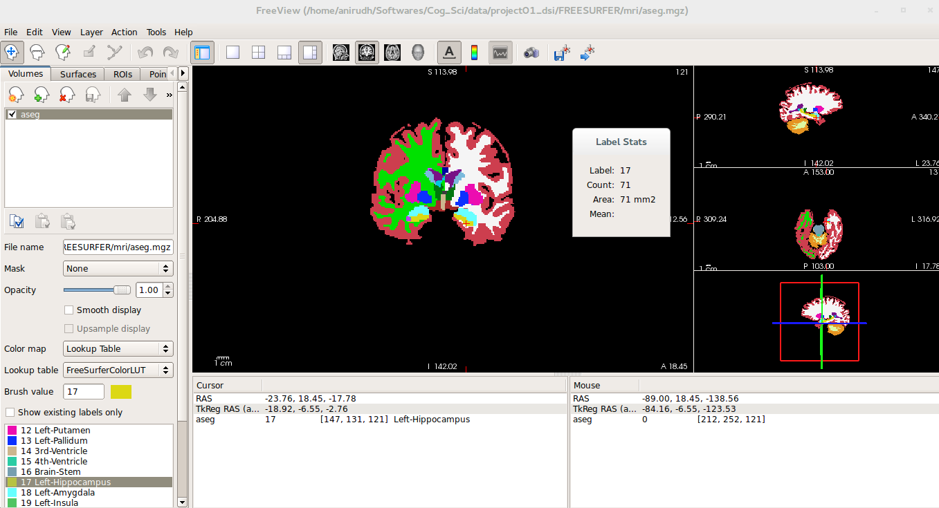
1) Surface areas of labels displayed in freeview doesn't match with subcortical structures' areas obtained by running mri_tesselate and mris_info. For example the attached screenshot shows that area of left-hippocampus is 71 mm^2 where as mri_tessellate followed by mris_info found the area for left-hippocampus to be 3586 (PFA the output of mris_tessellate and mris_info). Why is there a mismatch?
2) Based on your reply I thought area stats are not calculated for subcortical structures during "recon-all". If that is the case, how is freeview displaying area stats for subcortical areas? Also which file does freeview use to display these stats? Is it stats/[l/r]h.aparc.stats? I ask because even cortical structures' areas displayed by freeview are not matching with what is stored in these files.
Please let me know. Thank you.
Regards, Anirudh
Hi Bruce,
Sorry I forgot to attach the files mentioned in my last email. PFA the files here. Thank you.
Regards, Anirudh
On Wed, 11 Nov 2015 at 16:18 anirudh nihalani anirudhnihalani@gmail.com wrote:
Hi Bruce,
Thank you very much for your quick reply. Sorry I hadn't been able toreply faster as I was trying to figure out the below mentioned issue from two days.
I have computed surface area of left-hippocampus using the commands mri_tessellate and mris_info. However I found that in freeview by selecting Tools->show label stats, I am able to display area of any label (PFA a screenshot) but these area stats do not match with the areas obtained using mri_tessellate and mris_info. I have two queries regarding this:
- Surface areas of labels displayed in freeview doesn't match with
subcortical structures' areas obtained by running mri_tesselate and mris_info. For example the attached screenshot shows that area of left-hippocampus is 71 mm^2 where as mri_tessellate followed by mris_info found the area for left-hippocampus to be 3586 (PFA the output of mris_tessellate and mris_info). Why is there a mismatch?
- Based on your reply I thought area stats are not calculated for
subcortical structures during "recon-all". If that is the case, how is freeview displaying area stats for subcortical areas? Also which file does freeview use to display these stats? Is it stats/[l/r]h.aparc.stats? I ask because even cortical structures' areas displayed by freeview are not matching with what is stored in these files.
Please let me know. Thank you.
Regards, Anirudh

Probably Ruopeng will have to weigh in on freeview.
On 11/11/15 5:57 AM, anirudh nihalani wrote:
Hi Bruce,
Sorry I forgot to attach the files mentioned in my last email. PFA the files here. Thank you.
Regards, Anirudh
On Wed, 11 Nov 2015 at 16:18 anirudh nihalani <anirudhnihalani@gmail.com mailto:anirudhnihalani@gmail.com> wrote:
Hi Bruce, Thank you very much for your quick reply. Sorry I hadn't been able to reply faster as I was trying to figure out the below mentioned issue from two days. I have computed surface area of left-hippocampus using the commands mri_tessellate and mris_info. However I found that in freeview by selecting Tools->show label stats, I am able to display area of any label (PFA a screenshot) but these area stats do not match with the areas obtained using mri_tessellate and mris_info. I have two queries regarding this: 1) Surface areas of labels displayed in freeview doesn't match with subcortical structures' areas obtained by running mri_tesselate and mris_info. For example the attached screenshot shows that area of left-hippocampus is 71 mm^2 where as mri_tessellate followed by mris_info found the area for left-hippocampus to be 3586 (PFA the output of mris_tessellate and mris_info). Why is there a mismatch? 2) Based on your reply I thought area stats are not calculated for subcortical structures during "recon-all". If that is the case, how is freeview displaying area stats for subcortical areas? Also which file does freeview use to display these stats? Is it stats/[l/r]h.aparc.stats? I ask because even cortical structures' areas displayed by freeview are not matching with what is stored in these files. Please let me know. Thank you. Regards, Anirudh
Freesurfer mailing list Freesurfer@nmr.mgh.harvard.edu https://mail.nmr.mgh.harvard.edu/mailman/listinfo/freesurfer

Hi Anirudh,
Label Stats just shows the area of the label in that slice, not surface area of the entire label (17, Left Hippocampus in your case). So for your slice in the screenshot, there were 71 voxels labeled 17, so the area is 71. The mri_tessellate then mris_info commands give the surface area of the entire left hippocampus (not just the area of the 2D slice).
Best, Lee
On Wed, 11 Nov 2015, Douglas Greve wrote:
Probably Ruopeng will have to weigh in on freeview.
On 11/11/15 5:57 AM, anirudh nihalani wrote: Hi Bruce, Sorry I forgot to attach the files mentioned in my last email. PFA the files here. Thank you.
Regards, Anirudh
On Wed, 11 Nov 2015 at 16:18 anirudh nihalani anirudhnihalani@gmail.com wrote: Hi Bruce, Thank you very much for your quick reply. Sorry I hadn't been able to reply faster as I was trying to figure out the below mentioned issue from two days.
I have computed surface area of left-hippocampus using the commands mri_tessellate and mris_info. However I found that in freeview by selecting Tools->show label stats, I am able to display area of any label (PFA a screenshot) but these area stats do not match with the areas obtained using mri_tessellate and mris_info. I have two queries regarding this:
- Surface areas of labels displayed in freeview doesn't match with subcortical structures' areas obtained by running mri_tesselate and mris_info. For example the attached screenshot shows that area of left-hippocampus is
71 mm^2 where as mri_tessellate followed by mris_info found the area for left-hippocampus to be 3586 (PFA the output of mris_tessellate and mris_info). Why is there a mismatch?
- Based on your reply I thought area stats are not calculated for subcortical structures during "recon-all". If that is the case, how is freeview displaying area stats for subcortical areas? Also which file does freeview
use to display these stats? Is it stats/[l/r]h.aparc.stats? I ask because even cortical structures' areas displayed by freeview are not matching with what is stored in these files.
Please let me know. Thank you.
Regards, Anirudh
Freesurfer mailing list Freesurfer@nmr.mgh.harvard.edu https://mail.nmr.mgh.harvard.edu/mailman/listinfo/freesurfer

Hi Anirudh,
Lee was right. Label stats shown in freeview is for volume labels. It shows the stats of the underlying volume slice in that label. It should not be compared with surface area.
Best, Ruopeng
On Nov 11, 2015, at 5:57 AM, anirudh nihalani anirudhnihalani@gmail.com wrote:
Hi Bruce,
Sorry I forgot to attach the files mentioned in my last email. PFA the files here. Thank you.
Regards, Anirudh
On Wed, 11 Nov 2015 at 16:18 anirudh nihalani <anirudhnihalani@gmail.com mailto:anirudhnihalani@gmail.com> wrote: Hi Bruce,
Thank you very much for your quick reply. Sorry I hadn't been able to reply faster as I was trying to figure out the below mentioned issue from two days.I have computed surface area of left-hippocampus using the commands mri_tessellate and mris_info. However I found that in freeview by selecting Tools->show label stats, I am able to display area of any label (PFA a screenshot) but these area stats do not match with the areas obtained using mri_tessellate and mris_info. I have two queries regarding this:
Surface areas of labels displayed in freeview doesn't match with subcortical structures' areas obtained by running mri_tesselate and mris_info. For example the attached screenshot shows that area of left-hippocampus is 71 mm^2 where as mri_tessellate followed by mris_info found the area for left-hippocampus to be 3586 (PFA the output of mris_tessellate and mris_info). Why is there a mismatch?
Based on your reply I thought area stats are not calculated for subcortical structures during "recon-all". If that is the case, how is freeview displaying area stats for subcortical areas? Also which file does freeview use to display these stats? Is it stats/[l/r]h.aparc.stats? I ask because even cortical structures' areas displayed by freeview are not matching with what is stored in these files.
Please let me know. Thank you.
Regards, Anirudh <subcort_area.txt><freeview_labelstats.png>_______________________________________________ Freesurfer mailing list Freesurfer@nmr.mgh.harvard.edu mailto:Freesurfer@nmr.mgh.harvard.edu https://mail.nmr.mgh.harvard.edu/mailman/listinfo/freesurfer https://mail.nmr.mgh.harvard.edu/mailman/listinfo/freesurfer
Hi Lee/Ruopeng,
Thank you for the information.
Regards, Anirudh
On Thu, 12 Nov 2015 at 19:12 Ruopeng Wang rpwang@nmr.mgh.harvard.edu wrote:
Hi Anirudh,
Lee was right. Label stats shown in freeview is for volume labels. It shows the stats of the underlying volume slice in that label. It should not be compared with surface area.
Best, Ruopeng
On Nov 11, 2015, at 5:57 AM, anirudh nihalani anirudhnihalani@gmail.com wrote:
Hi Bruce,
Sorry I forgot to attach the files mentioned in my last email. PFA the files here. Thank you.
Regards, Anirudh
On Wed, 11 Nov 2015 at 16:18 anirudh nihalani anirudhnihalani@gmail.com wrote:
Hi Bruce,
Thank you very much for your quick reply. Sorry I hadn't been ableto reply faster as I was trying to figure out the below mentioned issue from two days.
I have computed surface area of left-hippocampus using the commands mri_tessellate and mris_info. However I found that in freeview by selecting Tools->show label stats, I am able to display area of any label (PFA a screenshot) but these area stats do not match with the areas obtained using mri_tessellate and mris_info. I have two queries regarding this:
- Surface areas of labels displayed in freeview doesn't match with
subcortical structures' areas obtained by running mri_tesselate and mris_info. For example the attached screenshot shows that area of left-hippocampus is 71 mm^2 where as mri_tessellate followed by mris_info found the area for left-hippocampus to be 3586 (PFA the output of mris_tessellate and mris_info). Why is there a mismatch?
- Based on your reply I thought area stats are not calculated for
subcortical structures during "recon-all". If that is the case, how is freeview displaying area stats for subcortical areas? Also which file does freeview use to display these stats? Is it stats/[l/r]h.aparc.stats? I ask because even cortical structures' areas displayed by freeview are not matching with what is stored in these files.
Please let me know. Thank you.
Regards, Anirudh
<subcort_area.txt><freeview_labelstats.png> _______________________________________________
Freesurfer mailing list Freesurfer@nmr.mgh.harvard.edu https://mail.nmr.mgh.harvard.edu/mailman/listinfo/freesurfer
Freesurfer mailing list Freesurfer@nmr.mgh.harvard.edu https://mail.nmr.mgh.harvard.edu/mailman/listinfo/freesurfer
The information in this e-mail is intended only for the person to whom it is addressed. If you believe this e-mail was sent to you in error and the e-mail contains patient information, please contact the Partners Compliance HelpLine at http://www.partners.org/complianceline . If the e-mail was sent to you in error but does not contain patient information, please contact the sender and properly dispose of the e-mail.

Hi Anirudh
sorry, I'm not sure what/how freeview computes things. Maybe Ruopeng can answer?
Bruce
On Wed, 11 Nov 2015, anirudh nihalani wrote:
Hi Bruce, Thank you very much for your quick reply. Sorry I hadn't been able to reply faster as I was trying to figure out the below mentioned issue from two days.
I have computed surface area of left-hippocampus using the commands mri_tessellate and mris_info. However I found that in freeview by selecting Tools->show label stats, I am able to display area of any label (PFA a screenshot) but these area stats do not match with the areas obtained using mri_tessellate and mris_info. I have two queries regarding this:
- Surface areas of labels displayed in freeview doesn't match with subcortical structures' areas obtained
by running mri_tesselate and mris_info. For example the attached screenshot shows that area of left-hippocampus is 71 mm^2 where as mri_tessellate followed by mris_info found the area for left-hippocampus to be 3586 (PFA the output of mris_tessellate and mris_info). Why is there a mismatch?
- Based on your reply I thought area stats are not calculated for subcortical structures during
"recon-all". If that is the case, how is freeview displaying area stats for subcortical areas? Also which file does freeview use to display these stats? Is it stats/[l/r]h.aparc.stats? I ask because even cortical structures' areas displayed by freeview are not matching with what is stored in these files.
Please let me know. Thank you.
Regards, Anirudh
freesurfer@nmr.mgh.harvard.edu
-
 anirudh nihalani
 -
 Bruce Fischl
 -
 Douglas Greve
 -
 Lee Tirrell
 -
 Ruopeng Wang