
External Email - Use Caution
Hi Freesurfer expert,
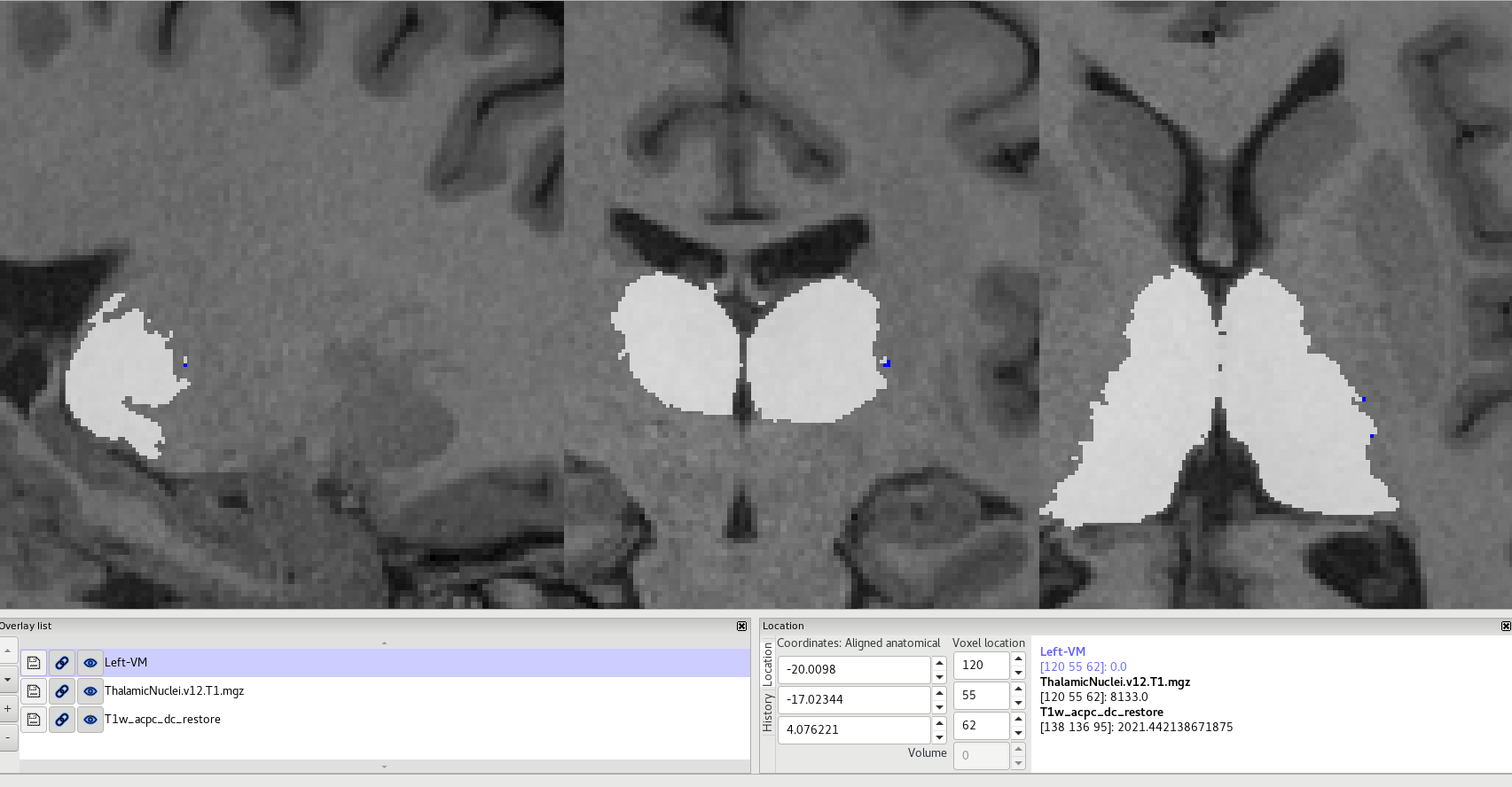
I have a question regarding the segmentation of thalamic nuclei. I processed 15 subjects with freesurfer v6 (recon-all) and additionally, ran the thalamic segmentation implemented in FreeSurfer. Then, I wanted to use the label of the thalamic nuclei to select an ROI in the VM, VPL and in other nuclei to perform the ROI analysis in SPM. Therefore, I converted the outputs of the segmentation (ThalamicNuclei.v12.T1.mgz) into nifti files by using the command "mri_convert .mgz .nii". Additionally, using ImgCal in the SPM, I created a mask for each nuclei separately. Most nuclei look great and correspond to their anatomical position. Unfortunately, the position of some nuclei is completely wrong. For example, the size of the VM (left) is definitely too small and the position seems to be on the border / outside of thalamus (attached files and link). I do not see the right-VM at all. This issue affects all my data as well as data analyzed in another institute.
In the attached picture (Thalamic_segmentation_VMleft.png), blue represents the VM. We can clearly see that the position as well as the size is completely incorrect compared to the known anatomical position or shown in your publication. Could I ask for an explanation and help in solving the problem.
The parameter for T1 and T2 scans: T1: 208slices, voxel size=0.8mm isotropic, TE=3.16ms, TR=2400ms, BW=220Hz/px, ES=7.5ms
T2: 208 slices, voxel size=0.8mm isotropic, TE=5.63ms, TR=3200ms, BW=744Hz/px, ES=3.52ms
Here you can find all the files: https://secure-web.cisco.com/1KtaGTX0Nfx2jwZl-X1QkJstv6OQtmYKCMAYA5rzu-lXHKl...
Best regards, Edyta Leks
Edyta Leks
High Field MR Center Max Planck Institute for Biological Cybernetics Max-Planck-Ring 11 72076 Tübingen Germany ________________________________________________ office: 4.B.06 tel: +49 7071 601 918 e:mail: edyta.leks@tuebingen.mpg.de

Dear Edyta,
The smaller nuclei are definitely less accurate than the bigger ones in the atlas.
Is it only the VM giving you trouble? Is it in every case, or only on this case?
Cheers,
Eugenio
Juan Eugenio Iglesias Senior research fellow CMIC (UCL), MGH (HMS) and CSAIL (MIT) http://www.jeiglesias.com
On 14 Oct 2021, at 10:18, Edyta Leks edyta.leks@tuebingen.mpg.de wrote:
External Email - Use CautionHi Freesurfer expert,
I have a question regarding the segmentation of thalamic nuclei. I processed 15 subjects with freesurfer v6 (recon-all) and additionally, ran the thalamic segmentation implemented in FreeSurfer. Then, I wanted to use the label of the thalamic nuclei to select an ROI in the VM, VPL and in other nuclei to perform the ROI analysis in SPM. Therefore, I converted the outputs of the segmentation (ThalamicNuclei.v12.T1.mgz) into nifti files by using the command "mri_convert .mgz .nii". Additionally, using ImgCal in the SPM, I created a mask for each nuclei separately. Most nuclei look great and correspond to their anatomical position. Unfortunately, the position of some nuclei is completely wrong. For example, the size of the VM (left) is definitely too small and the position seems to be on the border / outside of thalamus (attached files and link). I do not see the right-VM at all. This issue affects all my data as well as data analyzed in another institute.
In the attached picture (Thalamic_segmentation_VMleft.png), blue represents the VM. We can clearly see that the position as well as the size is completely incorrect compared to the known anatomical position or shown in your publication. Could I ask for an explanation and help in solving the problem.
The parameter for T1 and T2 scans: T1: 208slices, voxel size=0.8mm isotropic, TE=3.16ms, TR=2400ms, BW=220Hz/px, ES=7.5ms
T2: 208 slices, voxel size=0.8mm isotropic, TE=5.63ms, TR=3200ms, BW=744Hz/px, ES=3.52ms
Here you can find all the files: https://secure-web.cisco.com/1KtaGTX0Nfx2jwZl-X1QkJstv6OQtmYKCMAYA5rzu-lXHKl...
Best regards, Edyta Leks
Edyta Leks
High Field MR Center Max Planck Institute for Biological Cybernetics Max-Planck-Ring 11 72076 Tübingen Germany ________________________________________________ office: 4.B.06 tel: +49 7071 601 918 e:mail: edyta.leks@tuebingen.mpg.de <Thalamic_segmentation_VMleft.png>_______________________________________________ Freesurfer mailing list Freesurfer@nmr.mgh.harvard.edu https://mail.nmr.mgh.harvard.edu/mailman/listinfo/freesurfer
freesurfer@nmr.mgh.harvard.edu
-
 Edyta Leks
 -
 Iglesias Gonzalez, Juan E.