
Dear freesurfers,
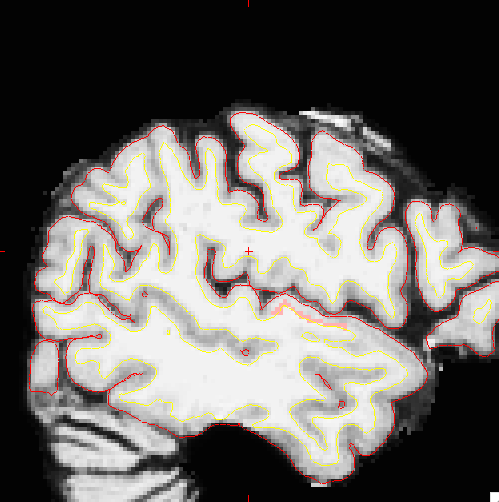
1) I have a little question concerning Freeview: As attachment there are 3 screenshots, which show a label of the 2009 atlas (Primary auditory cortex). I used brain.mgz as "input" volume and chose the label in the ROI tab in freeview - but the screenshots look like as if there is a vast part of the labeled cortex not colored. I'm wondering why the other cortical voxels - which definitely belong to Heschl-Gyrus (A1) - are not marked? I thought that a label marks all the pixels/voxels (also in 2D view) between the white matter surface and the pial surface that belong to the chosen cortical structure. Am I mistaken by interpreting the meaning of a label in this way?
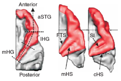
2) Actually I got stuck when trying to find a good 3D-view of Heschl'Gyrus (lh.G_temp_sup-G_T_transv.label) cause it is hidden in the deep Sylvian fissure. Is there a possibility to get a better view on this label (e.g. transparent view of the surrounding brain structures)? I attached another screenshot (Schneider, Cortex) which was taken from a colleague of mine working with brainvoyager - is it possible to create something like this in freesurfer/freeview?
Looking forward to answers, thanks a lot...
Holger
freesurfer@nmr.mgh.harvard.edu
-
 Klein, Holger