
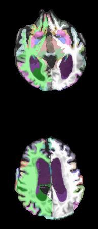
We have a T1 image that has been problematic to process. This patient has thicker skull and extensive hypointensities in the white matter along with big vents.
Skullstripping by modifying the watershed parameter either leaves too much skull or removes entire chunks of the brain in this Siemens image; unfortunately there seems to be no middle ground.
In recon-all FreeSurfer has been unable to discern the gray matter boundary in the posterior of the brain and omits folding detail of the sulci. I am attaching a screenshot overview of the "best" attempt so far, which finished this morning (there is a bit of skull left over on this run; on previous runs where the skull is cleanly removed this problem persists). As a last-ditch effort I had re-sliced the axis-reformat T1 images as additional images (002.mgz, 003.mgz, 004.mgz) (any feedback on the feasibility of this approach is welcomed also).
Any suggestions to improve the processing on these cortical problem areas are very much appreciated (ventricles are as good as I can hope for today; that's matter for a whole other post!).
Much thanks,
Eve LoCastro Senior Research Assistant IDEAL, Radiology Department Weill Cornell Medical College 515 E. 71st St S118 Tel: 212-746-1289 Fax: 212-746-4189 http://ideal-cornell.com/http://ideal-cornell.com

Hi Eve
if you upload the subject we will take a look Bruce On Fri, 6 Dec 2013, Eve M. LoCastro wrote:
We have a T1 image that has been problematic to process. This patient has thicker skull and extensive hypointensities in the white matter along with big vents.
Skullstripping by modifying the watershed parameter either leaves too much skull or removes entire chunks of the brain in this Siemens image; unfortunately there seems to be no middle ground.
In recon-all FreeSurfer has been unable to discern the gray matter boundary in the posterior of the brain and omits folding detail of the sulci. I am attaching a screenshot overview of the "best" attempt so far, which finished this morning (there is a bit of skull left over on this run; on previous runs where the skull is cleanly removed this problem persists). As a last-ditch effort I had re-sliced the axis-reformat T1 images as additional images (002.mgz, 003.mgz, 004.mgz) (any feedback on the feasibility of this approach is welcomed also).
Any suggestions to improve the processing on these cortical problem areas are very much appreciated (ventricles are as good as I can hope for today; that's matter for a whole other post!).
Much thanks,
Eve LoCastro Senior Research Assistant IDEAL, Radiology Department Weill Cornell Medical College 515 E. 71st St S118 Tel: 212-746-1289 Fax: 212-746-4189 http://ideal-cornell.com/
Thank you, Bruce. I have uploaded the 001.mgz here:
http://gate.nmr.mgh.harvard.edu/filedrop2/?p=8xy0v6sj50o
And the segmentation (aparc+aseg.mgz).
http://gate.nmr.mgh.harvard.edu/filedrop2/?p=7ibj59tsn6p
If there are any other files of interest, please let me know.
Much thanks,
Eve LoCastro Senior Research Assistant IDEAL, Radiology Department Weill Cornell Medical College 515 E. 71st St S118 Tel: 212-746-1289 Fax: 212-746-4189 http://ideal-cornell.com/
________________________________________ From: Bruce Fischl [fischl@nmr.mgh.harvard.edu] Sent: Friday, December 06, 2013 4:28 PM To: Eve M. LoCastro Cc: freesurfer@nmr.mgh.harvard.edu Subject: Re: [Freesurfer] Segmentation misses sulci in posterior
Hi Eve
if you upload the subject we will take a look Bruce On Fri, 6 Dec 2013, Eve M. LoCastro wrote:
We have a T1 image that has been problematic to process. This patient has thicker skull and extensive hypointensities in the white matter along with big vents.
Skullstripping by modifying the watershed parameter either leaves too much skull or removes entire chunks of the brain in this Siemens image; unfortunately there seems to be no middle ground.
In recon-all FreeSurfer has been unable to discern the gray matter boundary in the posterior of the brain and omits folding detail of the sulci. I am attaching a screenshot overview of the "best" attempt so far, which finished this morning (there is a bit of skull left over on this run; on previous runs where the skull is cleanly removed this problem persists). As a last-ditch effort I had re-sliced the axis-reformat T1 images as additional images (002.mgz, 003.mgz, 004.mgz) (any feedback on the feasibility of this approach is welcomed also).
Any suggestions to improve the processing on these cortical problem areas are very much appreciated (ventricles are as good as I can hope for today; that's matter for a whole other post!).
Much thanks,
Eve LoCastro Senior Research Assistant IDEAL, Radiology Department Weill Cornell Medical College 515 E. 71st St S118 Tel: 212-746-1289 Fax: 212-746-4189 http://ideal-cornell.com/
The information in this e-mail is intended only for the person to whom it is addressed. If you believe this e-mail was sent to you in error and the e-mail contains patient information, please contact the Partners Compliance HelpLine at http://www.partners.org/complianceline . If the e-mail was sent to you in error but does not contain patient information, please contact the sender and properly dispose of the e-mail.
ok, I'll take a look On Fri, 6 Dec 2013, Eve M. LoCastro wrote:
Thank you, Bruce. I have uploaded the 001.mgz here:
http://gate.nmr.mgh.harvard.edu/filedrop2/?p=8xy0v6sj50o
And the segmentation (aparc+aseg.mgz).
http://gate.nmr.mgh.harvard.edu/filedrop2/?p=7ibj59tsn6p
If there are any other files of interest, please let me know.
Much thanks,
Eve LoCastro Senior Research Assistant IDEAL, Radiology Department Weill Cornell Medical College 515 E. 71st St S118 Tel: 212-746-1289 Fax: 212-746-4189 http://ideal-cornell.com/
From: Bruce Fischl [fischl@nmr.mgh.harvard.edu] Sent: Friday, December 06, 2013 4:28 PM To: Eve M. LoCastro Cc: freesurfer@nmr.mgh.harvard.edu Subject: Re: [Freesurfer] Segmentation misses sulci in posterior
Hi Eve
if you upload the subject we will take a look Bruce On Fri, 6 Dec 2013, Eve M. LoCastro wrote:
We have a T1 image that has been problematic to process. This patient has thicker skull and extensive hypointensities in the white matter along with big vents.
Skullstripping by modifying the watershed parameter either leaves too much skull or removes entire chunks of the brain in this Siemens image; unfortunately there seems to be no middle ground.
In recon-all FreeSurfer has been unable to discern the gray matter boundary in the posterior of the brain and omits folding detail of the sulci. I am attaching a screenshot overview of the "best" attempt so far, which finished this morning (there is a bit of skull left over on this run; on previous runs where the skull is cleanly removed this problem persists). As a last-ditch effort I had re-sliced the axis-reformat T1 images as additional images (002.mgz, 003.mgz, 004.mgz) (any feedback on the feasibility of this approach is welcomed also).
Any suggestions to improve the processing on these cortical problem areas are very much appreciated (ventricles are as good as I can hope for today; that's matter for a whole other post!).
Much thanks,
Eve LoCastro Senior Research Assistant IDEAL, Radiology Department Weill Cornell Medical College 515 E. 71st St S118 Tel: 212-746-1289 Fax: 212-746-4189 http://ideal-cornell.com/
The information in this e-mail is intended only for the person to whom it is addressed. If you believe this e-mail was sent to you in error and the e-mail contains patient information, please contact the Partners Compliance HelpLine at http://www.partners.org/complianceline . If the e-mail was sent to you in error but does not contain patient information, please contact the sender and properly dispose of the e-mail.
Freesurfer mailing list Freesurfer@nmr.mgh.harvard.edu https://mail.nmr.mgh.harvard.edu/mailman/listinfo/freesurfer
I realize the files I included were rather limited. I zipped the entire output folder and uploaded it:
-> subj161.bz2 (477.45 MiB) http://gate.nmr.mgh.harvard.edu/filedrop2/?p=bmuvoddq6g1
Cheers,
Eve LoCastro Senior Research Assistant IDEAL, Radiology Department Weill Cornell Medical College 515 E. 71st St S118 Tel: 212-746-1289 Fax: 212-746-4189 http://ideal-cornell.com/
________________________________________ From: Bruce Fischl [fischl@nmr.mgh.harvard.edu] Sent: Friday, December 06, 2013 5:02 PM To: Eve M. LoCastro Cc: freesurfer@nmr.mgh.harvard.edu Subject: Re: [Freesurfer] Segmentation misses sulci in posterior
ok, I'll take a look On Fri, 6 Dec 2013, Eve M. LoCastro wrote:
Thank you, Bruce. I have uploaded the 001.mgz here:
http://gate.nmr.mgh.harvard.edu/filedrop2/?p=8xy0v6sj50o
And the segmentation (aparc+aseg.mgz).
http://gate.nmr.mgh.harvard.edu/filedrop2/?p=7ibj59tsn6p
If there are any other files of interest, please let me know.
Much thanks,
Eve LoCastro Senior Research Assistant IDEAL, Radiology Department Weill Cornell Medical College 515 E. 71st St S118 Tel: 212-746-1289 Fax: 212-746-4189 http://ideal-cornell.com/
From: Bruce Fischl [fischl@nmr.mgh.harvard.edu] Sent: Friday, December 06, 2013 4:28 PM To: Eve M. LoCastro Cc: freesurfer@nmr.mgh.harvard.edu Subject: Re: [Freesurfer] Segmentation misses sulci in posterior
Hi Eve
if you upload the subject we will take a look Bruce On Fri, 6 Dec 2013, Eve M. LoCastro wrote:
We have a T1 image that has been problematic to process. This patient has thicker skull and extensive hypointensities in the white matter along with big vents.
Skullstripping by modifying the watershed parameter either leaves too much skull or removes entire chunks of the brain in this Siemens image; unfortunately there seems to be no middle ground.
In recon-all FreeSurfer has been unable to discern the gray matter boundary in the posterior of the brain and omits folding detail of the sulci. I am attaching a screenshot overview of the "best" attempt so far, which finished this morning (there is a bit of skull left over on this run; on previous runs where the skull is cleanly removed this problem persists). As a last-ditch effort I had re-sliced the axis-reformat T1 images as additional images (002.mgz, 003.mgz, 004.mgz) (any feedback on the feasibility of this approach is welcomed also).
Any suggestions to improve the processing on these cortical problem areas are very much appreciated (ventricles are as good as I can hope for today; that's matter for a whole other post!).
Much thanks,
Eve LoCastro Senior Research Assistant IDEAL, Radiology Department Weill Cornell Medical College 515 E. 71st St S118 Tel: 212-746-1289 Fax: 212-746-4189 http://ideal-cornell.com/
The information in this e-mail is intended only for the person to whom it is addressed. If you believe this e-mail was sent to you in error and the e-mail contains patient information, please contact the Partners Compliance HelpLine at http://www.partners.org/complianceline . If the e-mail was sent to you in error but does not contain patient information, please contact the sender and properly dispose of the e-mail.
Freesurfer mailing list Freesurfer@nmr.mgh.harvard.edu https://mail.nmr.mgh.harvard.edu/mailman/listinfo/freesurfer
freesurfer@nmr.mgh.harvard.edu
-
 Bruce Fischl
 -
 Eve M. LoCastro