
Hi FreeSurfer community,
I am analyzing some SWI scans of some old subjects. For most of my subjects when a SWI was acquired, the resulting files that I would get would be a Magnitude image, a Phase image, a mIP image, and the SWI image. However, I have a couple of subjects for whom I get only the mIP and the SWI images, and the SWI image looks more blurry than it's supposed to be. I was wondering:
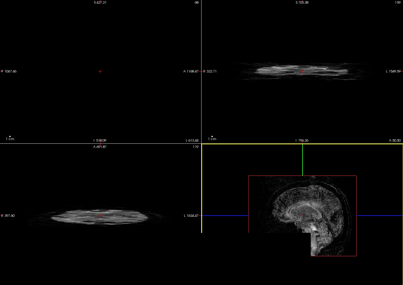
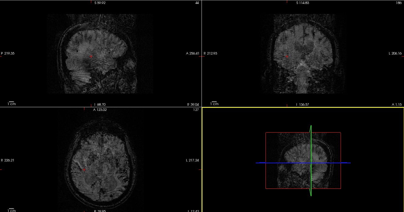
1) Why do I get only the mIP and the SWI images for those subjects and not the magnitude and phase images as well, 2) Is it normal for the mIP to look like that? Usually it looks different, and 3) Why is the final SWI image more blurry than usual? By looking at the image, I believe that maybe the head coil was not properly connected to the scanner at the time of the acquisition but I don't know whether that would be a sufficient explanation.
I have attached two attachments: One of the resulting mIP and one of the equivalent blurry SWI of the same subject.
Thank you in advance for your help and time!
Best, Panos
freesurfer@nmr.mgh.harvard.edu
-
 Fotiadis, Panagiotis