
Hi surfers,
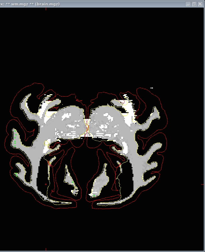
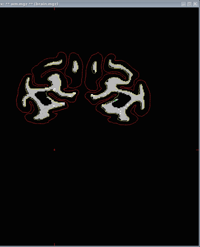
We met a problem when processing a monkey's anatomical brain. We got a very good white matter segmentation (wm.mgz) by manually editing the automatic segmentation. After recon-all, we found that at some regions, the yellow surface won't follow the wm.mgz. It is strange because the wm.mgz at these regions are very well defined and smooth. I would expect that the main surface would follow the wm.mgz tightly and smoothly. You can see the problem from the images I attached. These are coronal and horizontal views of one region.
Actually I posted the same type of problem for the other monkey's brain in January. We have solved the problem by filling all the ventricles and improving the smoothness of wm.mgz. However, we tried all methods that we learnt from the previous monkey on the second monkey, but all failed.
I am curious about how the main (yellow) surface is generated. Is it purelly reconstructed from wm.mgz, or it also need brainmask.mgz? I also edited the brainmask.mgz by darkening the gray matter surrounding the problematic regions, but it didn't work out either.
I think this problem might be quite common for the monkey community, because we met it on both of our monkeys.
Thanks,
Yang Liu

that means tere was a large topological defect that required a "fill". Look at the orig.nofix surface and you should see it following the wm.mgz much more closely. It's hard to tell from one slice, but this is probably caused by a spurious connection between the posterior horn of the ventricle and the fundus of the calcarine. There's a tiny strip of wm there that frequently gets lost, and even missing one or two voxels can create a giant topological defect.
cheers Bruce
On Fri, 18 Mar 2011, Yang Liu wrote:
Hi surfers,
We met a problem when processing a monkey's anatomical brain. We got a very good white matter segmentation (wm.mgz) by manually editing the automatic segmentation. After recon-all, we found that at some regions, the yellow surface won't follow the wm.mgz. It is strange because the wm.mgz at these regions are very well defined and smooth. I would expect that the main surface would follow the wm.mgz tightly and smoothly. You can see the problem from the images I attached. These are coronal and horizontal views of one region.
Actually I posted the same type of problem for the other monkey's brain in January. We have solved the problem by filling all the ventricles and improving the smoothness of wm.mgz. However, we tried all methods that we learnt from the previous monkey on the second monkey, but all failed.
I am curious about how the main (yellow) surface is generated. Is it purelly reconstructed from wm.mgz, or it also need brainmask.mgz? I also edited the brainmask.mgz by darkening the gray matter surrounding the problematic regions, but it didn't work out either.
I think this problem might be quite common for the monkey community, because we met it on both of our monkeys.
Thanks,
Yang Liu
freesurfer@nmr.mgh.harvard.edu
-
 Bruce Fischl
 -
 Yang Liu