
External Email - Use Caution
Hi Freesurfer devs,
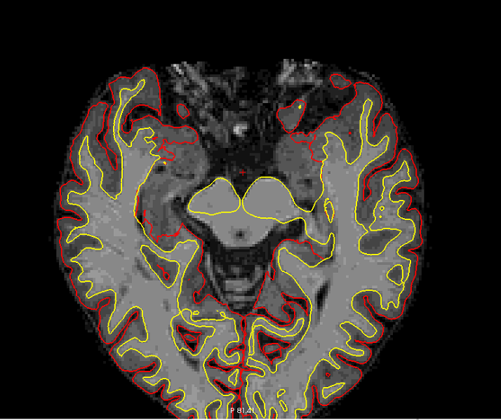
I´m doing a visual quality control of my pial and white matter segmentation using freeview and I noticed that several subjects could present an error in pial surface segmentation. I have attached two images.
Is this an pial surface error? in that case, How can I fix it? maybe adding control points in order to extend the pial surface limit?
Thanks
Cheers,

Hi Miguel,
not sure exactly where you mean, but the big chunk of gray that is missing from the surfaces is probably hippocampus (so not a problem, since it is not neocortex)
cheers Bruce
On Thu, 29 Nov 2018, Miguel Ángel Rivas Fernández wrote:
External Email - Use Caution
Hi Freesurfer devs,
I´m doing a visual quality control of my pial and white matter segmentation using freeview and I noticed that several subjects could present an error in pial surface segmentation. I have attached two images.
Is this an pial surface error? in that case, How can I fix it? maybe adding control points in order to extend the pial surface limit?
Thanks
Cheers,
-- Miguel Ángel Rivas Fernández
External Email - Use Caution
Hi Bruce,
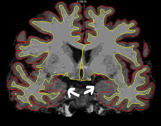
Effectively, I was referring to the big chunck of gray matter that is missing from surfaces (I attached another image). I have decided to revise the pial and white matter segmentation again because in the study that I am carrying out I found an "unexpected" or perhaps not very frequent result in the literature. I have compared general (eg BrainSegVolNotVent) and specific (eg Middle temporal, fusiform) measures of volume, thickness and area (Desikan-Killiany atlas) between a control group of healthy adults and two experimental groups, (single and multiple domain Mild Cognitive Impairment adults) using an ANCOVA (age, sex, years of education and eTIV as covariates) in external analysis (SPSS software). Results showed that control group present a higher volume, area and thickness than the multiple-domain MCI group (a result consistent with previous studies). However, no significant differences were observed between the control and the single-domain MCI groups. Specifically, the single-domain MCI group showed an equal or even higher mean volume, thickness and area than the control group in sevaral ROI´s. Given this situation, we have decided to review the recon-all's output again in order to verify that there is no error that is influencing this last result.
Could there be an error in the recon-all output that may be influencing these results? Could you suggest some procedure to check the output of the recon-all? I fixed skullstrip as well as pial and white matter surface errors when it was necessary. In the next few days I will analyze this data using QDEC.
Any recommendation will be very appreciated.
Thanks in advance,
Cheers,
El jue., 29 nov. 2018 a las 16:40, Bruce Fischl (fischl@nmr.mgh.harvard.edu) escribió:
Hi Miguel,
not sure exactly where you mean, but the big chunk of gray that is missing from the surfaces is probably hippocampus (so not a problem, since it is not neocortex)
cheers Bruce
On Thu, 29 Nov 2018, Miguel Ángel Rivas Fernández wrote:
External Email - Use CautionHi Freesurfer devs,
I´m doing a visual quality control of my pial and white matter
segmentation using freeview and I
noticed that several subjects could present an error in pial surface
segmentation. I have attached
two images.
Is this an pial surface error? in that case, How can I fix it? maybe
adding control points in order
to extend the pial surface limit?
Thanks
Cheers,
-- Miguel Ángel Rivas Fernández
Freesurfer mailing list Freesurfer@nmr.mgh.harvard.edu https://mail.nmr.mgh.harvard.edu/mailman/listinfo/freesurfer
Hi Miguel
the only way to rule out a consistent error that could induce a bias it to visually inspect the results. That said, the image you show looks fine. The white arrows are pointing to hippocampal gm, which is not supposed to be captured by the surfaces. We don't include these regions in our calculations of area/volume/thickness (except of course in hippocampal volume)
cheers Bruce
On Thu, 29 Nov 2018, Miguel Ángel Rivas Fernández wrote:
External Email - Use Caution
Hi Bruce,
Effectively, I was referring to the big chunck of gray matter that is missing from surfaces (I attached another image). I have decided to revise the pial and white matter segmentation again because in the study that I am carrying out I found an "unexpected" or perhaps not very frequent result in the literature. I have compared general (eg BrainSegVolNotVent) and specific (eg Middle temporal, fusiform) measures of volume, thickness and area (Desikan-Killiany atlas) between a control group of healthy adults and two experimental groups, (single and multiple domain Mild Cognitive Impairment adults) using an ANCOVA (age, sex, years of education and eTIV as covariates) in external analysis (SPSS software). Results showed that control group present a higher volume, area and thickness than the multiple-domain MCI group (a result consistent with previous studies). However, no significant differences were observed between the control and the single-domain MCI groups. Specifically, the single-domain MCI group showed an equal or even higher mean volume, thickness and area than the control group in sevaral ROI´s. Given this situation, we have decided to review the recon-all's output again in order to verify that there is no error that is influencing this last result.
Could there be an error in the recon-all output that may be influencing these results? Could you suggest some procedure to check the output of the recon-all? I fixed skullstrip as well as pial and white matter surface errors when it was necessary. In the next few days I will analyze this data using QDEC.
Any recommendation will be very appreciated.
Thanks in advance,
Cheers,
El jue., 29 nov. 2018 a las 16:40, Bruce Fischl (fischl@nmr.mgh.harvard.edu) escribió: Hi Miguel,
  not sure exactly where you mean, but the big chunk of gray that is
  missing from the surfaces is probably hippocampus (so not a problem, since
  it is not neocortex)

  cheers
  Bruce


  On Thu, 29 Nov 2018, Miguel Ángel Rivas Fernández
  wrote:

  >
  >         External Email - Use Caution
  >
  > Hi Freesurfer devs,
  >
  >  I´m doing a visual quality control of my pial and white matter segmentation using
  freeview and I
  > noticed that several subjects could present an error in pial surface segmentation. I
  have attached
  > two images.
  >
  > Is this an pial surface error? in that case, How can I fix it? maybe adding control
  points in order
  > to extend the pial surface limit?
  >
  >
  > Thanks
  >
  > Cheers,
  >
  >
  > --
  > Miguel Ángel Rivas Fernández
  >
  >_______________________________________________
  Freesurfer mailing list
  Freesurfer@nmr.mgh.harvard.edu
  https://mail.nmr.mgh.harvard.edu/mailman/listinfo/freesurfer
-- Miguel Ángel Rivas Fernández
freesurfer@nmr.mgh.harvard.edu
-
 Bruce Fischl
 -
 Miguel Ángel Rivas Fernández