
Hi,
I've started segmenting several structural scans from an MR-PET study conducted in Bay 7, and was wondering if someone has previous experience working with MPRAGE images acquired using the MR-PET coils?
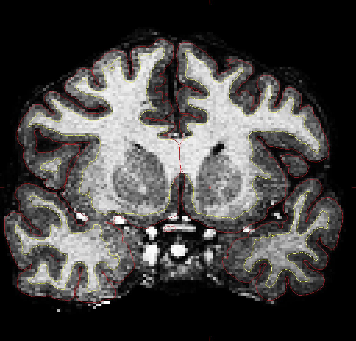
The scans have a very low SNR and I am trying to figure out if this could potentially affect the performance of the automated surface reconstructions. I've scrolled through the volumes and even though the surfaces seem normal, I am somewhat skeptical about their true quality given that intermediate stages, such as the intensity normalization, seem to be failing (please find an example from one of the brain masks attached).
Is it essential to filter the raw structurals before segmenting them, and if so, are there any readily available denoising tools? Furthermore, are there any additional issues I should take into consideration?
Thanks for your help!
Gabriel

  Hi Gabriel, I don't have experience with the MR-PET yet. Why do
  you think there is a problem? Try running wm-anat-snr on this
  subject. In general, you don't want to be smoothing/denoising
  the anatomical
  doug


  On 6/18/13 1:13 PM, obregon@nmr.mgh.harvard.edu wrote:
Hi,
I've started segmenting several structural scans from an MR-PET study conducted in Bay 7, and was wondering if someone has previous experience working with MPRAGE images acquired using the MR-PET coils?
The scans have a very low SNR and I am trying to figure out if this could potentially affect the performance of the automated surface reconstructions. I've scrolled through the volumes and even though the surfaces seem normal, I am somewhat skeptical about their true quality given that intermediate stages, such as the intensity normalization, seem to be failing (please find an example from one of the brain masks attached).
Is it essential to filter the raw structurals before segmenting them, and if so, are there any readily available denoising tools? Furthermore, are there any additional issues I should take into consideration?
Thanks for your help!
Gabriel
Freesurfer mailing list Freesurfer@nmr.mgh.harvard.edu https://mail.nmr.mgh.harvard.edu/mailman/listinfo/freesurfer
Hi Douglas,
The brainmask volumes are extremely grainy (similar to the raw structurals), i.e., dark voxels throughout the WM and high-intensity voxels across the gray matter. As far as I understood, all WM voxels (unless a lesion is present) should have a uniform intensity distribution after the normalization stages within the volume processing pipeline. Moreover, this makes the WM edits particularly challenging, for it's very difficult to trace the boundaries of some of the WM strands.
I don't know if this represents an issue but it's definitely something I hadn't encountered before. Any input would be really appreciated!
Thanks,
--G
On Jun 19, 2013, at 2:23 AM, Douglas Greve greve@nmr.mgh.harvard.edu wrote:
Hi Gabriel, I don't have experience with the MR-PET yet. Why do you think there is a problem? Try running wm-anat-snr on this subject. In general, you don't want to be smoothing/denoising the anatomical doug
On 6/18/13 1:13 PM, obregon@nmr.mgh.harvard.edu wrote:
Hi,
I've started segmenting several structural scans from an MR-PET study conducted in Bay 7, and was wondering if someone has previous experience working with MPRAGE images acquired using the MR-PET coils?
The scans have a very low SNR and I am trying to figure out if this could potentially affect the performance of the automated surface reconstructions. I've scrolled through the volumes and even though the surfaces seem normal, I am somewhat skeptical about their true quality given that intermediate stages, such as the intensity normalization, seem to be failing (please find an example from one of the brain masks attached).
Is it essential to filter the raw structurals before segmenting them, and if so, are there any readily available denoising tools? Furthermore, are there any additional issues I should take into consideration?
Thanks for your help!
Gabriel
Freesurfer mailing list Freesurfer@nmr.mgh.harvard.edu https://mail.nmr.mgh.harvard.edu/mailman/listinfo/freesurfer

Hi Gabriel
can you give us details of your acquisition? Why is it so noisy? Bruce
On Wed, 19 Jun 2013, Gabriel Obregon wrote:
Hi Douglas,
The brainmask volumes are extremely grainy (similar to the raw structurals), i.e., dark voxels throughout the WM and high-intensity voxels across the gray matter. As far as I understood, all WM voxels (unless a lesion is present) should have a uniform intensity distribution after the normalization stages within the volume processing pipeline. Moreover, this makes the WM edits particularly challenging, for it's very difficult to trace the boundaries of some of the WM strands.
I don't know if this represents an issue but it's definitely something I hadn't encountered before. Any input would be really appreciated!
Thanks,
--G
On Jun 19, 2013, at 2:23 AM, Douglas Greve greve@nmr.mgh.harvard.edu wrote:
Hi Gabriel, I don't have experience with the MR-PET yet. Why do you think there is a problem? Try running wm-anat-snr on this subject. In general, you don't want to be smoothing/denoising the anatomical doug On 6/18/13 1:13 PM, obregon@nmr.mgh.harvard.edu wrote:Hi,
I've started segmenting several structural scans from an MR-PET study conducted in Bay 7, and was wondering if someone has previous experience working with MPRAGE images acquired using the MR-PET coils?
The scans have a very low SNR and I am trying to figure out if this could potentially affect the performance of the automated surface reconstructions. I've scrolled through the volumes and even though the surfaces seem normal, I am somewhat skeptical about their true quality given that intermediate stages, such as the intensity normalization, seem to be failing (please find an example from one of the brain masks attached).
Is it essential to filter the raw structurals before segmenting them, and if so, are there any readily available denoising tools? Furthermore, are there any additional issues I should take into consideration?
Thanks for your help!
Gabriel
Freesurfer mailing list Freesurfer@nmr.mgh.harvard.edu https://mail.nmr.mgh.harvard.edu/mailman/listinfo/freesurfer
freesurfer@nmr.mgh.harvard.edu
-
 Bruce Fischl
 -
 Douglas Greve
 -
 Gabriel Obregon
 -
obregon@nmr.mgh.harvard.edu