
Dear Freesurfers,
I computed the functional activation in a sensory task using freesurfer's fsfast stream. In the cortex surface line like activated regions are present. Their extension is much larger than the extension of the expected regions. Therefore, I suppose that those regions are due to vessels. Is there a possibility in freesurfer to remove them. Ideally there would be something like a volume based mode which does not sxclude the cortex. In this mode the vessels couls be identified and removed. Any help is highly apprexiated.
Respectfully yours
pfannmoe

Hi Jorg, can you tell us a little more? A picture would be good. Also, describe how you have done your analysis (eg, on the surface or in the volume, how much smoothing). doug
On 10/15/2012 11:01 AM, Jörg Pfannmöller wrote:
Dear Freesurfers,
I computed the functional activation in a sensory task using freesurfer's fsfast stream. In the cortex surface line like activated regions are present. Their extension is much larger than the extension of the expected regions. Therefore, I suppose that those regions are due to vessels. Is there a possibility in freesurfer to remove them. Ideally there would be something like a volume based mode which does not sxclude the cortex. In this mode the vessels couls be identified and removed. Any help is highly apprexiated.
Respectfully yours
pfannmoe
Freesurfer mailing list Freesurfer@nmr.mgh.harvard.edu https://mail.nmr.mgh.harvard.edu/mailman/listinfo/freesurfer
Hi Doug,
the analysis is done on the cortex surface. The following commands are used for the analysis of the functional activation in the right hemisphere cortex:
preproc-sess -s sessionID -fsd bold -nostc -surface fsaverage lhrh -mni305 -fwhm 0 -per-run -nosmooth
plot-twf-sess -s sessionID -fsd bold -mc
tkregister-sess -s sessionID -fsd bold -per-run -bbr-sum
mkanalysis-sess -fsd bold -surface fsaverage rh -fwhm 0 -event-related -paradigm stim.par -nconditions 1 -spmhrf 0 -TR 2 -refeventdur 10 -nskip 5 -polyfit 2 -analysis stim.sm0.rh -force
mkcontrast-sess -analysis stim.sm0.rh -contrast stim-li-v-base -a 1
selxavg3-sess -s sessionID -analysis stim.sm0.rh -no-preproc
tksurfer-sess -s sessionID -analysis stim.sm0.rh -c stim-li-v-base -tcl ./label_01.tcl.
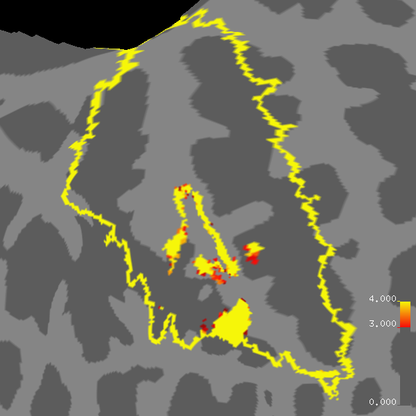
Left hemisphere and sub-cortical analysis are also carried out. Results are depicted on the flatted cortex surface in the primary somatosensory cortex. This label is a combination of the freesurfer areas BA1, BA2, BA3a and BA3b. The activation is masked to this label. An example of the line like pattern is uploaded on your ftp server using my email (pfannmoelj ... uni-greifswald.de, the at sign is replaced by dots) as the password. There is a snaking line in the middle of the image which is a candidate for a vessel. In other images the straight line like patterns or line like patterns with gaps are found. Is there a way to analyze the data in a volume based stream without differentiation between cortex and sub-cortical brain in which the distance of the line like pattern relative to the gray substance is visible? This could be used in order to classify between true activation and vessel activation. We have epi data with spatial resolution of 2x2x4mm^3 (used to generate attached image) and 1.5x1.5x2mm^3 which both show line like patterns. Structural data are taken using the single echo freesurfer protocol for the flash sequence, recommended for cortex. I hope those information are of help.
Sincerely yours
pfannmoe
On Mon, 15 Oct 2012 17:52:04 -0400 Douglas N Greve greve@nmr.mgh.harvard.edu wrote:
Hi Jorg, can you tell us a little more? A picture would be good. Also, describe how you have done your analysis (eg, on the surface or in the volume, how much smoothing). doug
On 10/15/2012 11:01 AM, wrote:
Dear Freesurfers,
I computed the functional activation in a sensory task using freesurfer's fsfast stream. In the cortex surface line like activated regions are present. Their extension is much larger than the extension of the expected regions. Therefore, I suppose that those regions are due to vessels. Is there a possibility in freesurfer to remove them. Ideally there would be something like a volume based mode which does not sxclude the cortex. In this mode the vessels couls be identified and removed. Any help is highly apprexiated.
Respectfully yours
pfannmoe
Freesurfer mailing list Freesurfer@nmr.mgh.harvard.edu https://mail.nmr.mgh.harvard.edu/mailman/listinfo/freesurfer
-- Douglas N. Greve, Ph.D. MGH-NMR Center greve@nmr.mgh.harvard.edu Phone Number: 617-724-2358 Fax: 617-726-7422
Bugs: surfer.nmr.mgh.harvard.edu/fswiki/BugReporting FileDrop: www.nmr.mgh.harvard.edu/facility/filedrop/index.html
Freesurfer mailing list Freesurfer@nmr.mgh.harvard.edu https://mail.nmr.mgh.harvard.edu/mailman/listinfo/freesurfer
The information in this e-mail is intended only for the person to whom it is addressed. If you believe this e-mail was sent to you in error and the e-mail contains patient information, please contact the Partners Compliance HelpLine at http://www.partners.org/complianceline . If the e-mail was sent to you in error but does not contain patient information, please contact the sender and properly dispose of the e-mail.
can you send a pic?
On 10/16/2012 05:44 AM, Jörg Pfannmöller wrote:
Hi Doug,
the analysis is done on the cortex surface. The following commands are used for the analysis of the functional activation in the right hemisphere cortex:
preproc-sess -s sessionID -fsd bold -nostc -surface fsaverage lhrh -mni305 -fwhm 0 -per-run -nosmooth
plot-twf-sess -s sessionID -fsd bold -mc
tkregister-sess -s sessionID -fsd bold -per-run -bbr-sum
mkanalysis-sess -fsd bold -surface fsaverage rh -fwhm 0 -event-related -paradigm stim.par -nconditions 1 -spmhrf 0 -TR 2 -refeventdur 10 -nskip 5 -polyfit 2 -analysis stim.sm0.rh -force
mkcontrast-sess -analysis stim.sm0.rh -contrast stim-li-v-base -a 1
selxavg3-sess -s sessionID -analysis stim.sm0.rh -no-preproc
tksurfer-sess -s sessionID -analysis stim.sm0.rh -c stim-li-v-base -tcl ./label_01.tcl.
Left hemisphere and sub-cortical analysis are also carried out. Results are depicted on the flatted cortex surface in the primary somatosensory cortex. This label is a combination of the freesurfer areas BA1, BA2, BA3a and BA3b. The activation is masked to this label. An example of the line like pattern is uploaded on your ftp server using my email (pfannmoelj ... uni-greifswald.de, the at sign is replaced by dots) as the password. There is a snaking line in the middle of the image which is a candidate for a vessel. In other images the straight line like patterns or line like patterns with gaps are found. Is there a way to analyze the data in a volume based stream without differentiation between cortex and sub-cortical brain in which the distance of the line like pattern relative to the gray substance is visible? This could be used in order to classify between true activation and vessel activation. We have epi data with spatial resolution of 2x2x4mm^3 (used to generate attached image) and 1.5x1.5x2mm^3 which both show line like patterns. Structural data are taken using the single echo freesurfer protocol for the flash sequence, recommended for cortex. I hope those information are of help.
Sincerely yours
pfannmoeOn Mon, 15 Oct 2012 17:52:04 -0400 Douglas N Grevegreve@nmr.mgh.harvard.edu wrote:
Hi Jorg, can you tell us a little more? A picture would be good. Also, describe how you have done your analysis (eg, on the surface or in the volume, how much smoothing). doug
On 10/15/2012 11:01 AM, wrote:
Dear Freesurfers,
I computed the functional activation in a sensory task using freesurfer's fsfast stream. In the cortex surface line like activated regions are present. Their extension is much larger than the extension of the expected regions. Therefore, I suppose that those regions are due to vessels. Is there a possibility in freesurfer to remove them. Ideally there would be something like a volume based mode which does not sxclude the cortex. In this mode the vessels couls be identified and removed. Any help is highly apprexiated.
Respectfully yours
pfannmoe
Freesurfer mailing list Freesurfer@nmr.mgh.harvard.edu https://mail.nmr.mgh.harvard.edu/mailman/listinfo/freesurfer
-- Douglas N. Greve, Ph.D. MGH-NMR Center greve@nmr.mgh.harvard.edu Phone Number: 617-724-2358 Fax: 617-726-7422
Bugs: surfer.nmr.mgh.harvard.edu/fswiki/BugReporting FileDrop: www.nmr.mgh.harvard.edu/facility/filedrop/index.html
Freesurfer mailing list Freesurfer@nmr.mgh.harvard.edu https://mail.nmr.mgh.harvard.edu/mailman/listinfo/freesurfer
The information in this e-mail is intended only for the person to whom it is addressed. If you believe this e-mail was sent to you in error and the e-mail contains patient information, please contact the Partners Compliance HelpLine at http://www.partners.org/complianceline . If the e-mail was sent to you in error but does not contain patient information, please contact the sender and properly dispose of the e-mail.
Freesurfer mailing list Freesurfer@nmr.mgh.harvard.edu https://mail.nmr.mgh.harvard.edu/mailman/listinfo/freesurfer
It is attached to this mail.
On Tue, 16 Oct 2012 16:16:54 -0400 Douglas N Greve greve@nmr.mgh.harvard.edu wrote:
can you send a pic?
On 10/16/2012 05:44 AM, Jörg Pfannmöller wrote:
Hi Doug,
the analysis is done on the cortex surface. The following commands are used for the analysis of the functional activation in the right hemisphere cortex:
preproc-sess -s sessionID -fsd bold -nostc -surface fsaverage lhrh -mni305 -fwhm 0 -per-run -nosmooth
plot-twf-sess -s sessionID -fsd bold -mc
tkregister-sess -s sessionID -fsd bold -per-run -bbr-sum
mkanalysis-sess -fsd bold -surface fsaverage rh -fwhm 0 -event-related -paradigm stim.par -nconditions 1 -spmhrf 0 -TR 2 -refeventdur 10 -nskip 5 -polyfit 2 -analysis stim.sm0.rh -force
mkcontrast-sess -analysis stim.sm0.rh -contrast stim-li-v-base -a 1
selxavg3-sess -s sessionID -analysis stim.sm0.rh -no-preproc
tksurfer-sess -s sessionID -analysis stim.sm0.rh -c stim-li-v-base -tcl ./label_01.tcl.
Left hemisphere and sub-cortical analysis are also carried out. Results are depicted on the flatted cortex surface in the primary somatosensory cortex. This label is a combination of the freesurfer areas BA1, BA2, BA3a and BA3b. The activation is masked to this label. An example of the line like pattern is uploaded on your ftp server using my email (pfannmoelj ... uni-greifswald.de, the at sign is replaced by dots) as the password. There is a snaking line in the middle of the image which is a candidate for a vessel. In other images the straight line like patterns or line like patterns with gaps are found. Is there a way to analyze the data in a volume based stream without differentiation between cortex and sub-cortical brain in which the distance of the line like pattern relative to the gray substance is visible? This could be used in order to classify between true activation and vessel activation. We have epi data with spatial resolution of 2x2x4mm^3 (used to generate attached image) and 1.5x1.5x2mm^3 which both show line like patterns. Structural data are taken using the single echo freesurfer protocol for the flash sequence, recommended for cortex. I hope those information are of help.
Sincerely yours
pfannmoeOn Mon, 15 Oct 2012 17:52:04 -0400 Douglas N Grevegreve@nmr.mgh.harvard.edu wrote:
Hi Jorg, can you tell us a little more? A picture would be good. Also, describe how you have done your analysis (eg, on the surface or in the volume, how much smoothing). doug
On 10/15/2012 11:01 AM, wrote:
Dear Freesurfers,
I computed the functional activation in a sensory task using freesurfer's fsfast stream. In the cortex surface line like activated regions are present. Their extension is much larger than the extension of the expected regions. Therefore, I suppose that those regions are due to vessels. Is there a possibility in freesurfer to remove them. Ideally there would be something like a volume based mode which does not sxclude the cortex. In this mode the vessels couls be identified and removed. Any help is highly apprexiated.
Respectfully yours
pfannmoe
Freesurfer mailing list Freesurfer@nmr.mgh.harvard.edu https://mail.nmr.mgh.harvard.edu/mailman/listinfo/freesurfer
-- Douglas N. Greve, Ph.D. MGH-NMR Center greve@nmr.mgh.harvard.edu Phone Number: 617-724-2358 Fax: 617-726-7422
Bugs: surfer.nmr.mgh.harvard.edu/fswiki/BugReporting FileDrop: www.nmr.mgh.harvard.edu/facility/filedrop/index.html
Freesurfer mailing list Freesurfer@nmr.mgh.harvard.edu https://mail.nmr.mgh.harvard.edu/mailman/listinfo/freesurfer
The information in this e-mail is intended only for the person to whom it is addressed. If you believe this e-mail was sent to you in error and the e-mail contains patient information, please contact the Partners Compliance HelpLine at http://www.partners.org/complianceline . If the e-mail was sent to you in error but does not contain patient information, please contact the sender and properly dispose of the e-mail.
Freesurfer mailing list Freesurfer@nmr.mgh.harvard.edu https://mail.nmr.mgh.harvard.edu/mailman/listinfo/freesurfer
-- Douglas N. Greve, Ph.D. MGH-NMR Center greve@nmr.mgh.harvard.edu Phone Number: 617-724-2358 Fax: 617-726-7422
Bugs: surfer.nmr.mgh.harvard.edu/fswiki/BugReporting FileDrop: www.nmr.mgh.harvard.edu/facility/filedrop/index.html
Freesurfer mailing list Freesurfer@nmr.mgh.harvard.edu https://mail.nmr.mgh.harvard.edu/mailman/listinfo/freesurfer
I just noticed that the data I am currently working with are from a data set in which the freesurfer protocol for the anatomical sequence has not been used. Instead an in-house protocol is applied.
On Wed, 17 Oct 2012 18:31:05 +0200 Jörg Pfannmöller pfannmoelj@uni-greifswald.de wrote:
It is attached to this mail.
On Tue, 16 Oct 2012 16:16:54 -0400 Douglas N Greve greve@nmr.mgh.harvard.edu wrote:
can you send a pic?
On 10/16/2012 05:44 AM, Jörg Pfannmöller wrote:
Hi Doug,
the analysis is done on the cortex surface. The following commands are used for the analysis of the functional activation in the right hemisphere cortex:
preproc-sess -s sessionID -fsd bold -nostc -surface fsaverage lhrh -mni305 -fwhm 0 -per-run -nosmooth
plot-twf-sess -s sessionID -fsd bold -mc
tkregister-sess -s sessionID -fsd bold -per-run -bbr-sum
mkanalysis-sess -fsd bold -surface fsaverage rh -fwhm 0 -event-related -paradigm stim.par -nconditions 1 -spmhrf 0 -TR 2 -refeventdur 10 -nskip 5 -polyfit 2 -analysis stim.sm0.rh -force
mkcontrast-sess -analysis stim.sm0.rh -contrast stim-li-v-base -a 1
selxavg3-sess -s sessionID -analysis stim.sm0.rh -no-preproc
tksurfer-sess -s sessionID -analysis stim.sm0.rh -c stim-li-v-base -tcl ./label_01.tcl.
Left hemisphere and sub-cortical analysis are also carried out. Results are depicted on the flatted cortex surface in the primary somatosensory cortex. This label is a combination of the freesurfer areas BA1, BA2, BA3a and BA3b. The activation is masked to this label. An example of the line like pattern is uploaded on your ftp server using my email (pfannmoelj ... uni-greifswald.de, the at sign is replaced by dots) as the password. There is a snaking line in the middle of the image which is a candidate for a vessel. In other images the straight line like patterns or line like patterns with gaps are found. Is there a way to analyze the data in a volume based stream without differentiation between cortex and sub-cortical brain in which the distance of the line like pattern relative to the gray substance is visible? This could be used in order to classify between true activation and vessel activation. We have epi data with spatial resolution of 2x2x4mm^3 (used to generate attached image) and 1.5x1.5x2mm^3 which both show line like patterns. Structural data are taken using the single echo freesurfer protocol for the flash sequence, recommended for cortex. I hope those information are of help.
Sincerely yours
pfannmoeOn Mon, 15 Oct 2012 17:52:04 -0400 Douglas N Grevegreve@nmr.mgh.harvard.edu wrote:
Hi Jorg, can you tell us a little more? A picture would be good. Also, describe how you have done your analysis (eg, on the surface or in the volume, how much smoothing). doug
On 10/15/2012 11:01 AM, wrote:
Dear Freesurfers,
I computed the functional activation in a sensory task using freesurfer's fsfast stream. In the cortex surface line like activated regions are present. Their extension is much larger than the extension of the expected regions. Therefore, I suppose that those regions are due to vessels. Is there a possibility in freesurfer to remove them. Ideally there would be something like a volume based mode which does not sxclude the cortex. In this mode the vessels couls be identified and removed. Any help is highly apprexiated.
Respectfully yours
pfannmoe
Freesurfer mailing list Freesurfer@nmr.mgh.harvard.edu https://mail.nmr.mgh.harvard.edu/mailman/listinfo/freesurfer
-- Douglas N. Greve, Ph.D. MGH-NMR Center greve@nmr.mgh.harvard.edu Phone Number: 617-724-2358 Fax: 617-726-7422
Bugs: surfer.nmr.mgh.harvard.edu/fswiki/BugReporting FileDrop: www.nmr.mgh.harvard.edu/facility/filedrop/index.html
Freesurfer mailing list Freesurfer@nmr.mgh.harvard.edu https://mail.nmr.mgh.harvard.edu/mailman/listinfo/freesurfer
The information in this e-mail is intended only for the person to whom it is addressed. If you believe this e-mail was sent to you in error and the e-mail contains patient information, please contact the Partners Compliance HelpLine at http://www.partners.org/complianceline . If the e-mail was sent to you in error but does not contain patient information, please contact the sender and properly dispose of the e-mail.
Freesurfer mailing list Freesurfer@nmr.mgh.harvard.edu https://mail.nmr.mgh.harvard.edu/mailman/listinfo/freesurfer
-- Douglas N. Greve, Ph.D. MGH-NMR Center greve@nmr.mgh.harvard.edu Phone Number: 617-724-2358 Fax: 617-726-7422
Bugs: surfer.nmr.mgh.harvard.edu/fswiki/BugReporting FileDrop: www.nmr.mgh.harvard.edu/facility/filedrop/index.html
Freesurfer mailing list Freesurfer@nmr.mgh.harvard.edu https://mail.nmr.mgh.harvard.edu/mailman/listinfo/freesurfer
-- Jörg Pfannmöller pfannmoelj@uni-greifswald.de
It is too hard to tell what is going on here from just this image. I would not immediately conclude that it is a vessel. Does the activation otherwise look ok? What about other contrasts? Is the registration accurate? When trying to debug these things, it often makes sense to analyze the data in the volume. doug
On 10/17/2012 12:31 PM, Jörg Pfannmöller wrote:
It is attached to this mail.
On Tue, 16 Oct 2012 16:16:54 -0400 Douglas N Grevegreve@nmr.mgh.harvard.edu wrote:
can you send a pic?
On 10/16/2012 05:44 AM, Jörg Pfannmöller wrote:
Hi Doug,
the analysis is done on the cortex surface. The following commands are used for the analysis of the functional activation in the right hemisphere cortex:
preproc-sess -s sessionID -fsd bold -nostc -surface fsaverage lhrh -mni305 -fwhm 0 -per-run -nosmooth
plot-twf-sess -s sessionID -fsd bold -mc
tkregister-sess -s sessionID -fsd bold -per-run -bbr-sum
mkanalysis-sess -fsd bold -surface fsaverage rh -fwhm 0 -event-related -paradigm stim.par -nconditions 1 -spmhrf 0 -TR 2 -refeventdur 10 -nskip 5 -polyfit 2 -analysis stim.sm0.rh -force
mkcontrast-sess -analysis stim.sm0.rh -contrast stim-li-v-base -a 1
selxavg3-sess -s sessionID -analysis stim.sm0.rh -no-preproc
tksurfer-sess -s sessionID -analysis stim.sm0.rh -c stim-li-v-base -tcl ./label_01.tcl.
Left hemisphere and sub-cortical analysis are also carried out. Results are depicted on the flatted cortex surface in the primary somatosensory cortex. This label is a combination of the freesurfer areas BA1, BA2, BA3a and BA3b. The activation is masked to this label. An example of the line like pattern is uploaded on your ftp server using my email (pfannmoelj ... uni-greifswald.de, the at sign is replaced by dots) as the password. There is a snaking line in the middle of the image which is a candidate for a vessel. In other images the straight line like patterns or line like patterns with gaps are found. Is there a way to analyze the data in a volume based stream without differentiation between cortex and sub-cortical brain in which the distance of the line like pattern relative to the gray substance is visible? This could be used in order to classify between true activation and vessel activation. We have epi data with spatial resolution of 2x2x4mm^3 (used to generate attached image) and 1.5x1.5x2mm^3 which both show line like patterns. Structural data are taken using the single echo freesurfer protocol for the flash sequence, recommended for cortex. I hope those information are of help.
Sincerely yours
pfannmoeOn Mon, 15 Oct 2012 17:52:04 -0400 Douglas N Grevegreve@nmr.mgh.harvard.edu wrote:
Hi Jorg, can you tell us a little more? A picture would be good. Also, describe how you have done your analysis (eg, on the surface or in the volume, how much smoothing). doug
On 10/15/2012 11:01 AM, wrote:
Dear Freesurfers,
I computed the functional activation in a sensory task using freesurfer's fsfast stream. In the cortex surface line like activated regions are present. Their extension is much larger than the extension of the expected regions. Therefore, I suppose that those regions are due to vessels. Is there a possibility in freesurfer to remove them. Ideally there would be something like a volume based mode which does not sxclude the cortex. In this mode the vessels couls be identified and removed. Any help is highly apprexiated.
Respectfully yours
pfannmoe
Freesurfer mailing list Freesurfer@nmr.mgh.harvard.edu https://mail.nmr.mgh.harvard.edu/mailman/listinfo/freesurfer
-- Douglas N. Greve, Ph.D. MGH-NMR Center greve@nmr.mgh.harvard.edu Phone Number: 617-724-2358 Fax: 617-726-7422
Bugs: surfer.nmr.mgh.harvard.edu/fswiki/BugReporting FileDrop: www.nmr.mgh.harvard.edu/facility/filedrop/index.html
Freesurfer mailing list Freesurfer@nmr.mgh.harvard.edu https://mail.nmr.mgh.harvard.edu/mailman/listinfo/freesurfer
The information in this e-mail is intended only for the person to whom it is addressed. If you believe this e-mail was sent to you in error and the e-mail contains patient information, please contact the Partners Compliance HelpLine at http://www.partners.org/complianceline . If the e-mail was sent to you in error but does not contain patient information, please contact the sender and properly dispose of the e-mail.
Freesurfer mailing list Freesurfer@nmr.mgh.harvard.edu https://mail.nmr.mgh.harvard.edu/mailman/listinfo/freesurfer
-- Douglas N. Greve, Ph.D. MGH-NMR Center greve@nmr.mgh.harvard.edu Phone Number: 617-724-2358 Fax: 617-726-7422
Bugs: surfer.nmr.mgh.harvard.edu/fswiki/BugReporting FileDrop: www.nmr.mgh.harvard.edu/facility/filedrop/index.html
Freesurfer mailing list Freesurfer@nmr.mgh.harvard.edu https://mail.nmr.mgh.harvard.edu/mailman/listinfo/freesurfer
Freesurfer mailing list Freesurfer@nmr.mgh.harvard.edu https://mail.nmr.mgh.harvard.edu/mailman/listinfo/freesurfer
Hi Doug,
the preprocessing is fine the quaility factor for the registration between functional and anatomical is ~0.3. Have I to use feat to do the volume analysis?
Cheers pfannmoe
On Thu, 18 Oct 2012 11:50:38 -0400 Douglas N Greve greve@nmr.mgh.harvard.edu wrote:
It is too hard to tell what is going on here from just this image. I would not immediately conclude that it is a vessel. Does the activation otherwise look ok? What about other contrasts? Is the registration accurate? When trying to debug these things, it often makes sense to analyze the data in the volume. doug
On 10/17/2012 12:31 PM, Jörg Pfannmöller wrote:
It is attached to this mail.
On Tue, 16 Oct 2012 16:16:54 -0400 Douglas N Grevegreve@nmr.mgh.harvard.edu wrote:
can you send a pic?
On 10/16/2012 05:44 AM, Jörg Pfannmöller wrote:
Hi Doug,
the analysis is done on the cortex surface. The following commands are used for the analysis of the functional activation in the right hemisphere cortex:
preproc-sess -s sessionID -fsd bold -nostc -surface fsaverage lhrh -mni305 -fwhm 0 -per-run -nosmooth
plot-twf-sess -s sessionID -fsd bold -mc
tkregister-sess -s sessionID -fsd bold -per-run -bbr-sum
mkanalysis-sess -fsd bold -surface fsaverage rh -fwhm 0 -event-related -paradigm stim.par -nconditions 1 -spmhrf 0 -TR 2 -refeventdur 10 -nskip 5 -polyfit 2 -analysis stim.sm0.rh -force
mkcontrast-sess -analysis stim.sm0.rh -contrast stim-li-v-base -a 1
selxavg3-sess -s sessionID -analysis stim.sm0.rh -no-preproc
tksurfer-sess -s sessionID -analysis stim.sm0.rh -c stim-li-v-base -tcl ./label_01.tcl.
Left hemisphere and sub-cortical analysis are also carried out. Results are depicted on the flatted cortex surface in the primary somatosensory cortex. This label is a combination of the freesurfer areas BA1, BA2, BA3a and BA3b. The activation is masked to this label. An example of the line like pattern is uploaded on your ftp server using my email (pfannmoelj ... uni-greifswald.de, the at sign is replaced by dots) as the password. There is a snaking line in the middle of the image which is a candidate for a vessel. In other images the straight line like patterns or line like patterns with gaps are found. Is there a way to analyze the data in a volume based stream without differentiation between cortex and sub-cortical brain in which the distance of the line like pattern relative to the gray substance is visible? This could be used in order to classify between true activation and vessel activation. We have epi data with spatial resolution of 2x2x4mm^3 (used to generate attached image) and 1.5x1.5x2mm^3 which both show line like patterns. Structural data are taken using the single echo freesurfer protocol for the flash sequence, recommended for cortex. I hope those information are of help.
Sincerely yours
pfannmoeOn Mon, 15 Oct 2012 17:52:04 -0400 Douglas N Grevegreve@nmr.mgh.harvard.edu wrote:
Hi Jorg, can you tell us a little more? A picture would be good. Also, describe how you have done your analysis (eg, on the surface or in the volume, how much smoothing). doug
On 10/15/2012 11:01 AM, wrote:
Dear Freesurfers,
I computed the functional activation in a sensory task using freesurfer's fsfast stream. In the cortex surface line like activated regions are present. Their extension is much larger than the extension of the expected regions. Therefore, I suppose that those regions are due to vessels. Is there a possibility in freesurfer to remove them. Ideally there would be something like a volume based mode which does not sxclude the cortex. In this mode the vessels couls be identified and removed. Any help is highly apprexiated.
Respectfully yours
pfannmoe
Freesurfer mailing list Freesurfer@nmr.mgh.harvard.edu https://mail.nmr.mgh.harvard.edu/mailman/listinfo/freesurfer
-- Douglas N. Greve, Ph.D. MGH-NMR Center greve@nmr.mgh.harvard.edu Phone Number: 617-724-2358 Fax: 617-726-7422
Bugs: surfer.nmr.mgh.harvard.edu/fswiki/BugReporting FileDrop: www.nmr.mgh.harvard.edu/facility/filedrop/index.html
Freesurfer mailing list Freesurfer@nmr.mgh.harvard.edu https://mail.nmr.mgh.harvard.edu/mailman/listinfo/freesurfer
The information in this e-mail is intended only for the person to whom it is addressed. If you believe this e-mail was sent to you in error and the e-mail contains patient information, please contact the Partners Compliance HelpLine at http://www.partners.org/complianceline . If the e-mail was sent to you in error but does not contain patient information, please contact the sender and properly dispose of the e-mail.
Freesurfer mailing list Freesurfer@nmr.mgh.harvard.edu https://mail.nmr.mgh.harvard.edu/mailman/listinfo/freesurfer
-- Douglas N. Greve, Ph.D. MGH-NMR Center greve@nmr.mgh.harvard.edu Phone Number: 617-724-2358 Fax: 617-726-7422
Bugs: surfer.nmr.mgh.harvard.edu/fswiki/BugReporting FileDrop: www.nmr.mgh.harvard.edu/facility/filedrop/index.html
Freesurfer mailing list Freesurfer@nmr.mgh.harvard.edu https://mail.nmr.mgh.harvard.edu/mailman/listinfo/freesurfer
Freesurfer mailing list Freesurfer@nmr.mgh.harvard.edu https://mail.nmr.mgh.harvard.edu/mailman/listinfo/freesurfer
-- Douglas N. Greve, Ph.D. MGH-NMR Center greve@nmr.mgh.harvard.edu Phone Number: 617-724-2358 Fax: 617-726-7422
Bugs: surfer.nmr.mgh.harvard.edu/fswiki/BugReporting FileDrop: www.nmr.mgh.harvard.edu/facility/filedrop/index.html
Freesurfer mailing list Freesurfer@nmr.mgh.harvard.edu https://mail.nmr.mgh.harvard.edu/mailman/listinfo/freesurfer
You'll have to run preproc like: preproc-sess -per-session -fwhm 5 -fsd bold -s sess01 In this case, you are just specifiying -per-session and not specifying -mni305 or -surface
When you run mkanalysis-sess, specify -native instead of -mni305 or -surface, like: mkanalysis-sess -native -fwhm 5 ...
doug
On 10/19/2012 03:59 AM, Jörg Pfannmöller wrote:
Hi Doug,
the preprocessing is fine the quaility factor for the registration between functional and anatomical is ~0.3. Have I to use feat to do the volume analysis?
Cheers pfannmoe
On Thu, 18 Oct 2012 11:50:38 -0400 Douglas N Grevegreve@nmr.mgh.harvard.edu wrote:
It is too hard to tell what is going on here from just this image. I would not immediately conclude that it is a vessel. Does the activation otherwise look ok? What about other contrasts? Is the registration accurate? When trying to debug these things, it often makes sense to analyze the data in the volume. doug
On 10/17/2012 12:31 PM, Jörg Pfannmöller wrote:
It is attached to this mail.
On Tue, 16 Oct 2012 16:16:54 -0400 Douglas N Grevegreve@nmr.mgh.harvard.edu wrote:
can you send a pic?
On 10/16/2012 05:44 AM, Jörg Pfannmöller wrote:
Hi Doug,
the analysis is done on the cortex surface. The following commands are used for the analysis of the functional activation in the right hemisphere cortex:
preproc-sess -s sessionID -fsd bold -nostc -surface fsaverage lhrh -mni305 -fwhm 0 -per-run -nosmooth
plot-twf-sess -s sessionID -fsd bold -mc
tkregister-sess -s sessionID -fsd bold -per-run -bbr-sum
mkanalysis-sess -fsd bold -surface fsaverage rh -fwhm 0 -event-related -paradigm stim.par -nconditions 1 -spmhrf 0 -TR 2 -refeventdur 10 -nskip 5 -polyfit 2 -analysis stim.sm0.rh -force
mkcontrast-sess -analysis stim.sm0.rh -contrast stim-li-v-base -a 1
selxavg3-sess -s sessionID -analysis stim.sm0.rh -no-preproc
tksurfer-sess -s sessionID -analysis stim.sm0.rh -c stim-li-v-base -tcl ./label_01.tcl.
Left hemisphere and sub-cortical analysis are also carried out. Results are depicted on the flatted cortex surface in the primary somatosensory cortex. This label is a combination of the freesurfer areas BA1, BA2, BA3a and BA3b. The activation is masked to this label. An example of the line like pattern is uploaded on your ftp server using my email (pfannmoelj ... uni-greifswald.de, the at sign is replaced by dots) as the password. There is a snaking line in the middle of the image which is a candidate for a vessel. In other images the straight line like patterns or line like patterns with gaps are found. Is there a way to analyze the data in a volume based stream without differentiation between cortex and sub-cortical brain in which the distance of the line like pattern relative to the gray substance is visible? This could be used in order to classify between true activation and vessel activation. We have epi data with spatial resolution of 2x2x4mm^3 (used to generate attached image) and 1.5x1.5x2mm^3 which both show line like patterns. Structural data are taken using the single echo freesurfer protocol for the flash sequence, recommended for cortex. I hope those information are of help.
Sincerely yours
pfannmoeOn Mon, 15 Oct 2012 17:52:04 -0400 Douglas N Grevegreve@nmr.mgh.harvard.edu wrote:
Hi Jorg, can you tell us a little more? A picture would be good. Also, describe how you have done your analysis (eg, on the surface or in the volume, how much smoothing). doug
On 10/15/2012 11:01 AM, wrote: > Dear Freesurfers, > > I computed the functional activation in a sensory task using freesurfer's fsfast stream. In the cortex surface line like activated regions are present. Their extension is much larger than the extension of the expected regions. Therefore, I suppose that those regions are due to vessels. Is there a possibility in freesurfer to remove them. Ideally there would be something like a volume based mode which does not sxclude the cortex. In this mode the vessels couls be identified and removed. Any help is highly apprexiated. > > Respectfully yours > > pfannmoe > _______________________________________________ > Freesurfer mailing list > Freesurfer@nmr.mgh.harvard.edu > https://mail.nmr.mgh.harvard.edu/mailman/listinfo/freesurfer >
>
Douglas N. Greve, Ph.D. MGH-NMR Center greve@nmr.mgh.harvard.edu Phone Number: 617-724-2358 Fax: 617-726-7422
Bugs: surfer.nmr.mgh.harvard.edu/fswiki/BugReporting FileDrop: www.nmr.mgh.harvard.edu/facility/filedrop/index.html
Freesurfer mailing list Freesurfer@nmr.mgh.harvard.edu https://mail.nmr.mgh.harvard.edu/mailman/listinfo/freesurfer
The information in this e-mail is intended only for the person to whom it is addressed. If you believe this e-mail was sent to you in error and the e-mail contains patient information, please contact the Partners Compliance HelpLine at http://www.partners.org/complianceline . If the e-mail was sent to you in error but does not contain patient information, please contact the sender and properly dispose of the e-mail.
Freesurfer mailing list Freesurfer@nmr.mgh.harvard.edu https://mail.nmr.mgh.harvard.edu/mailman/listinfo/freesurfer
-- Douglas N. Greve, Ph.D. MGH-NMR Center greve@nmr.mgh.harvard.edu Phone Number: 617-724-2358 Fax: 617-726-7422
Bugs: surfer.nmr.mgh.harvard.edu/fswiki/BugReporting FileDrop: www.nmr.mgh.harvard.edu/facility/filedrop/index.html
Freesurfer mailing list Freesurfer@nmr.mgh.harvard.edu https://mail.nmr.mgh.harvard.edu/mailman/listinfo/freesurfer
Freesurfer mailing list Freesurfer@nmr.mgh.harvard.edu https://mail.nmr.mgh.harvard.edu/mailman/listinfo/freesurfer
-- Douglas N. Greve, Ph.D. MGH-NMR Center greve@nmr.mgh.harvard.edu Phone Number: 617-724-2358 Fax: 617-726-7422
Bugs: surfer.nmr.mgh.harvard.edu/fswiki/BugReporting FileDrop: www.nmr.mgh.harvard.edu/facility/filedrop/index.html
Freesurfer mailing list Freesurfer@nmr.mgh.harvard.edu https://mail.nmr.mgh.harvard.edu/mailman/listinfo/freesurfer
Thanks for your help. It works fine if one uses
preproc-sess -per-run -fwhm 5 -fsd bold -s sess01 ... .
On Fri, 19 Oct 2012 12:49:00 -0400 Douglas N Greve greve@nmr.mgh.harvard.edu wrote:
You'll have to run preproc like: preproc-sess -per-session -fwhm 5 -fsd bold -s sess01 In this case, you are just specifiying -per-session and not specifying -mni305 or -surface
When you run mkanalysis-sess, specify -native instead of -mni305 or -surface, like: mkanalysis-sess -native -fwhm 5 ...
doug
On 10/19/2012 03:59 AM, Jörg Pfannmöller wrote:
Hi Doug,
the preprocessing is fine the quaility factor for the registration between functional and anatomical is ~0.3. Have I to use feat to do the volume analysis?
Cheers pfannmoe
On Thu, 18 Oct 2012 11:50:38 -0400 Douglas N Grevegreve@nmr.mgh.harvard.edu wrote:
It is too hard to tell what is going on here from just this image. I would not immediately conclude that it is a vessel. Does the activation otherwise look ok? What about other contrasts? Is the registration accurate? When trying to debug these things, it often makes sense to analyze the data in the volume. doug
On 10/17/2012 12:31 PM, Jörg Pfannmöller wrote:
It is attached to this mail.
On Tue, 16 Oct 2012 16:16:54 -0400 Douglas N Grevegreve@nmr.mgh.harvard.edu wrote:
can you send a pic?
On 10/16/2012 05:44 AM, Jörg Pfannmöller wrote:
Hi Doug,
the analysis is done on the cortex surface. The following commands are used for the analysis of the functional activation in the right hemisphere cortex:
preproc-sess -s sessionID -fsd bold -nostc -surface fsaverage lhrh -mni305 -fwhm 0 -per-run -nosmooth
plot-twf-sess -s sessionID -fsd bold -mc
tkregister-sess -s sessionID -fsd bold -per-run -bbr-sum
mkanalysis-sess -fsd bold -surface fsaverage rh -fwhm 0 -event-related -paradigm stim.par -nconditions 1 -spmhrf 0 -TR 2 -refeventdur 10 -nskip 5 -polyfit 2 -analysis stim.sm0.rh -force
mkcontrast-sess -analysis stim.sm0.rh -contrast stim-li-v-base -a 1
selxavg3-sess -s sessionID -analysis stim.sm0.rh -no-preproc
tksurfer-sess -s sessionID -analysis stim.sm0.rh -c stim-li-v-base -tcl ./label_01.tcl.
Left hemisphere and sub-cortical analysis are also carried out. Results are depicted on the flatted cortex surface in the primary somatosensory cortex. This label is a combination of the freesurfer areas BA1, BA2, BA3a and BA3b. The activation is masked to this label. An example of the line like pattern is uploaded on your ftp server using my email (pfannmoelj ... uni-greifswald.de, the at sign is replaced by dots) as the password. There is a snaking line in the middle of the image which is a candidate for a vessel. In other images the straight line like patterns or line like patterns with gaps are found. Is there a way to analyze the data in a volume based stream without differentiation between cortex and sub-cortical brain in which the distance of the line like pattern relative to the gray substance is visible? This could be used in order to classify between true activation and vessel activation. We have epi data with spatial resolution of 2x2x4mm^3 (used to generate attached image) and 1.5x1.5x2mm^3 which both show line like patterns. Structural data are taken using the single echo freesurfer protocol for the flash sequence, recommended for cortex. I hope those information are of help.
Sincerely yours
pfannmoeOn Mon, 15 Oct 2012 17:52:04 -0400 Douglas N Grevegreve@nmr.mgh.harvard.edu wrote:
> Hi Jorg, can you tell us a little more? A picture would be good. Also, > describe how you have done your analysis (eg, on the surface or in the > volume, how much smoothing). > doug > > On 10/15/2012 11:01 AM, wrote: >> Dear Freesurfers, >> >> I computed the functional activation in a sensory task using freesurfer's fsfast stream. In the cortex surface line like activated regions are present. Their extension is much larger than the extension of the expected regions. Therefore, I suppose that those regions are due to vessels. Is there a possibility in freesurfer to remove them. Ideally there would be something like a volume based mode which does not sxclude the cortex. In this mode the vessels couls be identified and removed. Any help is highly apprexiated. >> >> Respectfully yours >> >> pfannmoe >> _______________________________________________ >> Freesurfer mailing list >> Freesurfer@nmr.mgh.harvard.edu >> https://mail.nmr.mgh.harvard.edu/mailman/listinfo/freesurfer >> >> > -- > Douglas N. Greve, Ph.D. > MGH-NMR Center > greve@nmr.mgh.harvard.edu > Phone Number: 617-724-2358 > Fax: 617-726-7422 > > Bugs: surfer.nmr.mgh.harvard.edu/fswiki/BugReporting > FileDrop: www.nmr.mgh.harvard.edu/facility/filedrop/index.html > > _______________________________________________ > Freesurfer mailing list > Freesurfer@nmr.mgh.harvard.edu > https://mail.nmr.mgh.harvard.edu/mailman/listinfo/freesurfer > > > The information in this e-mail is intended only for the person to whom it is > addressed. If you believe this e-mail was sent to you in error and the e-mail > contains patient information, please contact the Partners Compliance HelpLine at > http://www.partners.org/complianceline . If the e-mail was sent to you in error > but does not contain patient information, please contact the sender and properly > dispose of the e-mail. > _______________________________________________ Freesurfer mailing list Freesurfer@nmr.mgh.harvard.edu https://mail.nmr.mgh.harvard.edu/mailman/listinfo/freesurfer
-- Douglas N. Greve, Ph.D. MGH-NMR Center greve@nmr.mgh.harvard.edu Phone Number: 617-724-2358 Fax: 617-726-7422
Bugs: surfer.nmr.mgh.harvard.edu/fswiki/BugReporting FileDrop: www.nmr.mgh.harvard.edu/facility/filedrop/index.html
Freesurfer mailing list Freesurfer@nmr.mgh.harvard.edu https://mail.nmr.mgh.harvard.edu/mailman/listinfo/freesurfer
Freesurfer mailing list Freesurfer@nmr.mgh.harvard.edu https://mail.nmr.mgh.harvard.edu/mailman/listinfo/freesurfer
-- Douglas N. Greve, Ph.D. MGH-NMR Center greve@nmr.mgh.harvard.edu Phone Number: 617-724-2358 Fax: 617-726-7422
Bugs: surfer.nmr.mgh.harvard.edu/fswiki/BugReporting FileDrop: www.nmr.mgh.harvard.edu/facility/filedrop/index.html
Freesurfer mailing list Freesurfer@nmr.mgh.harvard.edu https://mail.nmr.mgh.harvard.edu/mailman/listinfo/freesurfer
Sorry, I have to cancel my last contribution. Another Problem crossed my way. If I have more than one run, only the first one is preprocessed if i use -per-session. What can I do about it? If I use -per-run all runs are preprocessed, but selxavg3-sess tells me that it can not determine the file format of the fmc files. Actually those files are called fmcpr, if I use -per-run. How can do I have to proceed if there is more than one run?
On Mon, 6 May 2013 10:54:54 +0200 Jörg Pfannmöller pfannmoelj@uni-greifswald.de wrote:
Thanks for your help. It works fine if one uses
preproc-sess -per-run -fwhm 5 -fsd bold -s sess01 ... .
On Fri, 19 Oct 2012 12:49:00 -0400 Douglas N Greve greve@nmr.mgh.harvard.edu wrote:
You'll have to run preproc like: preproc-sess -per-session -fwhm 5 -fsd bold -s sess01 In this case, you are just specifiying -per-session and not specifying -mni305 or -surface
When you run mkanalysis-sess, specify -native instead of -mni305 or -surface, like: mkanalysis-sess -native -fwhm 5 ...
doug
On 10/19/2012 03:59 AM, Jörg Pfannmöller wrote:
Hi Doug,
the preprocessing is fine the quaility factor for the registration between functional and anatomical is ~0.3. Have I to use feat to do the volume analysis?
Cheers pfannmoe
On Thu, 18 Oct 2012 11:50:38 -0400 Douglas N Grevegreve@nmr.mgh.harvard.edu wrote:
It is too hard to tell what is going on here from just this image. I would not immediately conclude that it is a vessel. Does the activation otherwise look ok? What about other contrasts? Is the registration accurate? When trying to debug these things, it often makes sense to analyze the data in the volume. doug
On 10/17/2012 12:31 PM, Jörg Pfannmöller wrote:
It is attached to this mail.
On Tue, 16 Oct 2012 16:16:54 -0400 Douglas N Grevegreve@nmr.mgh.harvard.edu wrote:
can you send a pic?
On 10/16/2012 05:44 AM, Jörg Pfannmöller wrote: > Hi Doug, > > the analysis is done on the cortex surface. The following commands are used for the analysis of the functional activation in the right hemisphere cortex: > > preproc-sess -s sessionID -fsd bold -nostc -surface fsaverage lhrh -mni305 -fwhm 0 -per-run -nosmooth > > plot-twf-sess -s sessionID -fsd bold -mc > > tkregister-sess -s sessionID -fsd bold -per-run -bbr-sum > > mkanalysis-sess -fsd bold -surface fsaverage rh -fwhm 0 -event-related -paradigm stim.par -nconditions 1 -spmhrf 0 -TR 2 -refeventdur 10 -nskip 5 -polyfit 2 -analysis stim.sm0.rh -force > > mkcontrast-sess -analysis stim.sm0.rh -contrast stim-li-v-base -a 1 > > selxavg3-sess -s sessionID -analysis stim.sm0.rh -no-preproc > > tksurfer-sess -s sessionID -analysis stim.sm0.rh -c stim-li-v-base -tcl ./label_01.tcl. > > Left hemisphere and sub-cortical analysis are also carried out. Results are depicted on the flatted cortex surface in the primary somatosensory cortex. > This label is a combination of the freesurfer areas BA1, BA2, BA3a and BA3b. The activation is masked to this label. An example of the line like pattern > is uploaded on your ftp server using my email (pfannmoelj ... uni-greifswald.de, the at sign is replaced by dots) as the password. There is a snaking > line in the middle of the image which is a candidate for a vessel. In other images the straight line like patterns or line like patterns with gaps are > found. Is there a way to analyze the data in a volume based stream without differentiation between cortex and sub-cortical brain in which the distance > of the line like pattern relative to the gray substance is visible? This could be used in order to classify between true activation and vessel activation. > We have epi data with spatial resolution of 2x2x4mm^3 (used to generate attached image) and 1.5x1.5x2mm^3 which both show line like patterns. Structural > data are taken using the single echo freesurfer protocol for the flash sequence, recommended for cortex. I hope those information are of help. > > Sincerely yours > > pfannmoe > > > On Mon, 15 Oct 2012 17:52:04 -0400 > Douglas N Grevegreve@nmr.mgh.harvard.edu wrote: > >> Hi Jorg, can you tell us a little more? A picture would be good. Also, >> describe how you have done your analysis (eg, on the surface or in the >> volume, how much smoothing). >> doug >> >> On 10/15/2012 11:01 AM, wrote: >>> Dear Freesurfers, >>> >>> I computed the functional activation in a sensory task using freesurfer's fsfast stream. In the cortex surface line like activated regions are present. Their extension is much larger than the extension of the expected regions. Therefore, I suppose that those regions are due to vessels. Is there a possibility in freesurfer to remove them. Ideally there would be something like a volume based mode which does not sxclude the cortex. In this mode the vessels couls be identified and removed. Any help is highly apprexiated. >>> >>> Respectfully yours >>> >>> pfannmoe >>> _______________________________________________ >>> Freesurfer mailing list >>> Freesurfer@nmr.mgh.harvard.edu >>> https://mail.nmr.mgh.harvard.edu/mailman/listinfo/freesurfer >>> >>> >> -- >> Douglas N. Greve, Ph.D. >> MGH-NMR Center >> greve@nmr.mgh.harvard.edu >> Phone Number: 617-724-2358 >> Fax: 617-726-7422 >> >> Bugs: surfer.nmr.mgh.harvard.edu/fswiki/BugReporting >> FileDrop: www.nmr.mgh.harvard.edu/facility/filedrop/index.html >> >> _______________________________________________ >> Freesurfer mailing list >> Freesurfer@nmr.mgh.harvard.edu >> https://mail.nmr.mgh.harvard.edu/mailman/listinfo/freesurfer >> >> >> The information in this e-mail is intended only for the person to whom it is >> addressed. If you believe this e-mail was sent to you in error and the e-mail >> contains patient information, please contact the Partners Compliance HelpLine at >> http://www.partners.org/complianceline . If the e-mail was sent to you in error >> but does not contain patient information, please contact the sender and properly >> dispose of the e-mail. >> > _______________________________________________ > Freesurfer mailing list > Freesurfer@nmr.mgh.harvard.edu > https://mail.nmr.mgh.harvard.edu/mailman/listinfo/freesurfer >
>
Douglas N. Greve, Ph.D. MGH-NMR Center greve@nmr.mgh.harvard.edu Phone Number: 617-724-2358 Fax: 617-726-7422
Bugs: surfer.nmr.mgh.harvard.edu/fswiki/BugReporting FileDrop: www.nmr.mgh.harvard.edu/facility/filedrop/index.html
Freesurfer mailing list Freesurfer@nmr.mgh.harvard.edu https://mail.nmr.mgh.harvard.edu/mailman/listinfo/freesurfer
Freesurfer mailing list Freesurfer@nmr.mgh.harvard.edu https://mail.nmr.mgh.harvard.edu/mailman/listinfo/freesurfer
-- Douglas N. Greve, Ph.D. MGH-NMR Center greve@nmr.mgh.harvard.edu Phone Number: 617-724-2358 Fax: 617-726-7422
Bugs: surfer.nmr.mgh.harvard.edu/fswiki/BugReporting FileDrop: www.nmr.mgh.harvard.edu/facility/filedrop/index.html
Freesurfer mailing list Freesurfer@nmr.mgh.harvard.edu https://mail.nmr.mgh.harvard.edu/mailman/listinfo/freesurfer
-- pfannmoelj@uni-greifswald.de
Freesurfer mailing list Freesurfer@nmr.mgh.harvard.edu https://mail.nmr.mgh.harvard.edu/mailman/listinfo/freesurfer
If you use -per-session, all of them should be processed. Why do you think they are not? Also, just saying something is not working is not very informative. Send command lines and terminal output of the error. thanks! doug
On 05/06/2013 05:46 AM, Jörg Pfannmöller wrote:
Sorry, I have to cancel my last contribution. Another Problem crossed my way. If I have more than one run, only the first one is preprocessed if i use -per-session. What can I do about it? If I use -per-run all runs are preprocessed, but selxavg3-sess tells me that it can not determine the file format of the fmc files. Actually those files are called fmcpr, if I use -per-run. How can do I have to proceed if there is more than one run?
On Mon, 6 May 2013 10:54:54 +0200 Jörg Pfannmöller pfannmoelj@uni-greifswald.de wrote:
Thanks for your help. It works fine if one uses
preproc-sess -per-run -fwhm 5 -fsd bold -s sess01 ... .
On Fri, 19 Oct 2012 12:49:00 -0400 Douglas N Greve greve@nmr.mgh.harvard.edu wrote:
You'll have to run preproc like: preproc-sess -per-session -fwhm 5 -fsd bold -s sess01 In this case, you are just specifiying -per-session and not specifying -mni305 or -surface
When you run mkanalysis-sess, specify -native instead of -mni305 or -surface, like: mkanalysis-sess -native -fwhm 5 ...
doug
On 10/19/2012 03:59 AM, Jörg Pfannmöller wrote:
Hi Doug,
the preprocessing is fine the quaility factor for the registration between functional and anatomical is ~0.3. Have I to use feat to do the volume analysis?
Cheers pfannmoe
On Thu, 18 Oct 2012 11:50:38 -0400 Douglas N Grevegreve@nmr.mgh.harvard.edu wrote:
It is too hard to tell what is going on here from just this image. I would not immediately conclude that it is a vessel. Does the activation otherwise look ok? What about other contrasts? Is the registration accurate? When trying to debug these things, it often makes sense to analyze the data in the volume. doug
On 10/17/2012 12:31 PM, Jörg Pfannmöller wrote:
It is attached to this mail.
On Tue, 16 Oct 2012 16:16:54 -0400 Douglas N Grevegreve@nmr.mgh.harvard.edu wrote:
> can you send a pic? > > > On 10/16/2012 05:44 AM, Jörg Pfannmöller wrote: >> Hi Doug, >> >> the analysis is done on the cortex surface. The following commands are used for the analysis of the functional activation in the right hemisphere cortex: >> >> preproc-sess -s sessionID -fsd bold -nostc -surface fsaverage lhrh -mni305 -fwhm 0 -per-run -nosmooth >> >> plot-twf-sess -s sessionID -fsd bold -mc >> >> tkregister-sess -s sessionID -fsd bold -per-run -bbr-sum >> >> mkanalysis-sess -fsd bold -surface fsaverage rh -fwhm 0 -event-related -paradigm stim.par -nconditions 1 -spmhrf 0 -TR 2 -refeventdur 10 -nskip 5 -polyfit 2 -analysis stim.sm0.rh -force >> >> mkcontrast-sess -analysis stim.sm0.rh -contrast stim-li-v-base -a 1 >> >> selxavg3-sess -s sessionID -analysis stim.sm0.rh -no-preproc >> >> tksurfer-sess -s sessionID -analysis stim.sm0.rh -c stim-li-v-base -tcl ./label_01.tcl. >> >> Left hemisphere and sub-cortical analysis are also carried out. Results are depicted on the flatted cortex surface in the primary somatosensory cortex. >> This label is a combination of the freesurfer areas BA1, BA2, BA3a and BA3b. The activation is masked to this label. An example of the line like pattern >> is uploaded on your ftp server using my email (pfannmoelj ... uni-greifswald.de, the at sign is replaced by dots) as the password. There is a snaking >> line in the middle of the image which is a candidate for a vessel. In other images the straight line like patterns or line like patterns with gaps are >> found. Is there a way to analyze the data in a volume based stream without differentiation between cortex and sub-cortical brain in which the distance >> of the line like pattern relative to the gray substance is visible? This could be used in order to classify between true activation and vessel activation. >> We have epi data with spatial resolution of 2x2x4mm^3 (used to generate attached image) and 1.5x1.5x2mm^3 which both show line like patterns. Structural >> data are taken using the single echo freesurfer protocol for the flash sequence, recommended for cortex. I hope those information are of help. >> >> Sincerely yours >> >> pfannmoe >> >> >> On Mon, 15 Oct 2012 17:52:04 -0400 >> Douglas N Grevegreve@nmr.mgh.harvard.edu wrote: >> >>> Hi Jorg, can you tell us a little more? A picture would be good. Also, >>> describe how you have done your analysis (eg, on the surface or in the >>> volume, how much smoothing). >>> doug >>> >>> On 10/15/2012 11:01 AM, wrote: >>>> Dear Freesurfers, >>>> >>>> I computed the functional activation in a sensory task using freesurfer's fsfast stream. In the cortex surface line like activated regions are present. Their extension is much larger than the extension of the expected regions. Therefore, I suppose that those regions are due to vessels. Is there a possibility in freesurfer to remove them. Ideally there would be something like a volume based mode which does not sxclude the cortex. In this mode the vessels couls be identified and removed. Any help is highly apprexiated. >>>> >>>> Respectfully yours >>>> >>>> pfannmoe >>>> _______________________________________________ >>>> Freesurfer mailing list >>>> Freesurfer@nmr.mgh.harvard.edu >>>> https://mail.nmr.mgh.harvard.edu/mailman/listinfo/freesurfer >>>> >>>> >>> -- >>> Douglas N. Greve, Ph.D. >>> MGH-NMR Center >>> greve@nmr.mgh.harvard.edu >>> Phone Number: 617-724-2358 >>> Fax: 617-726-7422 >>> >>> Bugs: surfer.nmr.mgh.harvard.edu/fswiki/BugReporting >>> FileDrop: www.nmr.mgh.harvard.edu/facility/filedrop/index.html >>> >>> _______________________________________________ >>> Freesurfer mailing list >>> Freesurfer@nmr.mgh.harvard.edu >>> https://mail.nmr.mgh.harvard.edu/mailman/listinfo/freesurfer >>> >>> >>> The information in this e-mail is intended only for the person to whom it is >>> addressed. If you believe this e-mail was sent to you in error and the e-mail >>> contains patient information, please contact the Partners Compliance HelpLine at >>> http://www.partners.org/complianceline . If the e-mail was sent to you in error >>> but does not contain patient information, please contact the sender and properly >>> dispose of the e-mail. >>> >> _______________________________________________ >> Freesurfer mailing list >> Freesurfer@nmr.mgh.harvard.edu >> https://mail.nmr.mgh.harvard.edu/mailman/listinfo/freesurfer >> >> > -- > Douglas N. Greve, Ph.D. > MGH-NMR Center > greve@nmr.mgh.harvard.edu > Phone Number: 617-724-2358 > Fax: 617-726-7422 > > Bugs: surfer.nmr.mgh.harvard.edu/fswiki/BugReporting > FileDrop: www.nmr.mgh.harvard.edu/facility/filedrop/index.html > > _______________________________________________ > Freesurfer mailing list > Freesurfer@nmr.mgh.harvard.edu > https://mail.nmr.mgh.harvard.edu/mailman/listinfo/freesurfer
Freesurfer mailing list Freesurfer@nmr.mgh.harvard.edu https://mail.nmr.mgh.harvard.edu/mailman/listinfo/freesurfer
-- Douglas N. Greve, Ph.D. MGH-NMR Center greve@nmr.mgh.harvard.edu Phone Number: 617-724-2358 Fax: 617-726-7422
Bugs: surfer.nmr.mgh.harvard.edu/fswiki/BugReporting FileDrop: www.nmr.mgh.harvard.edu/facility/filedrop/index.html
Freesurfer mailing list Freesurfer@nmr.mgh.harvard.edu https://mail.nmr.mgh.harvard.edu/mailman/listinfo/freesurfer
-- pfannmoelj@uni-greifswald.de
Freesurfer mailing list Freesurfer@nmr.mgh.harvard.edu https://mail.nmr.mgh.harvard.edu/mailman/listinfo/freesurfer
Hi, the problem is that there is only one register.dof6.dat in the bold folder. I expected a register.dof6.dat in each run folder. I did not see an error message, but have attached a log file to this mail. A solution of this problem would help me to simplify my workflow and improve the analysis. Thanks in advance. Joerg
On Mon, 06 May 2013 10:48:14 -0400 Douglas N Greve greve@nmr.mgh.harvard.edu wrote:
If you use -per-session, all of them should be processed. Why do you think they are not? Also, just saying something is not working is not very informative. Send command lines and terminal output of the error. thanks! doug
On 05/06/2013 05:46 AM, Jörg Pfannmöller wrote:
Sorry, I have to cancel my last contribution. Another Problem crossed my way. If I have more than one run, only the first one is preprocessed if i use -per-session. What can I do about it? If I use -per-run all runs are preprocessed, but selxavg3-sess tells me that it can not determine the file format of the fmc files. Actually those files are called fmcpr, if I use -per-run. How can do I have to proceed if there is more than one run?
On Mon, 6 May 2013 10:54:54 +0200 Jörg Pfannmöller pfannmoelj@uni-greifswald.de wrote:
Thanks for your help. It works fine if one uses
preproc-sess -per-run -fwhm 5 -fsd bold -s sess01 ... .
On Fri, 19 Oct 2012 12:49:00 -0400 Douglas N Greve greve@nmr.mgh.harvard.edu wrote:
You'll have to run preproc like: preproc-sess -per-session -fwhm 5 -fsd bold -s sess01 In this case, you are just specifiying -per-session and not specifying -mni305 or -surface
When you run mkanalysis-sess, specify -native instead of -mni305 or -surface, like: mkanalysis-sess -native -fwhm 5 ...
doug
On 10/19/2012 03:59 AM, Jörg Pfannmöller wrote:
Hi Doug,
the preprocessing is fine the quaility factor for the registration between functional and anatomical is ~0.3. Have I to use feat to do the volume analysis?
Cheers pfannmoe
On Thu, 18 Oct 2012 11:50:38 -0400 Douglas N Grevegreve@nmr.mgh.harvard.edu wrote:
It is too hard to tell what is going on here from just this image. I would not immediately conclude that it is a vessel. Does the activation otherwise look ok? What about other contrasts? Is the registration accurate? When trying to debug these things, it often makes sense to analyze the data in the volume. doug
On 10/17/2012 12:31 PM, Jörg Pfannmöller wrote: > It is attached to this mail. > > > > On Tue, 16 Oct 2012 16:16:54 -0400 > Douglas N Grevegreve@nmr.mgh.harvard.edu wrote: > >> can you send a pic? >> >> >> On 10/16/2012 05:44 AM, Jörg Pfannmöller wrote: >>> Hi Doug, >>> >>> the analysis is done on the cortex surface. The following commands are used for the analysis of the functional activation in the right hemisphere cortex: >>> >>> preproc-sess -s sessionID -fsd bold -nostc -surface fsaverage lhrh -mni305 -fwhm 0 -per-run -nosmooth >>> >>> plot-twf-sess -s sessionID -fsd bold -mc >>> >>> tkregister-sess -s sessionID -fsd bold -per-run -bbr-sum >>> >>> mkanalysis-sess -fsd bold -surface fsaverage rh -fwhm 0 -event-related -paradigm stim.par -nconditions 1 -spmhrf 0 -TR 2 -refeventdur 10 -nskip 5 -polyfit 2 -analysis stim.sm0.rh -force >>> >>> mkcontrast-sess -analysis stim.sm0.rh -contrast stim-li-v-base -a 1 >>> >>> selxavg3-sess -s sessionID -analysis stim.sm0.rh -no-preproc >>> >>> tksurfer-sess -s sessionID -analysis stim.sm0.rh -c stim-li-v-base -tcl ./label_01.tcl. >>> >>> Left hemisphere and sub-cortical analysis are also carried out. Results are depicted on the flatted cortex surface in the primary somatosensory cortex. >>> This label is a combination of the freesurfer areas BA1, BA2, BA3a and BA3b. The activation is masked to this label. An example of the line like pattern >>> is uploaded on your ftp server using my email (pfannmoelj ... uni-greifswald.de, the at sign is replaced by dots) as the password. There is a snaking >>> line in the middle of the image which is a candidate for a vessel. In other images the straight line like patterns or line like patterns with gaps are >>> found. Is there a way to analyze the data in a volume based stream without differentiation between cortex and sub-cortical brain in which the distance >>> of the line like pattern relative to the gray substance is visible? This could be used in order to classify between true activation and vessel activation. >>> We have epi data with spatial resolution of 2x2x4mm^3 (used to generate attached image) and 1.5x1.5x2mm^3 which both show line like patterns. Structural >>> data are taken using the single echo freesurfer protocol for the flash sequence, recommended for cortex. I hope those information are of help. >>> >>> Sincerely yours >>> >>> pfannmoe >>> >>> >>> On Mon, 15 Oct 2012 17:52:04 -0400 >>> Douglas N Grevegreve@nmr.mgh.harvard.edu wrote: >>> >>>> Hi Jorg, can you tell us a little more? A picture would be good. Also, >>>> describe how you have done your analysis (eg, on the surface or in the >>>> volume, how much smoothing). >>>> doug >>>> >>>> On 10/15/2012 11:01 AM, wrote: >>>>> Dear Freesurfers, >>>>> >>>>> I computed the functional activation in a sensory task using freesurfer's fsfast stream. In the cortex surface line like activated regions are present. Their extension is much larger than the extension of the expected regions. Therefore, I suppose that those regions are due to vessels. Is there a possibility in freesurfer to remove them. Ideally there would be something like a volume based mode which does not sxclude the cortex. In this mode the vessels couls be identified and removed. Any help is highly apprexiated. >>>>> >>>>> Respectfully yours >>>>> >>>>> pfannmoe >>>>> _______________________________________________ >>>>> Freesurfer mailing list >>>>> Freesurfer@nmr.mgh.harvard.edu >>>>> https://mail.nmr.mgh.harvard.edu/mailman/listinfo/freesurfer >>>>> >>>>> >>>> -- >>>> Douglas N. Greve, Ph.D. >>>> MGH-NMR Center >>>> greve@nmr.mgh.harvard.edu >>>> Phone Number: 617-724-2358 >>>> Fax: 617-726-7422 >>>> >>>> Bugs: surfer.nmr.mgh.harvard.edu/fswiki/BugReporting >>>> FileDrop: www.nmr.mgh.harvard.edu/facility/filedrop/index.html >>>> >>>> _______________________________________________ >>>> Freesurfer mailing list >>>> Freesurfer@nmr.mgh.harvard.edu >>>> https://mail.nmr.mgh.harvard.edu/mailman/listinfo/freesurfer >>>> >>>> >>>> The information in this e-mail is intended only for the person to whom it is >>>> addressed. If you believe this e-mail was sent to you in error and the e-mail >>>> contains patient information, please contact the Partners Compliance HelpLine at >>>> http://www.partners.org/complianceline . If the e-mail was sent to you in error >>>> but does not contain patient information, please contact the sender and properly >>>> dispose of the e-mail. >>>> >>> _______________________________________________ >>> Freesurfer mailing list >>> Freesurfer@nmr.mgh.harvard.edu >>> https://mail.nmr.mgh.harvard.edu/mailman/listinfo/freesurfer >>> >>> >> -- >> Douglas N. Greve, Ph.D. >> MGH-NMR Center >> greve@nmr.mgh.harvard.edu >> Phone Number: 617-724-2358 >> Fax: 617-726-7422 >> >> Bugs: surfer.nmr.mgh.harvard.edu/fswiki/BugReporting >> FileDrop: www.nmr.mgh.harvard.edu/facility/filedrop/index.html >> >> _______________________________________________ >> Freesurfer mailing list >> Freesurfer@nmr.mgh.harvard.edu >> https://mail.nmr.mgh.harvard.edu/mailman/listinfo/freesurfer > > _______________________________________________ > Freesurfer mailing list > Freesurfer@nmr.mgh.harvard.edu
> https://mail.nmr.mgh.harvard.edu/mailman/listinfo/freesurfer
Douglas N. Greve, Ph.D. MGH-NMR Center greve@nmr.mgh.harvard.edu Phone Number: 617-724-2358 Fax: 617-726-7422
Bugs: surfer.nmr.mgh.harvard.edu/fswiki/BugReporting FileDrop: www.nmr.mgh.harvard.edu/facility/filedrop/index.html
Freesurfer mailing list Freesurfer@nmr.mgh.harvard.edu https://mail.nmr.mgh.harvard.edu/mailman/listinfo/freesurfer
-- Douglas N. Greve, Ph.D. MGH-NMR Center greve@nmr.mgh.harvard.edu Phone Number: 617-724-2358 Fax: 617-726-7422
Bugs: surfer.nmr.mgh.harvard.edu/fswiki/BugReporting FileDrop: www.nmr.mgh.harvard.edu/facility/filedrop/index.html
Freesurfer mailing list Freesurfer@nmr.mgh.harvard.edu https://mail.nmr.mgh.harvard.edu/mailman/listinfo/freesurfer
-- pfannmoelj@uni-greifswald.de
Freesurfer mailing list Freesurfer@nmr.mgh.harvard.edu https://mail.nmr.mgh.harvard.edu/mailman/listinfo/freesurfer
-- Douglas N. Greve, Ph.D. MGH-NMR Center greve@nmr.mgh.harvard.edu Phone Number: 617-724-2358 Fax: 617-726-7422
Bugs: surfer.nmr.mgh.harvard.edu/fswiki/BugReporting FileDrop: https://gate.nmr.mgh.harvard.edu/filedrop2 www.nmr.mgh.harvard.edu/facility/filedrop/index.html Outgoing: ftp://surfer.nmr.mgh.harvard.edu/transfer/outgoing/flat/greve/
Freesurfer mailing list Freesurfer@nmr.mgh.harvard.edu https://mail.nmr.mgh.harvard.edu/mailman/listinfo/freesurfer
When you use -per-session, it uses the first image of the first run as the template. All runs are motion-corrected to this template, and the template is registered to the anatomical. In this way, all images get aligned to the anatomical. The -per-session command means that there is only one template and so only one register.dof6.dat. If you want one for each run you would use -per-run.
doug
On 05/07/2013 10:51 AM, Jörg Pfannmöller wrote:
Hi, the problem is that there is only one register.dof6.dat in the bold folder. I expected a register.dof6.dat in each run folder. I did not see an error message, but have attached a log file to this mail. A solution of this problem would help me to simplify my workflow and improve the analysis. Thanks in advance. Joerg
On Mon, 06 May 2013 10:48:14 -0400 Douglas N Greve greve@nmr.mgh.harvard.edu wrote:
If you use -per-session, all of them should be processed. Why do you think they are not? Also, just saying something is not working is not very informative. Send command lines and terminal output of the error. thanks! doug
On 05/06/2013 05:46 AM, Jörg Pfannmöller wrote:
Sorry, I have to cancel my last contribution. Another Problem crossed my way. If I have more than one run, only the first one is preprocessed if i use -per-session. What can I do about it? If I use -per-run all runs are preprocessed, but selxavg3-sess tells me that it can not determine the file format of the fmc files. Actually those files are called fmcpr, if I use -per-run. How can do I have to proceed if there is more than one run?
On Mon, 6 May 2013 10:54:54 +0200 Jörg Pfannmöller pfannmoelj@uni-greifswald.de wrote:
Thanks for your help. It works fine if one uses
preproc-sess -per-run -fwhm 5 -fsd bold -s sess01 ... .
On Fri, 19 Oct 2012 12:49:00 -0400 Douglas N Greve greve@nmr.mgh.harvard.edu wrote:
You'll have to run preproc like: preproc-sess -per-session -fwhm 5 -fsd bold -s sess01 In this case, you are just specifiying -per-session and not specifying -mni305 or -surface
When you run mkanalysis-sess, specify -native instead of -mni305 or -surface, like: mkanalysis-sess -native -fwhm 5 ...
doug
On 10/19/2012 03:59 AM, Jörg Pfannmöller wrote:
Hi Doug,
the preprocessing is fine the quaility factor for the registration between functional and anatomical is ~0.3. Have I to use feat to do the volume analysis?
Cheers pfannmoe
On Thu, 18 Oct 2012 11:50:38 -0400 Douglas N Grevegreve@nmr.mgh.harvard.edu wrote:
> It is too hard to tell what is going on here from just this image. I > would not immediately conclude that it is a vessel. Does the activation > otherwise look ok? What about other contrasts? Is the registration > accurate? When trying to debug these things, it often makes sense to > analyze the data in the volume. > doug > > On 10/17/2012 12:31 PM, Jörg Pfannmöller wrote: >> It is attached to this mail. >> >> >> >> On Tue, 16 Oct 2012 16:16:54 -0400 >> Douglas N Grevegreve@nmr.mgh.harvard.edu wrote: >> >>> can you send a pic? >>> >>> >>> On 10/16/2012 05:44 AM, Jörg Pfannmöller wrote: >>>> Hi Doug, >>>> >>>> the analysis is done on the cortex surface. The following commands are used for the analysis of the functional activation in the right hemisphere cortex: >>>> >>>> preproc-sess -s sessionID -fsd bold -nostc -surface fsaverage lhrh -mni305 -fwhm 0 -per-run -nosmooth >>>> >>>> plot-twf-sess -s sessionID -fsd bold -mc >>>> >>>> tkregister-sess -s sessionID -fsd bold -per-run -bbr-sum >>>> >>>> mkanalysis-sess -fsd bold -surface fsaverage rh -fwhm 0 -event-related -paradigm stim.par -nconditions 1 -spmhrf 0 -TR 2 -refeventdur 10 -nskip 5 -polyfit 2 -analysis stim.sm0.rh -force >>>> >>>> mkcontrast-sess -analysis stim.sm0.rh -contrast stim-li-v-base -a 1 >>>> >>>> selxavg3-sess -s sessionID -analysis stim.sm0.rh -no-preproc >>>> >>>> tksurfer-sess -s sessionID -analysis stim.sm0.rh -c stim-li-v-base -tcl ./label_01.tcl. >>>> >>>> Left hemisphere and sub-cortical analysis are also carried out. Results are depicted on the flatted cortex surface in the primary somatosensory cortex. >>>> This label is a combination of the freesurfer areas BA1, BA2, BA3a and BA3b. The activation is masked to this label. An example of the line like pattern >>>> is uploaded on your ftp server using my email (pfannmoelj ... uni-greifswald.de, the at sign is replaced by dots) as the password. There is a snaking >>>> line in the middle of the image which is a candidate for a vessel. In other images the straight line like patterns or line like patterns with gaps are >>>> found. Is there a way to analyze the data in a volume based stream without differentiation between cortex and sub-cortical brain in which the distance >>>> of the line like pattern relative to the gray substance is visible? This could be used in order to classify between true activation and vessel activation. >>>> We have epi data with spatial resolution of 2x2x4mm^3 (used to generate attached image) and 1.5x1.5x2mm^3 which both show line like patterns. Structural >>>> data are taken using the single echo freesurfer protocol for the flash sequence, recommended for cortex. I hope those information are of help. >>>> >>>> Sincerely yours >>>> >>>> pfannmoe >>>> >>>> >>>> On Mon, 15 Oct 2012 17:52:04 -0400 >>>> Douglas N Grevegreve@nmr.mgh.harvard.edu wrote: >>>> >>>>> Hi Jorg, can you tell us a little more? A picture would be good. Also, >>>>> describe how you have done your analysis (eg, on the surface or in the >>>>> volume, how much smoothing). >>>>> doug >>>>> >>>>> On 10/15/2012 11:01 AM, wrote: >>>>>> Dear Freesurfers, >>>>>> >>>>>> I computed the functional activation in a sensory task using freesurfer's fsfast stream. In the cortex surface line like activated regions are present. Their extension is much larger than the extension of the expected regions. Therefore, I suppose that those regions are due to vessels. Is there a possibility in freesurfer to remove them. Ideally there would be something like a volume based mode which does not sxclude the cortex. In this mode the vessels couls be identified and removed. Any help is highly apprexiated. >>>>>> >>>>>> Respectfully yours >>>>>> >>>>>> pfannmoe >>>>>> _______________________________________________ >>>>>> Freesurfer mailing list >>>>>> Freesurfer@nmr.mgh.harvard.edu >>>>>> https://mail.nmr.mgh.harvard.edu/mailman/listinfo/freesurfer >>>>>> >>>>>> >>>>> -- >>>>> Douglas N. Greve, Ph.D. >>>>> MGH-NMR Center >>>>> greve@nmr.mgh.harvard.edu >>>>> Phone Number: 617-724-2358 >>>>> Fax: 617-726-7422 >>>>> >>>>> Bugs: surfer.nmr.mgh.harvard.edu/fswiki/BugReporting >>>>> FileDrop: www.nmr.mgh.harvard.edu/facility/filedrop/index.html >>>>> >>>>> _______________________________________________ >>>>> Freesurfer mailing list >>>>> Freesurfer@nmr.mgh.harvard.edu >>>>> https://mail.nmr.mgh.harvard.edu/mailman/listinfo/freesurfer >>>>> >>>>> >>>>> The information in this e-mail is intended only for the person to whom it is >>>>> addressed. If you believe this e-mail was sent to you in error and the e-mail >>>>> contains patient information, please contact the Partners Compliance HelpLine at >>>>> http://www.partners.org/complianceline . If the e-mail was sent to you in error >>>>> but does not contain patient information, please contact the sender and properly >>>>> dispose of the e-mail. >>>>> >>>> _______________________________________________ >>>> Freesurfer mailing list >>>> Freesurfer@nmr.mgh.harvard.edu >>>> https://mail.nmr.mgh.harvard.edu/mailman/listinfo/freesurfer >>>> >>>> >>> -- >>> Douglas N. Greve, Ph.D. >>> MGH-NMR Center >>> greve@nmr.mgh.harvard.edu >>> Phone Number: 617-724-2358 >>> Fax: 617-726-7422 >>> >>> Bugs: surfer.nmr.mgh.harvard.edu/fswiki/BugReporting >>> FileDrop: www.nmr.mgh.harvard.edu/facility/filedrop/index.html >>> >>> _______________________________________________ >>> Freesurfer mailing list >>> Freesurfer@nmr.mgh.harvard.edu >>> https://mail.nmr.mgh.harvard.edu/mailman/listinfo/freesurfer >> _______________________________________________ >> Freesurfer mailing list >> Freesurfer@nmr.mgh.harvard.edu >> https://mail.nmr.mgh.harvard.edu/mailman/listinfo/freesurfer > -- > Douglas N. Greve, Ph.D. > MGH-NMR Center > greve@nmr.mgh.harvard.edu > Phone Number: 617-724-2358 > Fax: 617-726-7422 > > Bugs: surfer.nmr.mgh.harvard.edu/fswiki/BugReporting > FileDrop: www.nmr.mgh.harvard.edu/facility/filedrop/index.html > > _______________________________________________ > Freesurfer mailing list > Freesurfer@nmr.mgh.harvard.edu > https://mail.nmr.mgh.harvard.edu/mailman/listinfo/freesurfer
-- Douglas N. Greve, Ph.D. MGH-NMR Center greve@nmr.mgh.harvard.edu Phone Number: 617-724-2358 Fax: 617-726-7422
Bugs: surfer.nmr.mgh.harvard.edu/fswiki/BugReporting FileDrop: www.nmr.mgh.harvard.edu/facility/filedrop/index.html
Freesurfer mailing list Freesurfer@nmr.mgh.harvard.edu https://mail.nmr.mgh.harvard.edu/mailman/listinfo/freesurfer
-- pfannmoelj@uni-greifswald.de
Freesurfer mailing list Freesurfer@nmr.mgh.harvard.edu https://mail.nmr.mgh.harvard.edu/mailman/listinfo/freesurfer
-- Douglas N. Greve, Ph.D. MGH-NMR Center greve@nmr.mgh.harvard.edu Phone Number: 617-724-2358 Fax: 617-726-7422
Bugs: surfer.nmr.mgh.harvard.edu/fswiki/BugReporting FileDrop: https://gate.nmr.mgh.harvard.edu/filedrop2 www.nmr.mgh.harvard.edu/facility/filedrop/index.html Outgoing: ftp://surfer.nmr.mgh.harvard.edu/transfer/outgoing/flat/greve/
Freesurfer mailing list Freesurfer@nmr.mgh.harvard.edu https://mail.nmr.mgh.harvard.edu/mailman/listinfo/freesurfer
Freesurfer mailing list Freesurfer@nmr.mgh.harvard.edu https://mail.nmr.mgh.harvard.edu/mailman/listinfo/freesurfer
freesurfer@nmr.mgh.harvard.edu
-
 Douglas N Greve
 -
 Jörg Pfannmöller