
Dear all,
I need to extract three surfaces from MRI data:
- GM-WM interface -> thus, lh.white and rh.white would be the answer. - Pial interface -> thus, lh.pial + rh.pial - CSF surface
For CSF, as far as I know, the best option would be to generate a binary volume merging the appropriate labels from aseg.mgz. Then use mri_mc to obtain the surface, right?
For white and pial I guess the best option would be to use the recon-all outcome, but I have two questions: * Is there any mean to merge both hemispheres? I've tried several methods but I get trouble with the consistency of the cells... the points are aggregated, but no work is done to merge cells and remove points. * What are the differences between the recon-all outcome (lh.pial,rh.pial,lh.white,rh.white) with respect obtaining masks from aseg.mgz and then mri_mc?
Thank you so much
Best regards, Oscar Esteban

Hi Oscar
what do you mean by "the CSF surface"? Outside the brain or in the ventricles? If outside, there is some code for creating boundary element models for EEG/MEG analysis.
There is no trivial way to create a single surface. The way I've done it in the past is to replace the lh or rh values in the filled.mgz with the other one and go from there, but it's not meant to work that way and you have to mess with it a fair amount.
cheers Bruce
On Tue, 4 Jun 2013, Oscar Esteban wrote:
Dear all, I need to extract three surfaces from MRI data:
- GM-WM interface -> thus, lh.white and rh.white would be the answer.
- Pial interface -> thus, lh.pial + rh.pial
- CSF surface
For CSF, as far as I know, the best option would be to generate a binary volume merging the appropriate labels from aseg.mgz. Then use mri_mc to obtain the surface, right?
For white and pial I guess the best option would be to use the recon-all outcome, but I have two questions:
- Is there any mean to merge both hemispheres? I've tried several methods but I get trouble with the
consistency of the cells... the points are aggregated, but no work is done to merge cells and remove points.
- What are the differences between the recon-all outcome (lh.pial,rh.pial,lh.white,rh.white) with respect
obtaining masks from aseg.mgz and then mri_mc?
Thank you so much
Best regards, Oscar Esteban
-- Oscar Esteban PhD Student / Researcher
Biomedical Image Technologies (BIT), UPM ETSI Telecomunicación Lab. C203, Av. Complutense s/n - E-28040 Madrid (Spain) +34 915 495 700 ext.4234
Signal Processing Laboratory (LTS5), EPFL-STI-IEL-LTS5 ELD 224 (Bâtiment ELD), Station 11, CH-1015 Lausanne, Switzerland
Thank you so much, Bruce.
Yes, I want the CSF inside the brain (e. g. the ventricles). Sorry for being unspecific.
I see that the way would be to start from filled.mgz and aseg.mgz until I get the desired ROIs to have the same label (one for white matter, another for ventricles ) and then run something like mri_mc, right? (Question 1)
Question 2: ok, if this is the way and regarding only white matter surface. Would there exist significant differences (per hemisphere) between the surface obtained with mri_mc and the surfaces from recon all? Because recon-all runs mri_tessellate, mris_smooth, mris_fix_topology & mris_make_surfaces to obtain the corresponding result... So just mri_mc does not seem so complete, right?
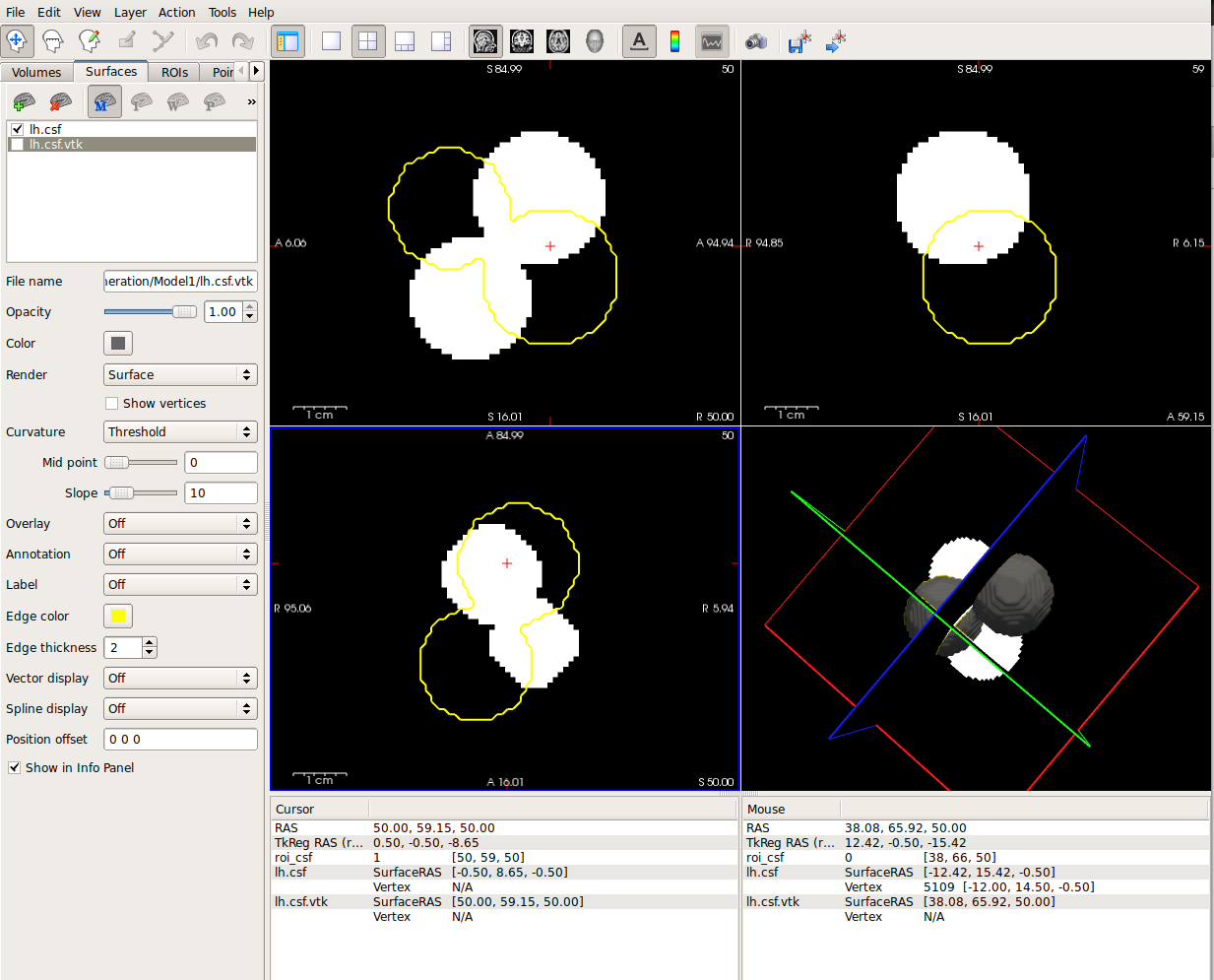
Q3: I've been trying one example: I have a binary mask with a grid of 101x101x101, 1mm^3 pixel size with the union of two spheres in nifti format. I run 'mri_mc mask.nii 1 out' and I get a very nice result, but I see problems regarding orientation. I attach a snapshot of freeview showing this and also the output of mri_info mask.nii.
Thanks again
Best regards, Oscar Esteban
[image: Inline image 1]
mri_info mask.nii:
Volume information for roi_csf.nii type: nii dimensions: 101 x 101 x 101 voxel sizes: 1.0000, 1.0000, 1.0000 type: FLOAT (3) fov: 101.000 dof: 0 xstart: -50.5, xend: 50.5 ystart: -50.5, yend: 50.5 zstart: -50.5, zend: 50.5 TR: 0.00 msec, TE: 0.00 msec, TI: 0.00 msec, flip angle: 0.00 degrees nframes: 1 PhEncDir: UNKNOWN ras xform present xform info: x_r = 1.0000, y_r = 0.0000, z_r = 0.0000, c_r = 50.5000 : x_a = 0.0000, y_a = 1.0000, z_a = 0.0000, c_a = 50.5000 : x_s = 0.0000, y_s = 0.0000, z_s = 1.0000, c_s = 50.5000 Orientation : RAS Primary Slice Direction: axial
voxel to ras transform: 1.0000 0.0000 0.0000 0.0000 0.0000 1.0000 0.0000 0.0000 0.0000 0.0000 1.0000 0.0000 0.0000 0.0000 0.0000 1.0000
voxel-to-ras determinant 1
ras to voxel transform: 1.0000 -0.0000 -0.0000 -0.0000 -0.0000 1.0000 -0.0000 -0.0000 -0.0000 -0.0000 1.0000 -0.0000 0.0000 0.0000 0.0000 1.0000
On Tue, Jun 4, 2013 at 3:08 PM, Bruce Fischl fischl@nmr.mgh.harvard.eduwrote:
Hi Oscar
what do you mean by "the CSF surface"? Outside the brain or in the ventricles? If outside, there is some code for creating boundary element models for EEG/MEG analysis.
There is no trivial way to create a single surface. The way I've done it in the past is to replace the lh or rh values in the filled.mgz with the other one and go from there, but it's not meant to work that way and you have to mess with it a fair amount.
cheers Bruce
On Tue, 4 Jun 2013, Oscar Esteban wrote:
Dear all,
I need to extract three surfaces from MRI data:
- GM-WM interface -> thus, lh.white and rh.white would be the answer.
- Pial interface -> thus, lh.pial + rh.pial
- CSF surface
For CSF, as far as I know, the best option would be to generate a binary volume merging the appropriate labels from aseg.mgz. Then use mri_mc to obtain the surface, right?
For white and pial I guess the best option would be to use the recon-all outcome, but I have two questions:
- Is there any mean to merge both hemispheres? I've tried several methods
but I get trouble with the consistency of the cells... the points are aggregated, but no work is done to merge cells and remove points.
- What are the differences between the recon-all outcome
(lh.pial,rh.pial,lh.white,rh.**white) with respect obtaining masks from aseg.mgz and then mri_mc?
Thank you so much
Best regards, Oscar Esteban
-- Oscar Esteban PhD Student / Researcher
Biomedical Image Technologies (BIT), UPM ETSI Telecomunicación Lab. C203, Av. Complutense s/n - E-28040 Madrid (Spain) +34 915 495 700 ext.4234
Signal Processing Laboratory (LTS5), EPFL-STI-IEL-LTS5 ELD 224 (Bâtiment ELD), Station 11, CH-1015 Lausanne, Switzerland
The information in this e-mail is intended only for the person to whom it is addressed. If you believe this e-mail was sent to you in error and the e-mail contains patient information, please contact the Partners Compliance HelpLine at http://www.partners.org/**compliancelinehttp://www.partners.org/complianceline. If the e-mail was sent to you in error but does not contain patient information, please contact the sender and properly dispose of the e-mail.
Hi Oscar
the mri_mc surfaces will no in general be topologically correct. The vertex spacing will also be less uniform
cheers Bruce On Wed, 5 Jun 2013, Oscar Esteban wrote:
Thank you so much, Bruce. Yes, I want the CSF inside the brain (e. g. the ventricles). Sorry for being unspecific.
I see that the way would be to start from filled.mgz and aseg.mgz until I get the desired ROIs to have the same label (one for white matter, another for ventricles ) and then run something like mri_mc, right? (Question 1)
Question 2: ok, if this is the way and regarding only white matter surface. Would there exist significant differences (per hemisphere) between the surface obtained with mri_mc and the surfaces from recon all? Because recon-all runs mri_tessellate, mris_smooth, mris_fix_topology & mris_make_surfaces to obtain the corresponding result... So just mri_mc does not seem so complete, right?
Q3: I've been trying one example: I have a binary mask with a grid of 101x101x101, 1mm^3 pixel size with the union of two spheres in nifti format. I run 'mri_mc mask.nii 1 out' and I get a very nice result, but I see problems regarding orientation. I attach a snapshot of freeview showing this and also the output of mri_info mask.nii.
Thanks again
Best regards, Oscar Esteban
Inline image 1
mri_info mask.nii:
Volume information for roi_csf.nii type: nii dimensions: 101 x 101 x 101 voxel sizes: 1.0000, 1.0000, 1.0000 type: FLOAT (3) fov: 101.000 dof: 0 xstart: -50.5, xend: 50.5 ystart: -50.5, yend: 50.5 zstart: -50.5, zend: 50.5 TR: 0.00 msec, TE: 0.00 msec, TI: 0.00 msec, flip angle: 0.00 degrees nframes: 1 PhEncDir: UNKNOWN ras xform present xform info: x_r = 1.0000, y_r = 0.0000, z_r = 0.0000, c_r = 50.5000 : x_a = 0.0000, y_a = 1.0000, z_a = 0.0000, c_a = 50.5000 : x_s = 0.0000, y_s = 0.0000, z_s = 1.0000, c_s = 50.5000 Orientation : RAS Primary Slice Direction: axial
voxel to ras transform: 1.0000 0.0000 0.0000 0.0000 0.0000 1.0000 0.0000 0.0000 0.0000 0.0000 1.0000 0.0000 0.0000 0.0000 0.0000 1.0000
voxel-to-ras determinant 1
ras to voxel transform: 1.0000 -0.0000 -0.0000 -0.0000 -0.0000 1.0000 -0.0000 -0.0000 -0.0000 -0.0000 1.0000 -0.0000 0.0000 0.0000 0.0000 1.0000
On Tue, Jun 4, 2013 at 3:08 PM, Bruce Fischl fischl@nmr.mgh.harvard.edu wrote: Hi Oscar
  what do you mean by "the CSF surface"? Outside the brain or in
  the ventricles? If outside, there is some code for creating
  boundary element models for EEG/MEG analysis.

  There is no trivial way to create a single surface. The way I've
  done it in the past is to replace the lh or rh values in the
  filled.mgz with the other one and go from there, but it's not
  meant to work that way and you have to mess with it a fair
  amount.

  cheers
  Bruce


  On Tue, 4 Jun 2013, Oscar Esteban wrote:

        Dear all,
        I need to extract three surfaces from MRI data:

        - GM-WM interface -> thus, lh.white and rh.white
        would be the answer.
        - Pial interface -> thus, lh.pial + rh.pial
        - CSF surface

        For CSF, as far as I know, the best option would be
        to generate a binary volume merging the appropriate
        labels
        from aseg.mgz. Then use mri_mc to obtain the
        surface, right?

        For white and pial I guess the best option would be
        to use the recon-all outcome, but I have two
        questions:
        * Is there any mean to merge both hemispheres? I've
        tried several methods but I get trouble with the
        consistency of the cells... the points are
        aggregated, but no work is done to merge cells and
        remove points.
        * What are the differences between the recon-all
        outcome (lh.pial,rh.pial,lh.white,rh.white) with
        respect
        obtaining masks from aseg.mgz and then mri_mc?

        Thank you so much

        Best regards,
        Oscar Esteban


        --
        Oscar Esteban
        PhD Student / Researcher

        Biomedical Image Technologies (BIT), UPM
        ETSI Telecomunicación Lab. C203, Av. Complutense s/n
        - E-28040 Madrid (Spain)
        +34 915 495 700 ext.4234

        Signal Processing Laboratory (LTS5),
        EPFL-STI-IEL-LTS5
        ELD 224 (Bâtiment ELD), Station 11, CH-1015
        Lausanne, Switzerland
The information in this e-mail is intended only for the person to whom it is addressed. If you believe this e-mail was sent to you in error and the e-mail contains patient information, please contact the Partners Compliance HelpLine at http://www.partners.org/complianceline . If the e-mail was sent to you in error but does not contain patient information, please contact the sender and properly dispose of the e-mail.
-- Oscar Esteban PhD Student / Researcher
Biomedical Image Technologies (BIT), UPM ETSI Telecomunicación Lab. C203, Av. Complutense s/n - E-28040 Madrid (Spain) +34 915 495 700 ext.4234
Signal Processing Laboratory (LTS5), EPFL-STI-IEL-LTS5 ELD 224 (Bâtiment ELD), Station 11, CH-1015 Lausanne, Switzerland
freesurfer@nmr.mgh.harvard.edu
-
 Bruce Fischl
 -
 Oscar Esteban