
Hey Falk,
Much appreciated for your reply on this! Fortunately I was able to correct the issue by contacting our MRI tech - he suggested we use his nifti files instead and the results turned out a lot better. I emailed Bruce but forgot to include the freesurfer mailing list in the CC.
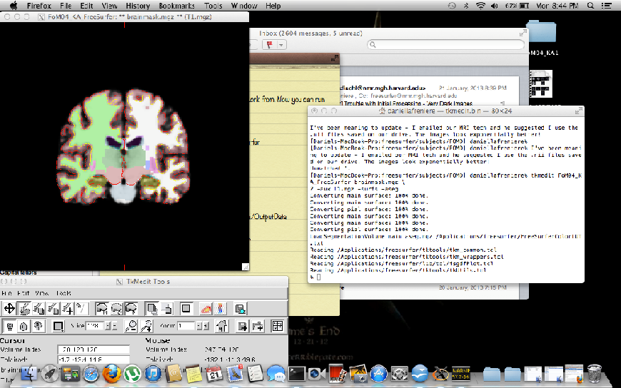
Here is an attached screenshot of my final results with the nifti files for the record. Thanks again for the replies! I'm sure I'll be contacting the experts down the road.
Cheers,
Dan
On Tue, Jan 22, 2013 at 4:06 AM, "Falk Lüsebrink" falk.luese@gmx.netwrote:
Hey Dan,
Bruce might be right about the SNR, but you could try to adjust the parameters of the inhomogeneity correction in the N3 algorithm.
You could either use the following command line: recon-all -autorecon1 -mprage -nuintensitycor-3t -i <data> -s <subject>
Or if this still isn't good enough, try changing the parameters of the N3 manually (e.g. distance and fwhm): SUBJ=ab11
recon-all -motioncor -talairach -tal-check -i <data> -s $SUBJ
mri_nu_correct.mni --i $SUBJECTS_DIR/$SUBJ/mri/orig.mgz --o $SUBJECTS_DIR/$SUBJ/mri/nu.mgz --proto-iters 1000 --distance 15 --fwhm 0.5 --n 1 --uchar $SUBJECTS_DIR/$SUBJ/mri/transforms/talairach.xfm
recon-all -mprage -normalization -skulstrip -s $SUBJ
If the skullstrip looks fine continue by using this: recon-all -mprage -autorecon2 -autorecon3 -s $SUBJ
Best, Falk
-------- Original-Nachricht --------
Datum: Mon, 21 Jan 2013 22:39:29 -0500 Von: Bruce Fischl fischl@nmr.mgh.harvard.edu An: Dan LaFreniere lafreniere.dj@gmail.com CC: "freesurfer@nmr.mgh.harvard.edu" freesurfer@nmr.mgh.harvard.edu Betreff: Re: [Freesurfer] Trouble with Initial Processing - Very Dark
Images
Wow, that's a huge bias field! Control points will help uniformity, but the snr may not be good enough for good results Bruce
On Jan 18, 2013, at 11:09 AM, Dan LaFreniere lafreniere.dj@gmail.com wrote:
Hey all,
I'm very new to Freesurfer and am having some issues with the initial
processing of our MPRAGE dicom files. So far, I'm able to get initial -autorecon1 steps to work but once this has finished and I open my
brainmask.mgz
files and the brain is incredibly dark in inferior regions. So much so
that
the skull strip appears to be removing large chunks of anterior temporal lobes. What remains of the temporal lobes is incredibly dark with grey
matter
almost indistinguishable from CSF.
I would really like to look into freesurfer for cortical thickness
analysis of subcortical regions since it seems like such an awesome
program but
I have really been struggling to get a decent looking brain. It's
probably
noteworthy that these brains have always been quite dark but I'm usually able to correct them to a point where they're pretty good in other
programs.
I have read about a few solutions to the problem but am having trouble
implementing them. One suggestion was to use the -mprage flag; unfortunately, I'm not sure how to run this or when. Another suggestion
was to use
control points but I don't yet have the red and blue segmentation
boundary lines
- I'm using tkmedit FoM04_KA brainmask.mgz -aux T1.mgz to view the
files.
For reference, this is the line I use to run autorecon1: Recon-all -autorecon1 -subjid FoM04_KA
Does anyone have any suggestions as to what I could do? It would be
greatly appreciated. I've attached a screenshot to illustrate. Also, we
used a
4.7T Varian scanner with 0.375/0.375/1mm voxels. I believe we used a gradient echo multislice sequence. In addition, all subjects' data have
been
obtained so there is no chance of changing sequences…
Best wishes,
Dan
Freesurfer mailing list Freesurfer@nmr.mgh.harvard.edu https://mail.nmr.mgh.harvard.edu/mailman/listinfo/freesurfer
Freesurfer mailing list Freesurfer@nmr.mgh.harvard.edu https://mail.nmr.mgh.harvard.edu/mailman/listinfo/freesurfer
The information in this e-mail is intended only for the person to whom it is addressed. If you believe this e-mail was sent to you in error and the e-mail contains patient information, please contact the Partners Compliance HelpLine at http://www.partners.org/complianceline . If the e-mail was sent to you
in
error but does not contain patient information, please contact the sender and properly dispose of the e-mail.

Dan: just to be clear this has nothing to do with nifti vs. dicom. It's likely that your tech ran prescan normalize and the nifti is the series that was normalized.
cheers Bruce
On Tue, 22 Jan 2013, Dan LaFreniere wrote:
Hey Falk,
Much appreciated for your reply on this! Fortunately I was able to correct the issue by contacting our MRI tech - he suggested we use his nifti files instead and the results turned out a lot better. I emailed Bruce but forgot to include the freesurfer mailing list in the CC.
Here is an attached screenshot of my final results with the nifti files for the record. Thanks again for the replies! I'm sure I'll be contacting the experts down the road.
Cheers,
Dan
On Tue, Jan 22, 2013 at 4:06 AM, "Falk Lüsebrink" falk.luese@gmx.net wrote: Hey Dan,
  Bruce might be right about the SNR, but you could try to adjust the parameters of the inhomogeneity correction in the N3
  algorithm.

  You could either use the following command line:
  recon-all -autorecon1 -mprage -nuintensitycor-3t -i <data> -s <subject>

  Or if this still isn't good enough, try changing the parameters of the N3 manually (e.g. distance and fwhm):
  SUBJ=ab11

  recon-all -motioncor -talairach -tal-check -i <data> -s $SUBJ

  mri_nu_correct.mni --i $SUBJECTS_DIR/$SUBJ/mri/orig.mgz --o $SUBJECTS_DIR/$SUBJ/mri/nu.mgz --proto-iters 1000 --distance 15
  --fwhm 0.5 --n 1 --uchar $SUBJECTS_DIR/$SUBJ/mri/transforms/talairach.xfm

  recon-all -mprage -normalization -skulstrip -s $SUBJ

  If the skullstrip looks fine continue by using this:
  recon-all -mprage -autorecon2 -autorecon3 -s $SUBJ

  Best,
  Falk


  -------- Original-Nachricht --------
  > Datum: Mon, 21 Jan 2013 22:39:29 -0500
  > Von: Bruce Fischl <fischl@nmr.mgh.harvard.edu>
  > An: Dan LaFreniere <lafreniere.dj@gmail.com>
  > CC: "freesurfer@nmr.mgh.harvard.edu" <freesurfer@nmr.mgh.harvard.edu>
  > Betreff: Re: [Freesurfer] Trouble with Initial Processing - Very Dark Images

  > Wow, that's a huge bias field! Control points will help uniformity, but
  > the snr may not be good enough for good results
  > Bruce
  >
  >
  >
  > On Jan 18, 2013, at 11:09 AM, Dan LaFreniere <lafreniere.dj@gmail.com>
  > wrote:
  >
  > > Hey all,
  > >
  > > I'm very new to Freesurfer and am having some issues with the initial
  > processing of our MPRAGE dicom files. So far, I'm able to get initial
  > -autorecon1 steps to work but once this has finished and I open my brainmask.mgz
  > files and the brain is incredibly dark in inferior regions. So much so that
  > the skull strip appears to be removing large chunks of anterior temporal
  > lobes. What remains of the temporal lobes is incredibly dark with grey matter
  > almost indistinguishable from CSF.
  > >
  > > I would really like to look into freesurfer for cortical thickness
  > analysis of subcortical regions since it seems like such an awesome program but
  > I have really been struggling to get a decent looking brain. It's probably
  > noteworthy that these brains have always been quite dark but I'm usually
  > able to correct them to a point where they're pretty good in other programs.
  > >
  > > I have read about a few solutions to the problem but am having trouble
  > implementing them. One suggestion was to use the -mprage flag;
  > unfortunately, I'm not sure how to run this or when. Another suggestion was to use
  > control points but I don't yet have the red and blue segmentation boundary lines
  > - I'm using tkmedit FoM04_KA brainmask.mgz -aux T1.mgz  to view the files.
  > For reference, this is the line I use to run autorecon1: Recon-all
  > -autorecon1 -subjid FoM04_KA
  > >
  > > Does anyone have any suggestions as to what I could do? It would be
  > greatly appreciated. I've attached a screenshot to illustrate. Also, we used a
  > 4.7T Varian scanner with 0.375/0.375/1mm voxels. I believe we used a
  > gradient echo multislice sequence. In addition, all subjects' data have been
  > obtained so there is no chance of changing sequences?
  > >
  > > Best wishes,
  > >
  > > Dan

  > > _______________________________________________
  > > Freesurfer mailing list
  > > Freesurfer@nmr.mgh.harvard.edu
  > > https://mail.nmr.mgh.harvard.edu/mailman/listinfo/freesurfer
  >
  > _______________________________________________
  > Freesurfer mailing list
  > Freesurfer@nmr.mgh.harvard.edu
  > https://mail.nmr.mgh.harvard.edu/mailman/listinfo/freesurfer
  >
  >
  > The information in this e-mail is intended only for the person to whom it
  > is
  > addressed. If you believe this e-mail was sent to you in error and the
  > e-mail
  > contains patient information, please contact the Partners Compliance
  > HelpLine at
  > http://www.partners.org/complianceline . If the e-mail was sent to you in
  > error
  > but does not contain patient information, please contact the sender and
  > properly
  > dispose of the e-mail.
That was exactly what he said as well; sorry that I wasn't more clear with my email.
Cheers guys,
Dan
On Tue, Jan 22, 2013 at 9:04 AM, Bruce Fischl fischl@nmr.mgh.harvard.eduwrote:
Dan: just to be clear this has nothing to do with nifti vs. dicom. It's likely that your tech ran prescan normalize and the nifti is the series that was normalized.
cheers Bruce
On Tue, 22 Jan 2013, Dan LaFreniere wrote:
Hey Falk,
Much appreciated for your reply on this! Fortunately I was able to correct the issue by contacting our MRI tech - he suggested we use his nifti files instead and the results turned out a lot better. I emailed Bruce but forgot to include the freesurfer mailing list in the CC.
Here is an attached screenshot of my final results with the nifti files for the record. Thanks again for the replies! I'm sure I'll be contacting the experts down the road.
Cheers,
Dan
On Tue, Jan 22, 2013 at 4:06 AM, "Falk Lüsebrink" falk.luese@gmx.net wrote: Hey Dan,
Bruce might be right about the SNR, but you could try to adjust theparameters of the inhomogeneity correction in the N3 algorithm.
  You could either use the following command line:
  recon-all -autorecon1 -mprage -nuintensitycor-3t -i <data> -s
<subject>
Or if this still isn't good enough, try changing the parameters ofthe N3 manually (e.g. distance and fwhm): SUBJ=ab11
  recon-all -motioncor -talairach -tal-check -i <data> -s $SUBJ

  mri_nu_correct.mni --i $SUBJECTS_DIR/$SUBJ/mri/orig.**mgz --o
$SUBJECTS_DIR/$SUBJ/mri/nu.mgz --proto-iters 1000 --distance 15 --fwhm 0.5 --n 1 --uchar $SUBJECTS_DIR/$SUBJ/mri/** transforms/talairach.xfm
  recon-all -mprage -normalization -skulstrip -s $SUBJ

  If the skullstrip looks fine continue by using this:
  recon-all -mprage -autorecon2 -autorecon3 -s $SUBJ

  Best,
  Falk


  -------- Original-Nachricht --------
  > Datum: Mon, 21 Jan 2013 22:39:29 -0500
  > Von: Bruce Fischl <fischl@nmr.mgh.harvard.edu>
  > An: Dan LaFreniere <lafreniere.dj@gmail.com>
  > CC: "freesurfer@nmr.mgh.harvard.**edu<freesurfer@nmr.mgh.harvard.edu>"
<freesurfer@nmr.mgh.harvard.**edu freesurfer@nmr.mgh.harvard.edu> > Betreff: Re: [Freesurfer] Trouble with Initial Processing - Very Dark Images
> Wow, that's a huge bias field! Control points will helpuniformity, but > the snr may not be good enough for good results > Bruce > > > > On Jan 18, 2013, at 11:09 AM, Dan LaFreniere < lafreniere.dj@gmail.com> > wrote: > > > Hey all, > > > > I'm very new to Freesurfer and am having some issues with the initial > processing of our MPRAGE dicom files. So far, I'm able to get initial > -autorecon1 steps to work but once this has finished and I open my brainmask.mgz > files and the brain is incredibly dark in inferior regions. So much so that > the skull strip appears to be removing large chunks of anterior temporal > lobes. What remains of the temporal lobes is incredibly dark with grey matter > almost indistinguishable from CSF. > > > > I would really like to look into freesurfer for cortical thickness > analysis of subcortical regions since it seems like such an awesome program but > I have really been struggling to get a decent looking brain. It's probably > noteworthy that these brains have always been quite dark but I'm usually > able to correct them to a point where they're pretty good in other programs. > > > > I have read about a few solutions to the problem but am having trouble > implementing them. One suggestion was to use the -mprage flag; > unfortunately, I'm not sure how to run this or when. Another suggestion was to use > control points but I don't yet have the red and blue segmentation boundary lines > - I'm using tkmedit FoM04_KA brainmask.mgz -aux T1.mgz to view the files. > For reference, this is the line I use to run autorecon1: Recon-all > -autorecon1 -subjid FoM04_KA > > > > Does anyone have any suggestions as to what I could do? It would be > greatly appreciated. I've attached a screenshot to illustrate. Also, we used a > 4.7T Varian scanner with 0.375/0.375/1mm voxels. I believe we used a > gradient echo multislice sequence. In addition, all subjects' data have been > obtained so there is no chance of changing sequences?
  > >
  > > Best wishes,
  > >
  > > Dan

  > > ______________________________**_________________
  > > Freesurfer mailing list
  > > Freesurfer@nmr.mgh.harvard.edu
  > > https://mail.nmr.mgh.harvard.**edu/mailman/listinfo/**
freesurfer https://mail.nmr.mgh.harvard.edu/mailman/listinfo/freesurfer > > ______________________________**_________________ > Freesurfer mailing list > Freesurfer@nmr.mgh.harvard.edu > https://mail.nmr.mgh.harvard.**edu/mailman/listinfo/**freesurferhttps://mail.nmr.mgh.harvard.edu/mailman/listinfo/freesurfer > > > The information in this e-mail is intended only for the person to whom it > is > addressed. If you believe this e-mail was sent to you in error and the > e-mail > contains patient information, please contact the Partners Compliance > HelpLine at > http://www.partners.org/**compliancelinehttp://www.partners.org/complianceline. If the e-mail was sent to you in > error > but does not contain patient information, please contact the sender and > properly > dispose of the e-mail.
freesurfer@nmr.mgh.harvard.edu
-
 Bruce Fischl
 -
 Dan LaFreniere