
Hey Freesurfers,
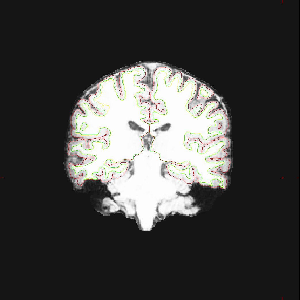
I have a question regarding segmentation in autorecon2. I have a bunch of sagittally-acquired MRI data -- Freesurfer has done a decent job after we manually added control points on the data and resegmented. The problem is that FS ends up leaving out some lateral areas of cortex. After control points, I am satisfied with the grey/white boundary but the pial surface could stand improvement. I am attaching an image to this email showing a segmentation in the coronal plane. Seems like a chunk or lateral temporal cortex is missing in the left hemi (and a little bit of missing cortex in right hemi too.) I turned the brightness up a bit so you could see the missing cortex in brainmask.mgz -- the lateral stuff as you can tell is not quite as intense as the more medial voxels, which is probably the root of the problem.
First of all, I want to get a sense of what is reasonable to expect. Does this look like a reasonable segmentation or is it reasonable to try and improve this? My PI is concerned about the missing lateral GM in any case.
Second, I was curious if any experienced users had advice regarding ways to get this missing cortex included in segmentation - specifically the situation in the left temporal lobe. Going off the website, since the white/grey surface looks correct, control points should not be used. (I don't know whether putting a few *right* outside the surface might help expand pial surface? I might try that more if you think it could help..) The guide would then suggest that pial surface edits must be made manually on every slice prior to re-running autorecon2-pial. This would take a long time for 1 person with 34 brains, but is certainly a possibility. Downside to this is it becomes somewhat subjective for someone with only a rudimentory understanding of neuroanatomy.
What if I could run the autorecon2 workflow step-by-step and try adjusting normalization parameters? There are two normalizations here, using mri_ca_normalize (-canorm) and mri_normalize (-normalization2). mri_ca_normalize has a flag -p, that specifies percentage of likely wm to use as control points (defaulting to 50%). Bumping this up may improve intial segmentations since we've had to extensively add control points, though I suppose the end result would be about the same (in theory). mri_normalize has a number of parameters that might effect this, as well as various programs called in the segmentation process.
Long story short, does anyone know of any parameter adjustment approach to this, or is manual editting of pial surface the only solution?
Thanks!
John Sheppard
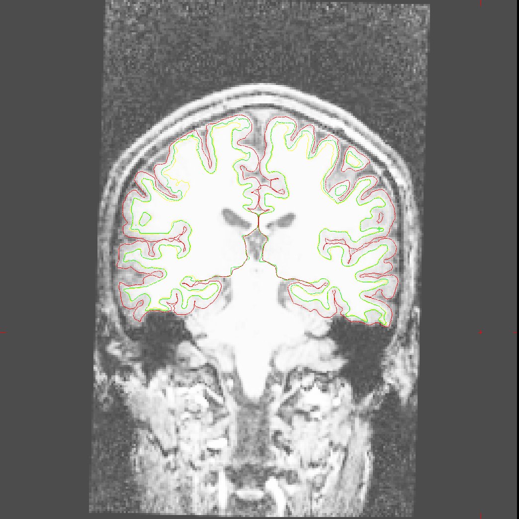
By the way, here is a matching picture of T1.mgz. Just to show you that there were really no problems that I could see with any of the skullstripping - thought FS did fine in that regard.
-John
On Tue, Oct 7, 2008 at 1:05 PM, John Sheppard < johnsheppard2007@u.northwestern.edu> wrote:
Hey Freesurfers,
I have a question regarding segmentation in autorecon2. I have a bunch of sagittally-acquired MRI data -- Freesurfer has done a decent job after we manually added control points on the data and resegmented. The problem is that FS ends up leaving out some lateral areas of cortex. After control points, I am satisfied with the grey/white boundary but the pial surface could stand improvement. I am attaching an image to this email showing a segmentation in the coronal plane. Seems like a chunk or lateral temporal cortex is missing in the left hemi (and a little bit of missing cortex in right hemi too.) I turned the brightness up a bit so you could see the missing cortex in brainmask.mgz -- the lateral stuff as you can tell is not quite as intense as the more medial voxels, which is probably the root of the problem.
First of all, I want to get a sense of what is reasonable to expect. Does this look like a reasonable segmentation or is it reasonable to try and improve this? My PI is concerned about the missing lateral GM in any case.
Second, I was curious if any experienced users had advice regarding ways to get this missing cortex included in segmentation - specifically the situation in the left temporal lobe. Going off the website, since the white/grey surface looks correct, control points should not be used. (I don't know whether putting a few *right* outside the surface might help expand pial surface? I might try that more if you think it could help..) The guide would then suggest that pial surface edits must be made manually on every slice prior to re-running autorecon2-pial. This would take a long time for 1 person with 34 brains, but is certainly a possibility. Downside to this is it becomes somewhat subjective for someone with only a rudimentory understanding of neuroanatomy.
What if I could run the autorecon2 workflow step-by-step and try adjusting normalization parameters? There are two normalizations here, using mri_ca_normalize (-canorm) and mri_normalize (-normalization2). mri_ca_normalize has a flag -p, that specifies percentage of likely wm to use as control points (defaulting to 50%). Bumping this up may improve intial segmentations since we've had to extensively add control points, though I suppose the end result would be about the same (in theory). mri_normalize has a number of parameters that might effect this, as well as various programs called in the segmentation process.
Long story short, does anyone know of any parameter adjustment approach to this, or is manual editting of pial surface the only solution?
Thanks!
John Sheppard

Hi John, To my knowledge, there is no mechanism of manual editing that can force the pial surface out further (provided that it wasn't a problem of missing pial tissue due to an overly aggressive skull-stripping).
cheers, Mike H.
By the way, here is a matching picture of T1.mgz. Just to show you that there were really no problems that I could see with any of the skullstripping - thought FS did fine in that regard.
-John
On Tue, Oct 7, 2008 at 1:05 PM, John Sheppard < johnsheppard2007@u.northwestern.edu> wrote:
Hey Freesurfers,
I have a question regarding segmentation in autorecon2. I have a bunch of sagittally-acquired MRI data -- Freesurfer has done a decent job after we manually added control points on the data and resegmented. The problem is that FS ends up leaving out some lateral areas of cortex. After control points, I am satisfied with the grey/white boundary but the pial surface could stand improvement. I am attaching an image to this email showing a segmentation in the coronal plane. Seems like a chunk or lateral temporal cortex is missing in the left hemi (and a little bit of missing cortex in right hemi too.) I turned the brightness up a bit so you could see the missing cortex in brainmask.mgz -- the lateral stuff as you can tell is not quite as intense as the more medial voxels, which is probably the root of the problem.
First of all, I want to get a sense of what is reasonable to expect. Does this look like a reasonable segmentation or is it reasonable to try and improve this? My PI is concerned about the missing lateral GM in any case.
Second, I was curious if any experienced users had advice regarding ways to get this missing cortex included in segmentation - specifically the situation in the left temporal lobe. Going off the website, since the white/grey surface looks correct, control points should not be used. (I don't know whether putting a few *right* outside the surface might help expand pial surface? I might try that more if you think it could help..) The guide would then suggest that pial surface edits must be made manually on every slice prior to re-running autorecon2-pial. This would take a long time for 1 person with 34 brains, but is certainly a possibility. Downside to this is it becomes somewhat subjective for someone with only a rudimentory understanding of neuroanatomy.
What if I could run the autorecon2 workflow step-by-step and try adjusting normalization parameters? There are two normalizations here, using mri_ca_normalize (-canorm) and mri_normalize (-normalization2). mri_ca_normalize has a flag -p, that specifies percentage of likely wm to use as control points (defaulting to 50%). Bumping this up may improve intial segmentations since we've had to extensively add control points, though I suppose the end result would be about the same (in theory). mri_normalize has a number of parameters that might effect this, as well as various programs called in the segmentation process.
Long story short, does anyone know of any parameter adjustment approach to this, or is manual editting of pial surface the only solution?
Thanks!
John Sheppard
Freesurfer mailing list Freesurfer@nmr.mgh.harvard.edu https://mail.nmr.mgh.harvard.edu/mailman/listinfo/freesurfer

usually we do this by putting control points in the underlying white matter. If the WM is significantly less than 110 this can help On Tue, 7 Oct 2008, Michael Harms wrote:
Hi John, To my knowledge, there is no mechanism of manual editing that can force the pial surface out further (provided that it wasn't a problem of missing pial tissue due to an overly aggressive skull-stripping).
cheers, Mike H.
By the way, here is a matching picture of T1.mgz. Just to show you that there were really no problems that I could see with any of the skullstripping - thought FS did fine in that regard.
-John
On Tue, Oct 7, 2008 at 1:05 PM, John Sheppard < johnsheppard2007@u.northwestern.edu> wrote:
Hey Freesurfers,
I have a question regarding segmentation in autorecon2. I have a bunch of sagittally-acquired MRI data -- Freesurfer has done a decent job after we manually added control points on the data and resegmented. The problem is that FS ends up leaving out some lateral areas of cortex. After control points, I am satisfied with the grey/white boundary but the pial surface could stand improvement. I am attaching an image to this email showing a segmentation in the coronal plane. Seems like a chunk or lateral temporal cortex is missing in the left hemi (and a little bit of missing cortex in right hemi too.) I turned the brightness up a bit so you could see the missing cortex in brainmask.mgz -- the lateral stuff as you can tell is not quite as intense as the more medial voxels, which is probably the root of the problem.
First of all, I want to get a sense of what is reasonable to expect. Does this look like a reasonable segmentation or is it reasonable to try and improve this? My PI is concerned about the missing lateral GM in any case.
Second, I was curious if any experienced users had advice regarding ways to get this missing cortex included in segmentation - specifically the situation in the left temporal lobe. Going off the website, since the white/grey surface looks correct, control points should not be used. (I don't know whether putting a few *right* outside the surface might help expand pial surface? I might try that more if you think it could help..) The guide would then suggest that pial surface edits must be made manually on every slice prior to re-running autorecon2-pial. This would take a long time for 1 person with 34 brains, but is certainly a possibility. Downside to this is it becomes somewhat subjective for someone with only a rudimentory understanding of neuroanatomy.
What if I could run the autorecon2 workflow step-by-step and try adjusting normalization parameters? There are two normalizations here, using mri_ca_normalize (-canorm) and mri_normalize (-normalization2). mri_ca_normalize has a flag -p, that specifies percentage of likely wm to use as control points (defaulting to 50%). Bumping this up may improve intial segmentations since we've had to extensively add control points, though I suppose the end result would be about the same (in theory). mri_normalize has a number of parameters that might effect this, as well as various programs called in the segmentation process.
Long story short, does anyone know of any parameter adjustment approach to this, or is manual editting of pial surface the only solution?
Thanks!
John Sheppard
Freesurfer mailing list Freesurfer@nmr.mgh.harvard.edu https://mail.nmr.mgh.harvard.edu/mailman/listinfo/freesurfer
Freesurfer mailing list Freesurfer@nmr.mgh.harvard.edu https://mail.nmr.mgh.harvard.edu/mailman/listinfo/freesurfer
freesurfer@nmr.mgh.harvard.edu
-
 Bruce Fischl
 -
 John Sheppard
 -
 Michael Harms