
Hello,
I have a couple of questions about lesions:
1. I would like to define cortical lesions and white matter lacunes in freesurfer using T1 and FLAIR images; maybe by combining the aseg results with the FLAIR images? I read some old threads about this topic and saw mention of something that the freesurfer team was working on to do this. Is this program now available? If not, has anyone tried to do this and had success?
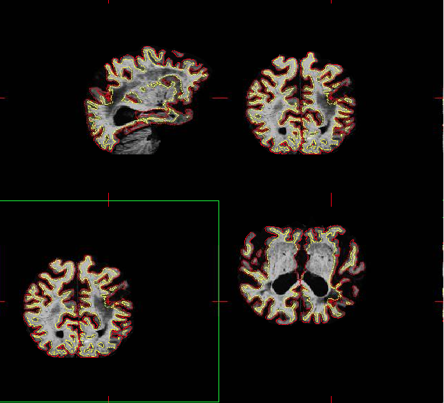
2. Some of the cortical lesions extend into the white matter, technically causing a discontinuation in the grey matter surface. Following the tutorial, I filled in the area of the stroke, but after rerunning recon2 freesurfer still represents a pial line around this area. Is there a way to fix this? I have attached a screenshot.
Thanks for your help,
Natalie

1. Koen Van Leemput (ccd) has been working on this - check with him. 2. Hmm, that's though. I guess we should really disable the surface deformation in regions labeled as lesion, which would mean you need to edit the aseg. If you want to try that we can build a version with lesion/hypointensities frozen.
cheers Bruce
On Tue, 8 Jun 2010, Natalie Marchant wrote:
Hello,
I have a couple of questions about lesions:
- I would like to define cortical lesions and white matter lacunes in
freesurfer using T1 and FLAIR images; maybe by combining the aseg results with the FLAIR images? I read some old threads about this topic and saw mention of something that the freesurfer team was working on to do this. Is this program now available? If not, has anyone tried to do this and had success?
- Some of the cortical lesions extend into the white matter, technically
causing a discontinuation in the grey matter surface. Following the tutorial, I filled in the area of the stroke, but after rerunning recon2 freesurfer still represents a pial line around this area. Is there a way to fix this? I have attached a screenshot.
Thanks for your help,
Natalie
freesurfer@nmr.mgh.harvard.edu
-
 Bruce Fischl
 -
 Natalie Marchant