
Hi
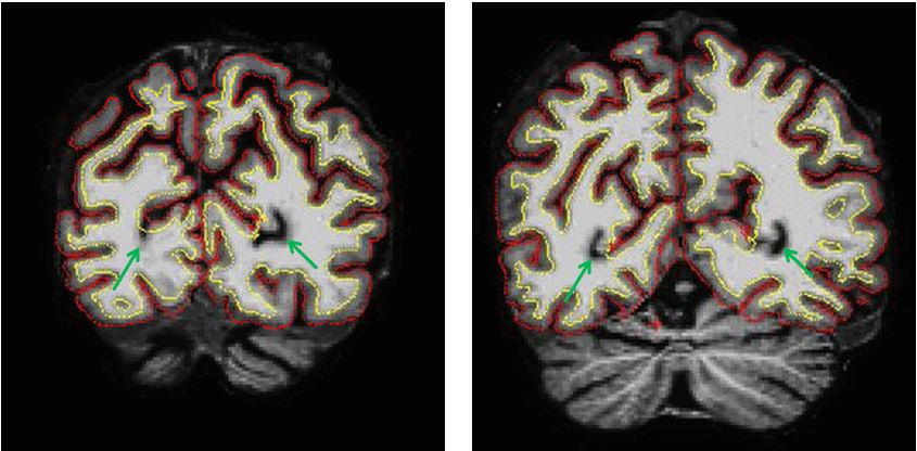
I have run into an issue that I'm not sure how to correct. What happens is that in some subjects the surfaces don't seem to properly track sulci coming off the medial part of temporo-occipital cortex. Please see green arrows on the attached image.
In fact the tutorial on topological defects shows an image with a similar issue, but fixing it is not discussed. http://surfer.nmr.mgh.harvard.edu/fswiki/FsTutorial/TopologicalDefect?action...
I have checked and these are sulci and not the occipital extent of the lateral ventricle.
Perhaps it is not a problem, or if it is then how to approach fixing it?
Thank you, Darren

Check the aseg and see if it was labeled ventricle (and make sure it really isn't). You may need to edit the aseg
On Sep 16, 2012, at 5:19 PM, Darren Gitelman d-gitelman@northwestern.edu wrote:
Hi
I have run into an issue that I'm not sure how to correct. What happens is that in some subjects the surfaces don't seem to properly track sulci coming off the medial part of temporo-occipital cortex. Please see green arrows on the attached image.
In fact the tutorial on topological defects shows an image with a similar issue, but fixing it is not discussed. http://surfer.nmr.mgh.harvard.edu/fswiki/FsTutorial/TopologicalDefect?action...
I have checked and these are sulci and not the occipital extent of the lateral ventricle.
Perhaps it is not a problem, or if it is then how to approach fixing it?
Thank you, Darren -- Darren Gitelman, MD Northwestern University 710 N. Lake Shore Dr. Abbott 11th Floor Chicago, IL 60611 Ph: (312) 908-8614 Fax: (312) 908-5073
<3809-error.png> _______________________________________________ Freesurfer mailing list Freesurfer@nmr.mgh.harvard.edu https://mail.nmr.mgh.harvard.edu/mailman/listinfo/freesurfer
freesurfer@nmr.mgh.harvard.edu
-
 Bruce Fischl
 -
 Darren Gitelman