
From: Christine Smith cnsmith@ucsd.edu Date: Thursday, January 23, 2014 3:58 PM To: Matt Glasser matt@ma-tea.com Subject: Re: [Freesurfer] poor grey/white distinction in superior part of scan
Please note that the left and right sides of the brain are flipped for freesurfer vs the dicom picture. Please find attached a dicom picture that is oriented the same way as freesurfer. Sorry for any confusion.
On Thu, Jan 23, 2014 at 1:56 PM, Christine Smith cnsmith@ucsd.edu wrote:
Dear Matt,
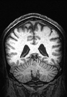
Please find attached two pictures. One is of the brain from reconstructed dicom files (using AFNI). The other one is a pic from tkmedit of the same subject and approximately the same slice of brain. Notice that the freesurfer image is very bright. Look at the white matter and pial lines (i.e., cortical thickness) in the upper left hand part of the image and see how thin the cortex appears. Notice also, that there is very little grey/white contrast in this same area in the brain image reconstructed from dicoms.
Please let me know if there are any other pictures you might desire or information you might need.
Christine
On Thu, Jan 23, 2014 at 11:33 AM, Matt Glasser matt@ma-tea.com wrote:
Some pictures would probably be helpful to know what the issue is.
Peace,
Matt.
From: Christine Smith cnsmith@ucsd.edu Date: Thursday, January 23, 2014 1:05 PM To: freesurfer@nmr.mgh.harvard.edu Subject: [Freesurfer] poor grey/white distinction in superior part of scan
Hello,
I am emailing to inquire about whether poor grey/white contrast in one part of a scan (i.e., the posterior and superior part of the scan; ~parietal cortex) can be addressed. The contrast in other parts of the scan looks good and freesurfer does a great job of distinguishing white from grey. For the parietal cortex area the cortex appears very thin. In addition, the entire brain appears 'white' or bright, even though the brain doesn't look this bright if you reconstruct it from dicoms.
We have now obtained 4 scans like this, so it isn't just one person with thinning cortex.
How can I make an adjustment to only this superior part of the brain and leave the rest of it alone? Or do I need to make an adjustment to the intensity early on for the entire scan and then basically start over with editing?
Best, Christine
-- Christine N. Smith, Ph.D. Department of Psychiatry University of California, San Diego _______________________________________________ Freesurfer mailing list Freesurfer@nmr.mgh.harvard.eduhttps://mail.nmr.mgh.harvard.edu/mailman/listin fo/freesurfer The information in this e-mail is intended only for the person to whom it is addressed. If you believe this e-mail was sent to you in error and the e-mail contains patient information, please contact the Partners Compliance HelpLine at http://www.partners.org/complianceline . If the e-mail was sent to you in error but does not contain patient information, please contact the sender and properly dispose of the e-mail.
-- Christine N. Smith, Ph.D. Department of Psychiatry University of California, San Diego

Hi Christine
this looks to me like a consequence of the high myelin content of motor cortex.If there really is as little contrast there as seems from your image there is not much you can do except change the acquisition. If you give us the details we can help with this.
cheers Bruce On Thu, 23 Jan 2014, Matt Glasser wrote:
From: Christine Smith cnsmith@ucsd.edu Date: Thursday, January 23, 2014 3:58 PM To: Matt Glasser matt@ma-tea.com Subject: Re: [Freesurfer] poor grey/white distinction in superior part of scan
Please note that the left and right sides of the brain are flipped for freesurfer vs the dicom picture. Please find attached a dicom picture that is oriented the same way as freesurfer. Sorry for any confusion.
On Thu, Jan 23, 2014 at 1:56 PM, Christine Smith cnsmith@ucsd.edu wrote: Dear Matt, Please find attached two pictures. One is of the brain from reconstructed dicom files (using AFNI). The other one is a pic from tkmedit of the same subject and approximately the same slice of brain. Notice that the freesurfer image is very bright. Look at the white matter and pial lines (i.e., cortical thickness) in the upper left hand part of the image and see how thin the cortex appears. Notice also, that there is very little grey/white contrast in this same area in the brain image reconstructed from dicoms.
Please let me know if there are any other pictures you might desire or information you might need.
Christine
On Thu, Jan 23, 2014 at 11:33 AM, Matt Glasser matt@ma-tea.com wrote: Some pictures would probably be helpful to know what the issue is.
Peace,
Matt.
From: Christine Smith cnsmith@ucsd.edu Date: Thursday, January 23, 2014 1:05 PM To: freesurfer@nmr.mgh.harvard.edu Subject: [Freesurfer] poor grey/white distinction in superior part of scan
Hello, I am emailing to inquire about whether poor grey/white contrast in one part of a scan (i.e., the posterior and superior part of the scan; ~parietal cortex) can be addressed. The contrast in other parts of the scan looks good and freesurfer does a great job of distinguishing white from grey. For the parietal cortex area the cortex appears very thin. In addition, the entire brain appears 'white' or bright, even though the brain doesn't look this bright if you reconstruct it from dicoms.
We have now obtained 4 scans like this, so it isn't just one person with thinning cortex.
How can I make an adjustment to only this superior part of the brain and leave the rest of it alone? Or do I need to make an adjustment to the intensity early on for the entire scan and then basically start over with editing?
Best, Christine
-- Christine N. Smith, Ph.D. Department of Psychiatry University of California, San Diego _______________________________________________ Freesurfer mailing listFreesurfer@nmr.mgh.harvard.eduhttps://mail.nmr.mgh.harvard.edu/mailman/list info/freesurfer The information in this e-mail is intended only for the person to whom it is addressed. If you believe this e-mail was sent to you in error and the e-mail contains patient information, please contact the Partners Compliance HelpLine at http://www.partners.org/complianceline . If the e-mail was sent to you in error but does not contain patient information, please contact the sender and properly dispose of the e-mail.
-- Christine N. Smith, Ph.D. Department of Psychiatry University of California, San Diego
-- Christine N. Smith, Ph.D. Department of Psychiatry University of California, San Diego
freesurfer@nmr.mgh.harvard.edu
-
 Bruce Fischl
 -
 Matt Glasser