
Please remember to post to the list ...
Try looking at your atlas on the volume first, eg, tkmeditfv ch2_template1mm nu.mgz -surfs -seg atlas_rh.mgz atlas_rh.mgz will show up as a segmentation and you can see how well it intersects with the surface. Also, you may need a different color table
On 10/30/2020 8:44 AM, Maron M. wrote:
External Email - Use Caution
Dear Douglas.
Thank you for your help.
I did follow your suggestions and performed
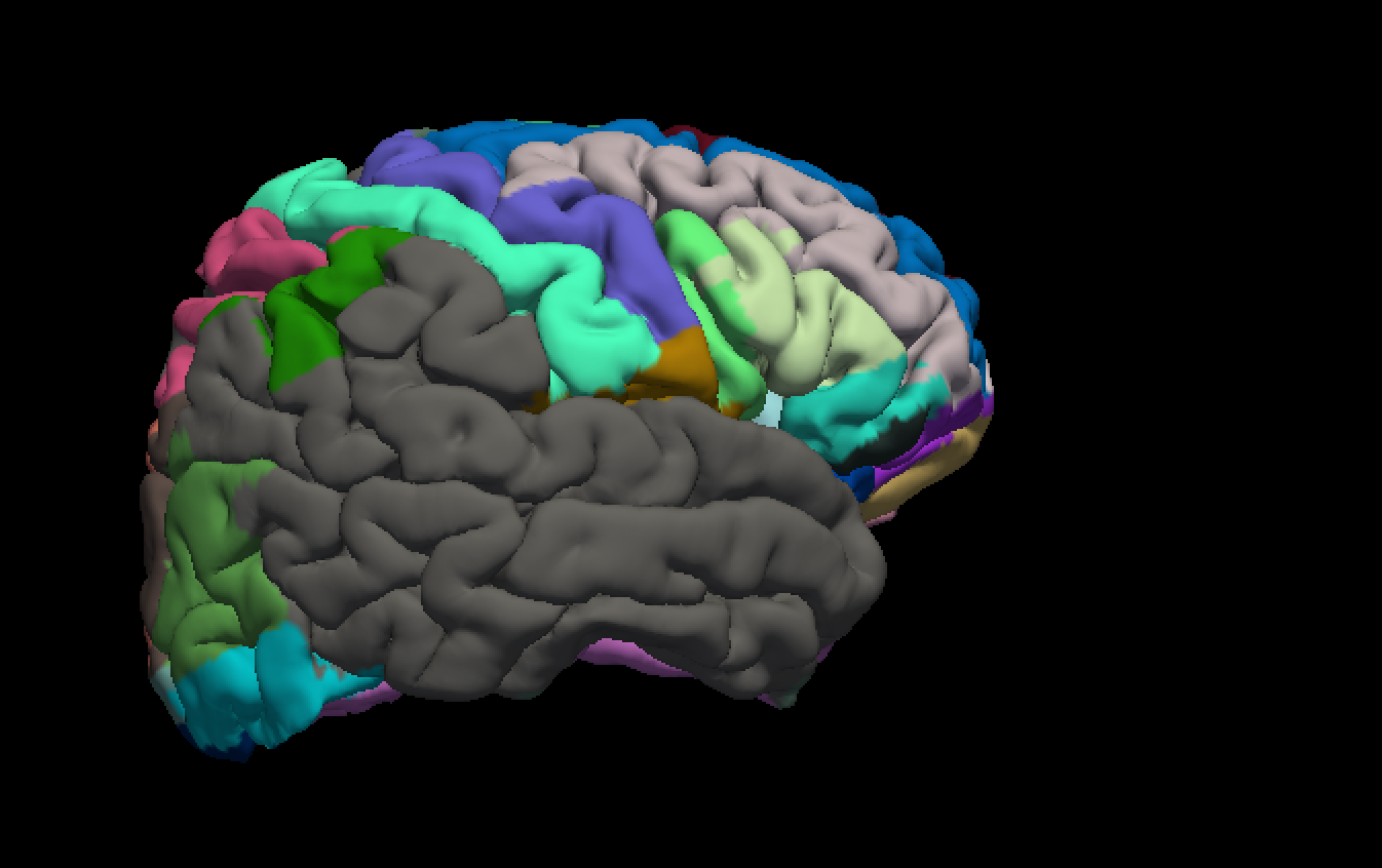
recon-all -all -i ch2.nii.gz -subjid ch2_template1mm
mri_vol2surf --src 1mm_atlas.nii --out atlas_rh.mgz --hemi rh --projfrac 0.5 --regheader ch2_template1mm
mris_seg2annot --seg atlas_rh.mgz --s ch2_template1mm --h rh --ctab-auto --o rh.atlasfile.annot
There were no errors.
However, testing with Freeview, I loaded the ch2_template1mm/surf/rh.pial.T1 file and overlaid with rh.atlasfile.annot. The result is not as anticipated and only covers frontal, parietal and parts of the visual cortex, missing most parts of the temporal cortex. I am not sure whether I can upload files here, but I will try to attach a screenshot. I tried different atlases, which resulted in different but similar (mismatching) results.
I also tested the ch2_template1mm/label/rh.aparc.a2009s.annot file, which looked flawless.
Any suggestions what I should do to fix this?
Thanks again!
freesurfer@nmr.mgh.harvard.edu
-
 Douglas N. Greve