
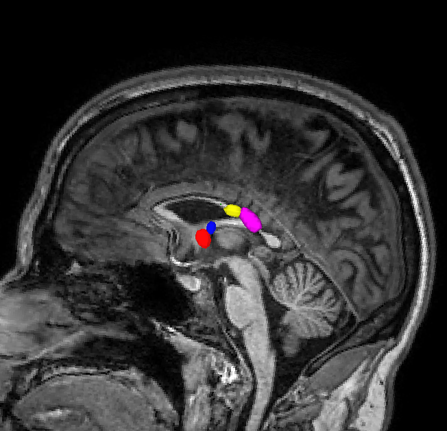
Hello all, I am examining diffusion tracking in the corpus callosum of individuals with periventricular leukomalacia (PVL). The corpus callosum is particularly small and the anterior and mid-anterior labels are being applied to the fornix (see attached Fornix_labelled_as_CC.png). The midposterior and posterior look ok, while the central is very small, maybe one voxel in size.
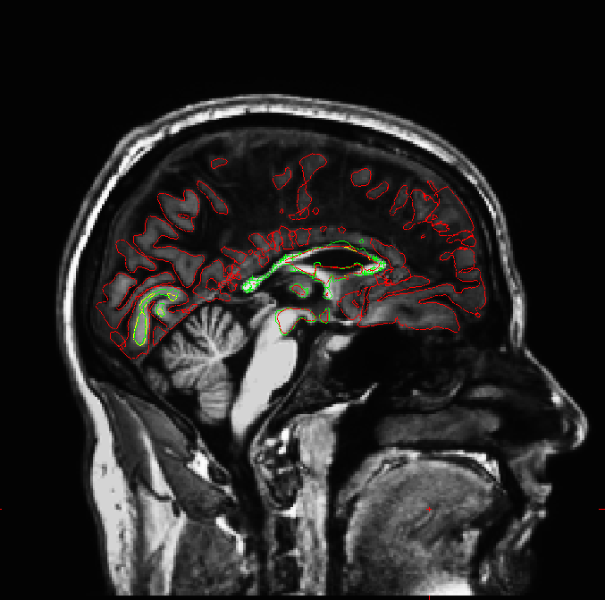
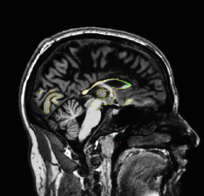
The wm segmentation, seen with ?l.white, also shows segmentation defaults with the anterior corpus callosum (see attached images). I tried to correct this using control points, which you can see did not fix the segmentation.
Any suggestions?
Corinna

Hi Corinna
how does the rest of the aseg.mgz look? Is it accurate? Bruce On Wed, 14 Aug 2013, Corinna Bauer wrote:
Hello all, I am examining diffusion tracking in the corpus callosum of individuals with periventricular leukomalacia (PVL). The corpus callosum is particularly small and the anterior and mid-anterior labels are being applied to the fornix (see attached Fornix_labelled_as_CC.png). The midposterior and posterior look ok, while the central is very small, maybe one voxel in size.
The wm segmentation, seen with ?l.white, also shows segmentation defaults with the anterior corpus callosum (see attached images). I tried to correct this using control points, which you can see did not fix the segmentation.
Any suggestions?
Corinna
freesurfer@nmr.mgh.harvard.edu
-
 Bruce Fischl
 -
 Corinna Bauer