
Hi all,
I’m working with a particularly stubborn data set whose defining characteristic is dura along the posterior of the brain. It’s become so consistent that I’m hoping to find a way to perhaps tweak recon-all so as to take care of the issue in a more automated manner and save many many man hours of manual work.
Is this possible?
jon

Hi Jon
do you want to send us an example image? Dura can be tough to distinguish. The best thing would be if you had a high res FLAIR or T2.
cheers Bruce
On Sun, 16 Mar 2014, Jonathan Holt wrote:
Hi all, I’m working with a particularly stubborn data set whose defining characteristic is dura along the posterior of the brain. It’s become so consistent that I’m hoping to find a way to perhaps tweak recon-all so as to take care of the issue in a more automated manner and save many many man hours of manual work.
Is this possible?
jon
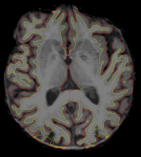
Here’s an image for reference, it is T1 unfortunately, but the problem area is always along the posterior in the same configuration.
On Mar 17, 2014, at 8:53 AM, Bruce Fischl fischl@nmr.mgh.harvard.edu wrote:
Hi Jon
do you want to send us an example image? Dura can be tough to distinguish. The best thing would be if you had a high res FLAIR or T2.
cheers Bruce
On Sun, 16 Mar 2014, Jonathan Holt wrote:
Hi all, I’m working with a particularly stubborn data set whose defining characteristic is dura along the posterior of the brain. It’s become so consistent that I’m hoping to find a way to perhaps tweak recon-all so as to take care of the issue in a more automated manner and save many many man hours of manual work. Is this possible? jon
The information in this e-mail is intended only for the person to whom it is addressed. If you believe this e-mail was sent to you in error and the e-mail contains patient information, please contact the Partners Compliance HelpLine at http://www.partners.org/complianceline . If the e-mail was sent to you in error but does not contain patient information, please contact the sender and properly dispose of the e-mail.
any idea why it looks so blurry? Is this the average of two or more? And what is the bandwidth of the acquisition? It looks like fat may have shifted into cortex, which is going to make it impossible to fix
On Mon, 17 Mar 2014, Jonathan Holt wrote:
Here’s an image for reference, it is T1 unfortunately, but the problem area is always along the posterior in the same configuration.
On Mar 17, 2014, at 8:53 AM, Bruce Fischl fischl@nmr.mgh.harvard.edu wrote:
Hi Jon
do you want to send us an example image? Dura can be tough to distinguish. The best thing would be if you had a high res FLAIR or T2.
cheers Bruce
On Sun, 16 Mar 2014, Jonathan Holt wrote:
Hi all, I’m working with a particularly stubborn data set whose defining characteristic is dura along the posterior of the brain. It’s become so consistent that I’m hoping to find a way to perhaps tweak recon-all so as to take care of the issue in a more automated manner and save many many man hours of manual work. Is this possible? jon
The information in this e-mail is intended only for the person to whom it is addressed. If you believe this e-mail was sent to you in error and the e-mail contains patient information, please contact the Partners Compliance HelpLine at http://www.partners.org/complianceline . If the e-mail was sent to you in error but does not contain patient information, please contact the sender and properly dispose of the e-mail.
It doesn’t seem any blurrier than other data I’ve worked with, so I’m not sure. I also cannot speak to the acquisition bandwidth,
what makes you think fat has shifted? I’m still wondering if there is any thing I can do to recon-all to avoid this, the data set is huge and it’s been an issue on every brain I’ve reconstructed so far, in the exact same areas.
jon On Mar 17, 2014, at 10:23 AM, Bruce Fischl fischl@nmr.mgh.harvard.edu wrote:
any idea why it looks so blurry? Is this the average of two or more? And what is the bandwidth of the acquisition? It looks like fat may have shifted into cortex, which is going to make it impossible to fix
On Mon, 17 Mar 2014, Jonathan Holt wrote:
Here’s an image for reference, it is T1 unfortunately, but the problem area is always along the posterior in the same configuration.
On Mar 17, 2014, at 8:53 AM, Bruce Fischl fischl@nmr.mgh.harvard.edu wrote:
Hi Jon
do you want to send us an example image? Dura can be tough to distinguish. The best thing would be if you had a high res FLAIR or T2.
cheers Bruce
On Sun, 16 Mar 2014, Jonathan Holt wrote:
Hi all, I’m working with a particularly stubborn data set whose defining characteristic is dura along the posterior of the brain. It’s become so consistent that I’m hoping to find a way to perhaps tweak recon-all so as to take care of the issue in a more automated manner and save many many man hours of manual work. Is this possible? jon
The information in this e-mail is intended only for the person to whom it is addressed. If you believe this e-mail was sent to you in error and the e-mail contains patient information, please contact the Partners Compliance HelpLine at http://www.partners.org/complianceline . If the e-mail was sent to you in error but does not contain patient information, please contact the sender and properly dispose of the e-mail.
why don't you upload one and we'll take a look? It looked either blurry or fat-shifted as in the image you sent I couldn't see any space between cortex and dura over a lot of the posterior brain On Mon, 17 Mar 2014, Jonathan Holt wrote:
It doesn’t seem any blurrier than other data I’ve worked with, so I’m not sure. I also cannot speak to the acquisition bandwidth,
what makes you think fat has shifted? I’m still wondering if there is any thing I can do to recon-all to avoid this, the data set is huge and it’s been an issue on every brain I’ve reconstructed so far, in the exact same areas.
jon On Mar 17, 2014, at 10:23 AM, Bruce Fischl fischl@nmr.mgh.harvard.edu wrote:
any idea why it looks so blurry? Is this the average of two or more? And what is the bandwidth of the acquisition? It looks like fat may have shifted into cortex, which is going to make it impossible to fix
On Mon, 17 Mar 2014, Jonathan Holt wrote:
Here’s an image for reference, it is T1 unfortunately, but the problem area is always along the posterior in the same configuration.
On Mar 17, 2014, at 8:53 AM, Bruce Fischl fischl@nmr.mgh.harvard.edu wrote:
Hi Jon
do you want to send us an example image? Dura can be tough to distinguish. The best thing would be if you had a high res FLAIR or T2.
cheers Bruce
On Sun, 16 Mar 2014, Jonathan Holt wrote:
Hi all, I’m working with a particularly stubborn data set whose defining characteristic is dura along the posterior of the brain. It’s become so consistent that I’m hoping to find a way to perhaps tweak recon-all so as to take care of the issue in a more automated manner and save many many man hours of manual work. Is this possible? jon
The information in this e-mail is intended only for the person to whom it is addressed. If you believe this e-mail was sent to you in error and the e-mail contains patient information, please contact the Partners Compliance HelpLine at http://www.partners.org/complianceline . If the e-mail was sent to you in error but does not contain patient information, please contact the sender and properly dispose of the e-mail.
It’s been uploaded as duraissues.tar
That lack of space between dura and cortex is the hallmark of this data set, those areas are near impossible to deal with and I often times end up removing the dura based on my best estimation. what is the best course of action in a situation like that On Mar 17, 2014, at 4:23 PM, Bruce Fischl fischl@nmr.mgh.harvard.edu wrote:
why don't you upload one and we'll take a look? It looked either blurry or fat-shifted as in the image you sent I couldn't see any space between cortex and dura over a lot of the posterior brain On Mon, 17 Mar 2014, Jonathan Holt wrote:
It doesn’t seem any blurrier than other data I’ve worked with, so I’m not sure. I also cannot speak to the acquisition bandwidth,
what makes you think fat has shifted? I’m still wondering if there is any thing I can do to recon-all to avoid this, the data set is huge and it’s been an issue on every brain I’ve reconstructed so far, in the exact same areas.
jon On Mar 17, 2014, at 10:23 AM, Bruce Fischl fischl@nmr.mgh.harvard.edu wrote:
any idea why it looks so blurry? Is this the average of two or more? And what is the bandwidth of the acquisition? It looks like fat may have shifted into cortex, which is going to make it impossible to fix
On Mon, 17 Mar 2014, Jonathan Holt wrote:
Here’s an image for reference, it is T1 unfortunately, but the problem area is always along the posterior in the same configuration.
On Mar 17, 2014, at 8:53 AM, Bruce Fischl fischl@nmr.mgh.harvard.edu wrote:
Hi Jon
do you want to send us an example image? Dura can be tough to distinguish. The best thing would be if you had a high res FLAIR or T2.
cheers Bruce
On Sun, 16 Mar 2014, Jonathan Holt wrote:
Hi all, I’m working with a particularly stubborn data set whose defining characteristic is dura along the posterior of the brain. It’s become so consistent that I’m hoping to find a way to perhaps tweak recon-all so as to take care of the issue in a more automated manner and save many many man hours of manual work. Is this possible? jon
The information in this e-mail is intended only for the person to whom it is addressed. If you believe this e-mail was sent to you in error and the e-mail contains patient information, please contact the Partners Compliance HelpLine at http://www.partners.org/complianceline . If the e-mail was sent to you in error but does not contain patient information, please contact the sender and properly dispose of the e-mail.
freesurfer@nmr.mgh.harvard.edu
-
 Bruce Fischl
 -
 Jonathan Holt