
I have questions regarding two issues we've run into processing dementia and age-matched healthy subjects:
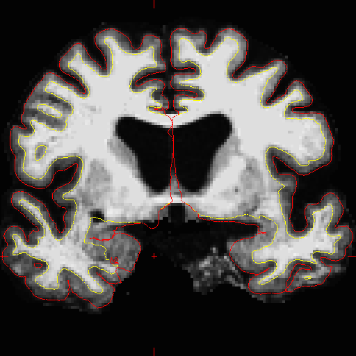
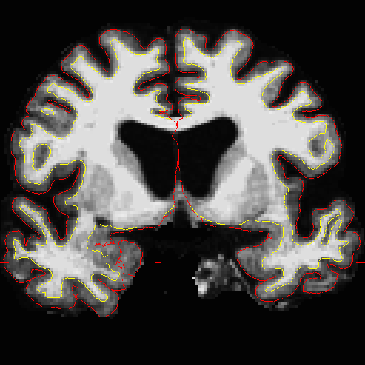
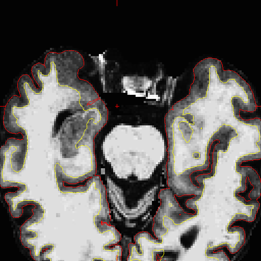
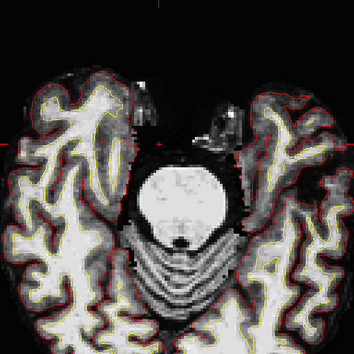
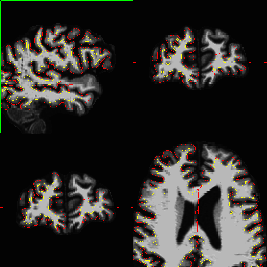
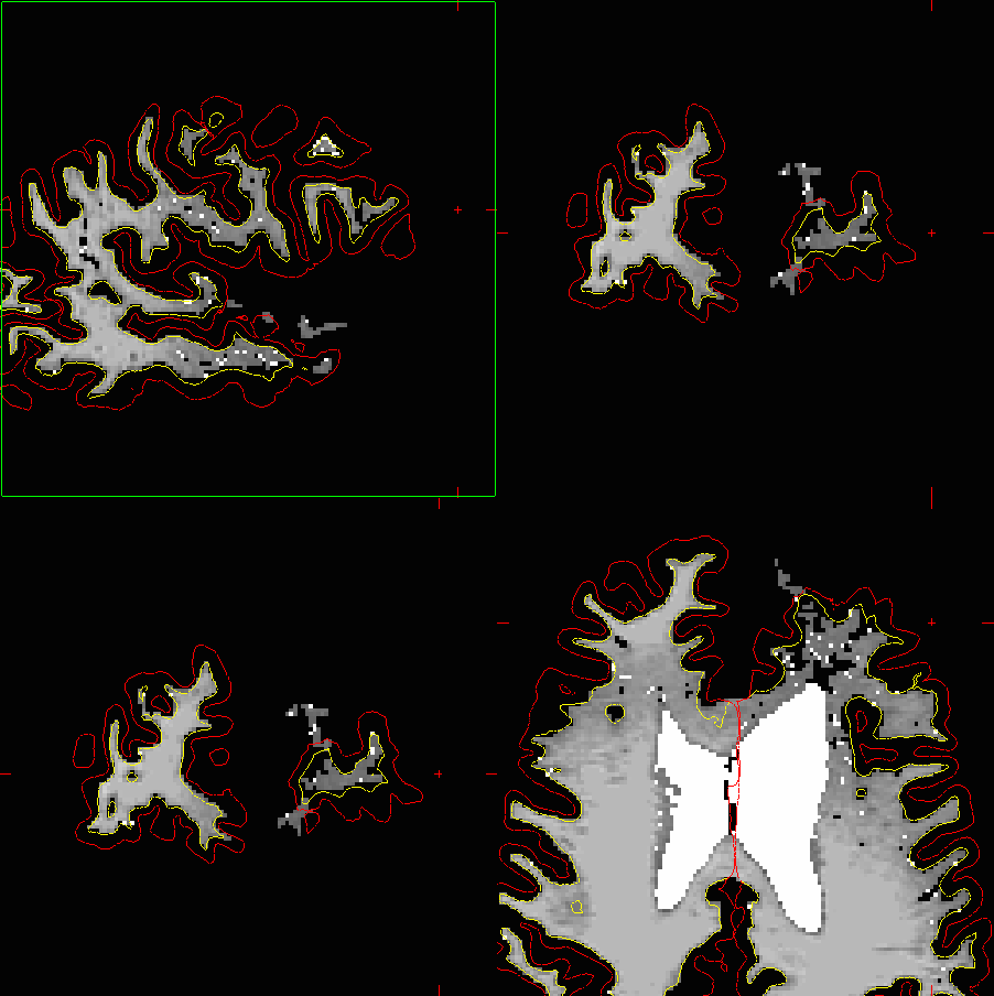
1. For several of our subjects, we've noticed that an isolated region of the entorhinal pial surface becomes 'indented' towards the white surface (see in RH in attached pics- P5b*). Not sure what's going, I've tried to correct it by removing any extraneous neighboring voxels or other 'anomalies' (no voxel gaps in the wm volumes from lesions, etc). It almost reminds me of the insula 'spike' a few versions back. Thoughts?
2. After re-running a subject for cp and wm edits, the resulting white (and consequently pial) surface has become inaccurate for large portions of the anterior temporal and frontal lobes, but just in the LH (attached pics- P8b*). To clarify, this was not the case after the data's initial run, only once edits were made and incorporated. We've run into this before, but only after editing a subject multiple times (3-4x, thus more recons), or 'updating' subjects to a new FS version (e.g., re-running v4.1.0 data with v4.5.0). Are edits changing the intensity threshold? It's odd that it's asymmetric.
Any help/thoughts would be appreciated, thanks.
-Derin

Hi Derin,
that is odd, not sure what's going on either with your wrinkles or the loss of surface after editing. Can you upload the pre and post edited datasets for us to look at? Bruce
On Thu, 1 Jul 2010, Derin Cobia wrote:
I have questions regarding two issues we've run into processing dementia and age-matched healthy subjects:
For several of our subjects, we've noticed that an isolated region of the entorhinal pial surface becomes 'indented' towards the white surface (see in RH in attached pics- P5b*). Not sure what's going, I've tried to correct it by removing any extraneous neighboring voxels or other 'anomalies' (no voxel gaps in the wm volumes from lesions, etc). It almost reminds me of the insula 'spike' a few versions back. Thoughts?
After re-running a subject for cp and wm edits, the resulting white (and consequently pial) surface has become inaccurate for large portions of the anterior temporal and frontal lobes, but just in the LH (attached pics- P8b*). To clarify, this was not the case after the data's initial run, only once edits were made and incorporated. We've run into this before, but only after editing a subject multiple times (3-4x, thus more recons), or 'updating' subjects to a new FS version (e.g., re-running v4.1.0 data with v4.5.0). Are edits changing the intensity threshold? It's odd that it's asymmetric.
Any help/thoughts would be appreciated, thanks.
-Derin
freesurfer@nmr.mgh.harvard.edu
-
 Bruce Fischl
 -
 Derin Cobia