
Dear All,
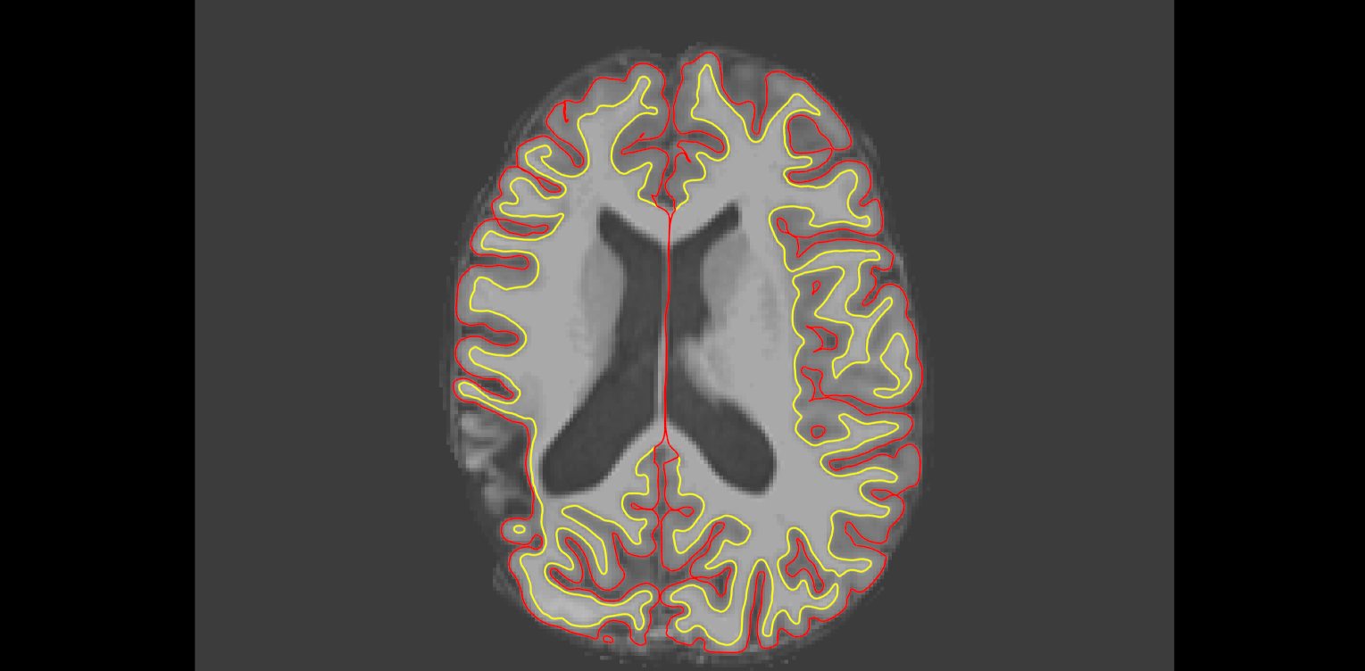
I am interested at getting as good pial/cortical reconstructions as possible on a series of epilepsy pt scans, some showing eccentric (cortical) lesions (focal encephalomalacias, infarcts, or after focal resections). One particularly difficult lesion type to get an accurate cortical surface is where there are prongs of grey matter bulging out (like a bag of worms), (mostly) without visible wm, see attached picture. These prongs are maintained in the brainmask.mgz and brain.finalsurfs.mgz, but not included in the pia with the default recon. Lesion voxel intensities vary from 30-90. I tried cp, wm edits (using brainmask or T1 as referece), both, did not matter, it did not work.
Since I am not intested in wm/subcortical segmentation but just in a good pia, I was wondering if I 'create' thin wm tracts through the center of these gm prongs to help pial segmentation. If this is a valid option, should I create those in brainmask, brain.finalsurfs or wm.mgz, and should I use 110 or 255 for the brush? Any other suggestions are appreciated. Nevertheless, I know there is a lot of tweaking of each lesion, and I try for more of a global approach to these kinds of lesions.
Thank you,
Octavian.
Dear All, any suggestion about this type of problem? I tried control points through the middle/axis of the atrophic gyri (intensity 30-90 in T1 or brainmask), wm.mgz edits through the same (adding from brainmask by cloning those voxels; painting 110; or 255 brush), and modifying brainmask directly by painting 110 internsity voxels through the same (gyri interiors, where wm is supposed to be). Did not work, the atrophic gyri continue to remain outside the pia. Again, this is an example of an eccentric lesion with gyral atrophy, without interruption in the gray matter ribbon but very faint wm signal through the base of the affected gyri. Thanks, Octavian
---------- Forwarded message ---------- From: Octavian Lie octavian.lie@gmail.com Date: Tue, Nov 18, 2014 at 10:50 AM Subject: Eccentric lesions To: "freesurfer@nmr.mgh.harvard.edu" freesurfer@nmr.mgh.harvard.edu
Dear All,
I am interested at getting as good pial/cortical reconstructions as possible on a series of epilepsy pt scans, some showing eccentric (cortical) lesions (focal encephalomalacias, infarcts, or after focal resections). One particularly difficult lesion type to get an accurate cortical surface is where there are prongs of grey matter bulging out (like a bag of worms), (mostly) without visible wm, see attached picture. These prongs are maintained in the brainmask.mgz and brain.finalsurfs.mgz, but not included in the pia with the default recon. Lesion voxel intensities vary from 30-90. I tried cp, wm edits (using brainmask or T1 as referece), both, did not matter, it did not work.
Since I am not intested in wm/subcortical segmentation but just in a good pia, I was wondering if I 'create' thin wm tracts through the center of these gm prongs to help pial segmentation. If this is a valid option, should I create those in brainmask, brain.finalsurfs or wm.mgz, and should I use 110 or 255 for the brush? Any other suggestions are appreciated. Nevertheless, I know there is a lot of tweaking of each lesion, and I try for more of a global approach to these kinds of lesions.
Thank you,
Octavian.

Hi Octavian
I wouldn't use control points since it really isn't healthy wm. The only thing you can do is edit the wm.mgz and possibly also the brainmask, but it's so abnormal looking you may not be able to get accurate surfaces. How does the aseg look? If it is ok there may be one other option.
cheers Bruce
On Tue, 25 Nov 2014, Octavian Lie wrote:
Dear All, any suggestion about this type of problem? I tried control points through the middle/axis of the atrophic gyri (intensity 30-90 in T1 or brainmask), wm.mgz edits through the same (adding from brainmask by cloning those voxels; painting 110; or 255 brush), and modifying brainmask directly by painting 110 internsity voxels through the same (gyri interiors, where wm is supposed to be). Did not work, the atrophic gyri continue to remain outside the pia. Again, this is an example of an eccentric lesion with gyral atrophy, without interruption in the gray matter ribbon but very faint wm signal through the base of the affected gyri. Thanks, Octavian
---------- Forwarded message ---------- From: Octavian Lie octavian.lie@gmail.com Date: Tue, Nov 18, 2014 at 10:50 AM Subject: Eccentric lesions To: "freesurfer@nmr.mgh.harvard.edu" freesurfer@nmr.mgh.harvard.edu
Dear All,
I am interested at getting as good pial/cortical reconstructions as possible on a series of epilepsy pt scans, some showing eccentric (cortical) lesions (focal encephalomalacias, infarcts, or after focal resections). One particularly difficult lesion type to get an accurate cortical surface is where there are prongs of grey matter bulging out (like a bag of worms), (mostly) without visible wm, see attached picture. These prongs are maintained in the brainmask.mgz and brain.finalsurfs.mgz, but not included in the pia with the default recon. Lesion voxel intensities vary from 30-90. I tried cp, wm edits (using brainmask or T1 as referece), both, did not matter, it did not work.
Since I am not intested in wm/subcortical segmentation but just in a good pia, I was wondering if I 'create' thin wm tracts through the center of these gm prongs to help pial segmentation. If this is a valid option, should I create those in brainmask, brain.finalsurfs or wm.mgz, and should I use 110 or 255 for the brush? Any other suggestions are appreciated. Nevertheless, I know there is a lot of tweaking of each lesion, and I try for more of a global approach to these kinds of lesions.
Thank you,
Octavian.
Dear Bruce,
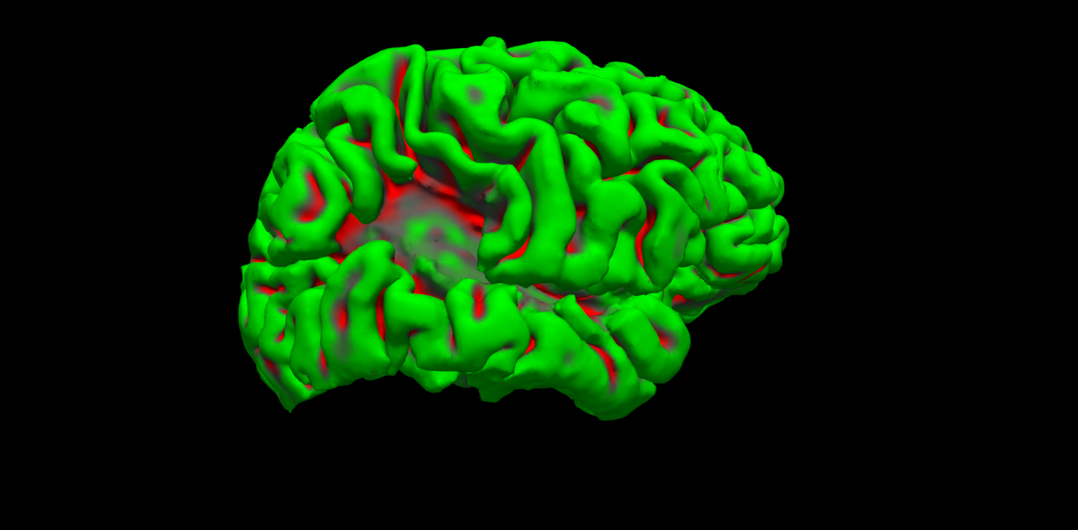
I am not sure how an ok aseg.mgz would look like for a lesion like that, but it looks like it includes one larger gyrus excluded by the pia, see attached, I would be happy to retrieve that one as it is the bulkiest of all the ones left out. Thank you,
Octavian
On Tue, Nov 25, 2014 at 7:38 AM, Bruce Fischl fischl@nmr.mgh.harvard.edu wrote:
Hi Octavian
I wouldn't use control points since it really isn't healthy wm. The only thing you can do is edit the wm.mgz and possibly also the brainmask, but it's so abnormal looking you may not be able to get accurate surfaces. How does the aseg look? If it is ok there may be one other option.
cheers Bruce
On Tue, 25 Nov 2014, Octavian Lie wrote:
Dear All, any suggestion about this type of problem? I tried control points through the middle/axis of the atrophic gyri (intensity 30-90 in T1 or
brainmask),
wm.mgz edits through the same (adding from brainmask by cloning those voxels; painting 110; or 255 brush), and modifying brainmask directly by painting 110 internsity voxels through the same (gyri interiors, where
wm is
supposed to be). Did not work, the atrophic gyri continue to remain
outside
the pia. Again, this is an example of an eccentric lesion with gyral atrophy, without interruption in the gray matter ribbon but very faint wm signal through the base of the affected gyri. Thanks, Octavian
---------- Forwarded message ---------- From: Octavian Lie octavian.lie@gmail.com Date: Tue, Nov 18, 2014 at 10:50 AM Subject: Eccentric lesions To: "freesurfer@nmr.mgh.harvard.edu" freesurfer@nmr.mgh.harvard.edu
Dear All,
I am interested at getting as good pial/cortical reconstructions as
possible
on a series of epilepsy pt scans, some showing eccentric (cortical)
lesions
(focal encephalomalacias, infarcts, or after focal resections). One particularly difficult lesion type to get an accurate cortical surface is where there are prongs of grey matter bulging out (like a bag of worms), (mostly) without visible wm, see attached picture. These prongs are maintained in the brainmask.mgz and brain.finalsurfs.mgz, but not
included
in the pia with the default recon. Lesion voxel intensities vary from
30-90.
I tried cp, wm edits (using brainmask or T1 as referece), both, did not matter, it did not work.
Since I am not intested in wm/subcortical segmentation but just in a good pia, I was wondering if I 'create' thin wm tracts through the center of these gm prongs to help pial segmentation. If this is a valid option,
should
I create those in brainmask, brain.finalsurfs or wm.mgz, and should I use 110 or 255 for the brush? Any other suggestions are appreciated. Nevertheless, I know there is a
lot
of tweaking of each lesion, and I try for more of a global approach to
these
kinds of lesions.
Thank you,
Octavian.
Freesurfer mailing list Freesurfer@nmr.mgh.harvard.edu https://mail.nmr.mgh.harvard.edu/mailman/listinfo/freesurfer
The information in this e-mail is intended only for the person to whom it is addressed. If you believe this e-mail was sent to you in error and the e-mail contains patient information, please contact the Partners Compliance HelpLine at http://www.partners.org/complianceline . If the e-mail was sent to you in error but does not contain patient information, please contact the sender and properly dispose of the e-mail.
so the GM is properly labeled by the aseg there? What about the faint wm? On Tue, 25 Nov 2014, Octavian Lie wrote:
Dear Bruce,
I am not sure how an ok aseg.mgz would look like for a lesion like that, but it looks like it includes one larger gyrus excluded by the pia, see attached, I would be happy to retrieve that one as it is the bulkiest of all the ones left out. Thank you,
Octavian
On Tue, Nov 25, 2014 at 7:38 AM, Bruce Fischl fischl@nmr.mgh.harvard.edu wrote: Hi Octavian
  I wouldn't use control points since it really isn't healthy wm.
  The only
  thing you can do is edit the wm.mgz and possibly also the
  brainmask, but
  it's so abnormal looking you may not be able to get accurate
  surfaces.
  How does the aseg look? If it is ok there may be one other
  option.

  cheers
  Bruce


  On Tue, 25 Nov 2014, Octavian Lie wrote:

  > Dear All,
  > any suggestion about this type of problem? I tried control
  points through
  > the middle/axis of the atrophic gyri (intensity 30-90 in T1 or
  brainmask),
  > wm.mgz edits through the same (adding from brainmask by
  cloning those
  > voxels; painting 110; or 255 brush), and modifying brainmask
  directly by
  > painting 110 internsity voxels through the same (gyri
  interiors, where wm is
  > supposed to be). Did not work, the atrophic gyri continue to
  remain outside
  > the pia. Again, this is an example of an eccentric lesion with
  gyral
  > atrophy, without interruption in the gray matter ribbon but
  very faint wm
  > signal through the base of the affected gyri.
  > Thanks,
  > Octavian
  >
  >
  >
  > ---------- Forwarded message ----------
  > From: Octavian Lie <octavian.lie@gmail.com>
  > Date: Tue, Nov 18, 2014 at 10:50 AM
  > Subject: Eccentric lesions
  > To: "freesurfer@nmr.mgh.harvard.edu"
  <freesurfer@nmr.mgh.harvard.edu>
  >
  >
  > Dear All,
  >
  > I am interested at getting as good pial/cortical
  reconstructions as possible
  > on a series of epilepsy pt scans, some showing eccentric
  (cortical) lesions
  > (focal encephalomalacias, infarcts, or after focal
  resections). One
  > particularly difficult lesion type to get an accurate cortical
  surface is
  > where there are prongs of grey matter bulging out (like a bag
  of worms),
  > (mostly) without visible wm, see attached picture. These
  prongs are
  > maintained in the brainmask.mgz and brain.finalsurfs.mgz, but
  not included
  > in the pia with the default recon. Lesion voxel intensities
  vary from 30-90.
  > I tried cp, wm edits (using brainmask or T1 as referece),
  both, did not
  > matter, it did not work.
  >
  > Since I am not intested in wm/subcortical segmentation but
  just in a good
  > pia, I was wondering if I 'create' thin wm tracts through the
  center of
  > these gm prongs to help pial segmentation. If this is a valid
  option, should
  > I create those in brainmask, brain.finalsurfs or wm.mgz, and
  should I use
  > 110 or 255 for the brush?
  > Any other suggestions are appreciated. Nevertheless, I know
  there is a lot
  > of tweaking of each lesion, and I try for more of a global
  approach to these
  > kinds of lesions.
  >
  > Thank you,
  >
  > Octavian.
  >
  >
  >
Freesurfer mailing list Freesurfer@nmr.mgh.harvard.edu https://mail.nmr.mgh.harvard.edu/mailman/listinfo/freesurfer
The information in this e-mail is intended only for the person to whom it is addressed. If you believe this e-mail was sent to you in error and the e-mail contains patient information, please contact the Partners Compliance HelpLine at http://www.partners.org/complianceline . If the e-mail was sent to you in error but does not contain patient information, please contact the sender and properly dispose of the e-mail.
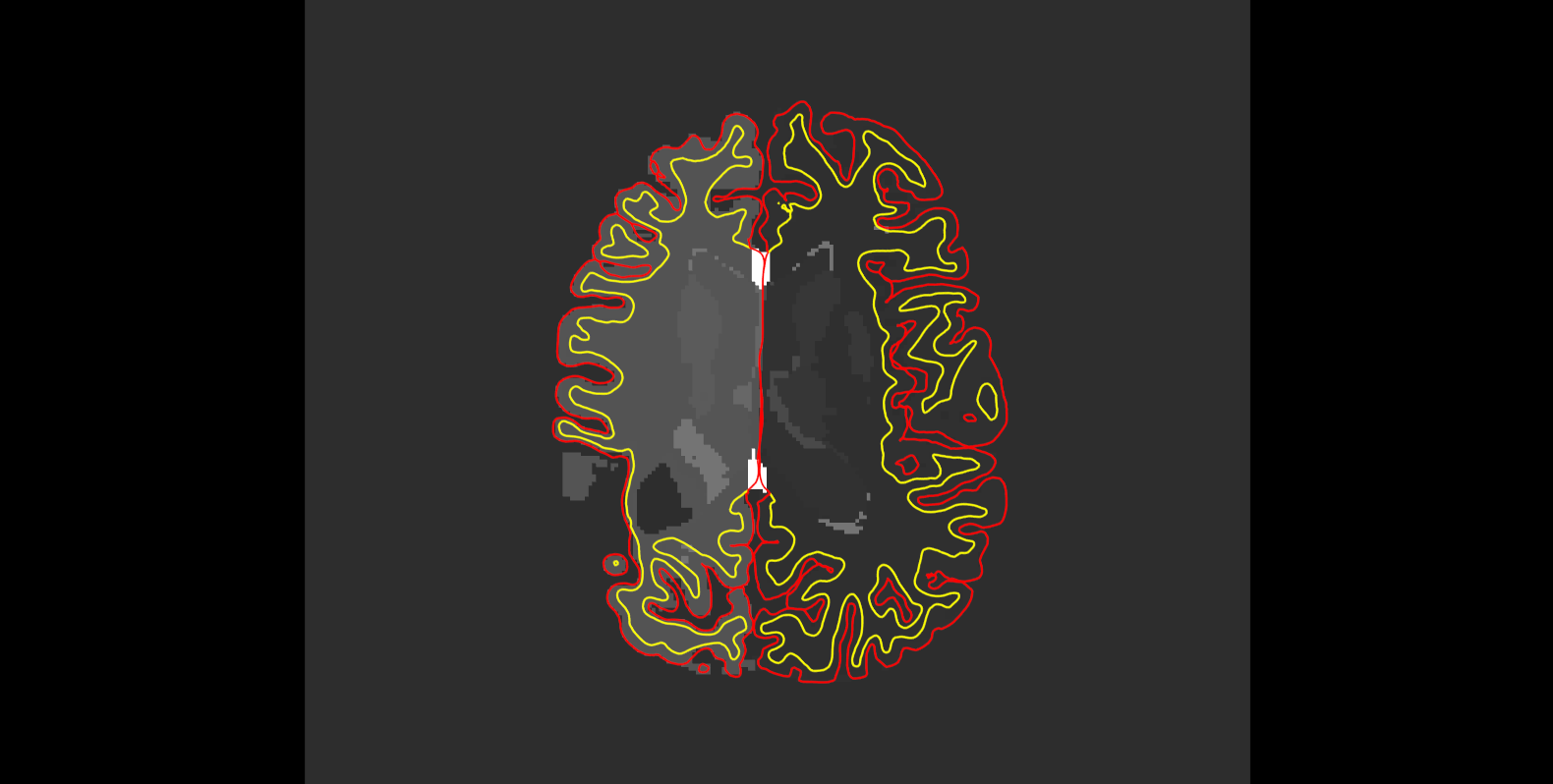
It looks like there is only gray matter (int 42), similar to cortical ribbon gray, no white matter in that gyrus in aseg, both in the original recon, and after painting gyral centers in brainmask. Octavian
On Tue, Nov 25, 2014 at 7:51 AM, Octavian Lie octavian.lie@gmail.com wrote:
Dear Bruce,
I am not sure how an ok aseg.mgz would look like for a lesion like that, but it looks like it includes one larger gyrus excluded by the pia, see attached, I would be happy to retrieve that one as it is the bulkiest of all the ones left out. Thank you,
Octavian
On Tue, Nov 25, 2014 at 7:38 AM, Bruce Fischl fischl@nmr.mgh.harvard.edu wrote:
Hi Octavian
I wouldn't use control points since it really isn't healthy wm. The only thing you can do is edit the wm.mgz and possibly also the brainmask, but it's so abnormal looking you may not be able to get accurate surfaces. How does the aseg look? If it is ok there may be one other option.
cheers Bruce
On Tue, 25 Nov 2014, Octavian Lie wrote:
Dear All, any suggestion about this type of problem? I tried control points
through
the middle/axis of the atrophic gyri (intensity 30-90 in T1 or
brainmask),
wm.mgz edits through the same (adding from brainmask by cloning those voxels; painting 110; or 255 brush), and modifying brainmask directly by painting 110 internsity voxels through the same (gyri interiors, where
wm is
supposed to be). Did not work, the atrophic gyri continue to remain
outside
the pia. Again, this is an example of an eccentric lesion with gyral atrophy, without interruption in the gray matter ribbon but very faint
wm
signal through the base of the affected gyri. Thanks, Octavian
---------- Forwarded message ---------- From: Octavian Lie octavian.lie@gmail.com Date: Tue, Nov 18, 2014 at 10:50 AM Subject: Eccentric lesions To: "freesurfer@nmr.mgh.harvard.edu" freesurfer@nmr.mgh.harvard.edu
Dear All,
I am interested at getting as good pial/cortical reconstructions as
possible
on a series of epilepsy pt scans, some showing eccentric (cortical)
lesions
(focal encephalomalacias, infarcts, or after focal resections). One particularly difficult lesion type to get an accurate cortical surface
is
where there are prongs of grey matter bulging out (like a bag of worms), (mostly) without visible wm, see attached picture. These prongs are maintained in the brainmask.mgz and brain.finalsurfs.mgz, but not
included
in the pia with the default recon. Lesion voxel intensities vary from
30-90.
I tried cp, wm edits (using brainmask or T1 as referece), both, did not matter, it did not work.
Since I am not intested in wm/subcortical segmentation but just in a
good
pia, I was wondering if I 'create' thin wm tracts through the center of these gm prongs to help pial segmentation. If this is a valid option,
should
I create those in brainmask, brain.finalsurfs or wm.mgz, and should I
use
110 or 255 for the brush? Any other suggestions are appreciated. Nevertheless, I know there is a
lot
of tweaking of each lesion, and I try for more of a global approach to
these
kinds of lesions.
Thank you,
Octavian.
Freesurfer mailing list Freesurfer@nmr.mgh.harvard.edu https://mail.nmr.mgh.harvard.edu/mailman/listinfo/freesurfer
The information in this e-mail is intended only for the person to whom it is addressed. If you believe this e-mail was sent to you in error and the e-mail contains patient information, please contact the Partners Compliance HelpLine at http://www.partners.org/complianceline . If the e-mail was sent to you in error but does not contain patient information, please contact the sender and properly dispose of the e-mail.
Dear Lilla, Here is a snapshot of the aseg volume, which rescues some gray matter not included in the pia. I am not sure if the aseg is ok, as the portion extending in the lesion is labeled gray matter (intensity 42). I guess this is not a problem. If I generate a surface from aseg as you suggested: a. I would need to generate a surface for each of the two hemispheres; b. If a is doable, is there a way to make it 'pial' in the sense that it gets registered with sphere and gets to be applied the DK atlas? A corrolary would be whether I can trick FS by labeling the new surface as smth.pial during the recon pipeline and further complete the rest of the processing steps to generate a true pia. Thank you for your assist,
Octavian

Hi, Could you use a colormap for your aseg? It is really hard to see the different ROIs on a grayscale image. Thanks, Lilla
On Wed, 26 Nov 2014, Octavian Lie wrote:
Dear Lilla, Here is a snapshot of the aseg volume, which rescues some gray matter not included in the pia. I am not sure if the aseg is ok, as the portion extending in the lesion is labeled gray matter (intensity 42). I guess this is not a problem. If I generate a surface from aseg as you suggested: a. I would need to generate a surface for each of the two hemispheres; b. If a is doable, is there a way to make it 'pial' in the sense that it gets registered with sphere and gets to be applied the DK atlas? A corrolary would be whether I can trick FS by labeling the new surface as smth.pial during the recon pipeline and further complete the rest of the processing steps to generate a true pia. Thank you for your assist, Octavian
Hi Octavian You should try it and see. It's hard to predict how it will do Cheers Bruce
On Nov 26, 2014, at 4:43 PM, Lilla Zollei lzollei@nmr.mgh.harvard.edu wrote:
Hi, Could you use a colormap for your aseg? It is really hard to see the different ROIs on a grayscale image. Thanks, Lilla
On Wed, 26 Nov 2014, Octavian Lie wrote:
Dear Lilla, Here is a snapshot of the aseg volume, which rescues some gray matter not included in the pia. I am not sure if the aseg is ok, as the portion extending in the lesion is labeled gray matter (intensity 42). I guess this is not a problem. If I generate a surface from aseg as you suggested: a. I would need to generate a surface for each of the two hemispheres; b. If a is doable, is there a way to make it 'pial' in the sense that it gets registered with sphere and gets to be applied the DK atlas? A corrolary would be whether I can trick FS by labeling the new surface as smth.pial during the recon pipeline and further complete the rest of the processing steps to generate a true pia. Thank you for your assist,
Octavian
Freesurfer mailing list Freesurfer@nmr.mgh.harvard.edu https://mail.nmr.mgh.harvard.edu/mailman/listinfo/freesurfer
I tried, here is the command:
mris_make_surfaces -cover_seg mri/aseg.mgz subjid lh
and the last 5 lines of the run:
... smoothing surface for 5 iterations... repositioning cortical surface to gray/csf boundary. smoothing T1 volume with sigma = 2.000 creating distance transform volume from segmentation ERROR: mris_make_surfaces-MRIcheckVolDims: volume1 height=256 != volume2 height=1.
If this is it and not working, I wonder that editing the aseg to exclude the nonlesional GM outside the pia, but leaving/ adding lesional GM of interest, either under the GM label or a custom label would allow create a 'proper' volume, followed by surface generation. I known this is doable, the question is whether the generated surface can be registered to sphere and whether DK atlas can be applied (this is a more general question, whether any surface generated outside the default recon-all pathway can be registered to sphere and in effect become "pia" or another classical boundary surface?)
example after labeling the lesion as 'o' (other):
% generate mask from aseg cd /usr/local/fs5.3/freesurfer/subjects/subjid mris_fill -c -r 0.1 surf/rh.pial mri/rh.pial.filled.mgz && \ mris_fill -c -r 0.1 surf/lh.pial mri/lh.pial.filled.mgz && \ mri_concat --combine --i mri/rh.pial.filled.mgz --i mri/lh.pial.filled.mgz --o mri/pial.filled.mgz && \ cd ./mri && \ mri_binarize --i aseg.mgz --match o --o lesion.mask.mgz && \ fscalc pial.filled.mgz add lesion.mask.mgz -o pialmask.mgz && \ mri_convert brain.mgz brain.float.mgz -odt float && \ mri_mask brain.float.mgz pialmask.mgz pialbrain.mgz
% generate new 'pial' surface: mri_tessellate mri/pialbrain.mgz -a surf/lh.pialbrain.pial
Please advise,
Octavian
freesurfer@nmr.mgh.harvard.edu
-
 Bruce Fischl
 -
 Lilla Zollei
 -
 Octavian Lie