
External Email - Use Caution
Dear FreeSurfer team,
I am currently using to Gray to White Matter Signal Ratio (GWR) to analyze our longitudinal AD/MCI MRI data. I feel a little confused about the interpretation after reading some recent papers like Putcha et al, 2022 or Jefferson et al, 2015 and some early papers like Salat et al, 2011 and I hope Freesurfer team could help me confirm if my understanding is correct.
1, Freesurfer "recon-all" generates the GWR map by default using "pctsurfcon" and the T1 image. The formula is 100*(W-G) GWR = --------- 0.5*(W+G)
The white matter is sampled 1mm below the white surface. The gray matter is sampled 30% the thickness into the cortex.
2, Gray to White Matter Signal Ratio (GWR) generated by "recon-all" is the same as Gray and White Matter tissue contrast. So Gray to white matter signal intensity ratio (GWR) declines across the lifespan? AD patients have smaller GWR than cognitively normal elders? Decrease of GWR is observed with increasing AD disease severity?
Thank you so much and best regards,
Xiao Da Senior Data Scientist Cognito Therapeutics, Inc. 1218 Massachusetts Ave, Cambridge, MA 02138 xda@cognitotx.commailto:xda@cognitotx.com [A picture containing text, sign Description automatically generated]
External Email - Use Caution
Dear FreeSurfer team,
I am currently using Gray to White Matter Signal Ratio (GWR) to analyze our longitudinal AD/MCI MRI data. I feel a little confused about the interpretation after reading some recent papers like Putcha et al, 2022 or Jefferson et al, 2015 and some early papers like Salat et al, 2011 and I hope Freesurfer team could help me confirm if my understanding is correct.
1, Freesurfer "recon-all" generates the GWR map by default using "pctsurfcon" and the T1 image. The formula is 100*(W-G) GWR = --------- 0.5*(W+G)
The white matter is sampled 1mm below the white surface. The gray matter is sampled 30% the thickness into the cortex.
2, Gray to White Matter Signal Ratio (GWR) generated by "recon-all" is the same as Gray and White Matter tissue contrast. So Gray to white matter signal intensity ratio (GWR) declines across the lifespan? AD patients have smaller GWR than cognitively normal elders? Decrease of GWR is observed with increasing AD disease severity?
Thank you so much and best regards,
Xiao Da Senior Data Scientist Cognito Therapeutics, Inc. 1218 Massachusetts Ave, Cambridge, MA 02138 xda@cognitotx.commailto:xda@cognitotx.com [A picture containing text, sign Description automatically generated]

On 1/10/2023 2:26 PM, Xiao Da wrote:
External Email - Use Caution
Dear FreeSurfer team,
I am currently using Gray to White Matter Signal Ratio (GWR) to analyze our longitudinal AD/MCI MRI data.
I feel a little confused about the interpretation after reading some recent papers like Putcha et al, 2022 or Jefferson et al, 2015 and some early papers like Salat et al, 2011 and I hope Freesurfer team could help me confirm if my understanding is correct.
1, Freesurfer “recon-all” generates the GWR map by default using “pctsurfcon” and the T1 image. The formula is
100*(W-G)
GWR = ---------
0.5*(W+G)
The white matter is sampled 1mm below the white surface. The gray matter is sampled 30% the thickness into the cortex.
This is correct. Is there a question?
2, Gray to White Matter Signal Ratio (GWR) generated by “recon-all” is the same as Gray and White Matter tissue contrast. So
Gray to white matter signal intensity ratio (GWR) declines across the lifespan?
AD patients have smaller GWR than cognitively normal elders?
Decrease of GWR is observed with increasing AD disease severity?
Aren't those questions addressed in the papers you mention above?
Thank you so much and best regards,
*Xiao Da*
Senior Data Scientist
Cognito Therapeutics, Inc.
1218 Massachusetts Ave, Cambridge, MA 02138
xda@cognitotx.com
A picture containing text, sign Description automatically generated
Freesurfer mailing list Freesurfer@nmr.mgh.harvard.edu https://mail.nmr.mgh.harvard.edu/mailman/listinfo/freesurfer
External Email - Use Caution
Hi Doug, Thank you so much for your reply. I got a little confused about the interpretation about GWR/Gray White Matter Tissue Contrast in the following papers and Freesurfer people co-authored all of them.
* In Salat et al., 2011, https://secure-web.cisco.com/13_aMxFfZEg3hEywalg973N7q-X0Q1aELufu834ksA4QfeD... "We specifically focused on the gray to white matter signal intensity ratio (GWR) at each point along the cortical surface to determine whether contrast properties were altered in a regionally specific manner throughout the brain. The GWR showed a considerable increase (towards a value of 1) with increasing severity of AD, demonstrating an overall decrease in the contrast between tissue classes."
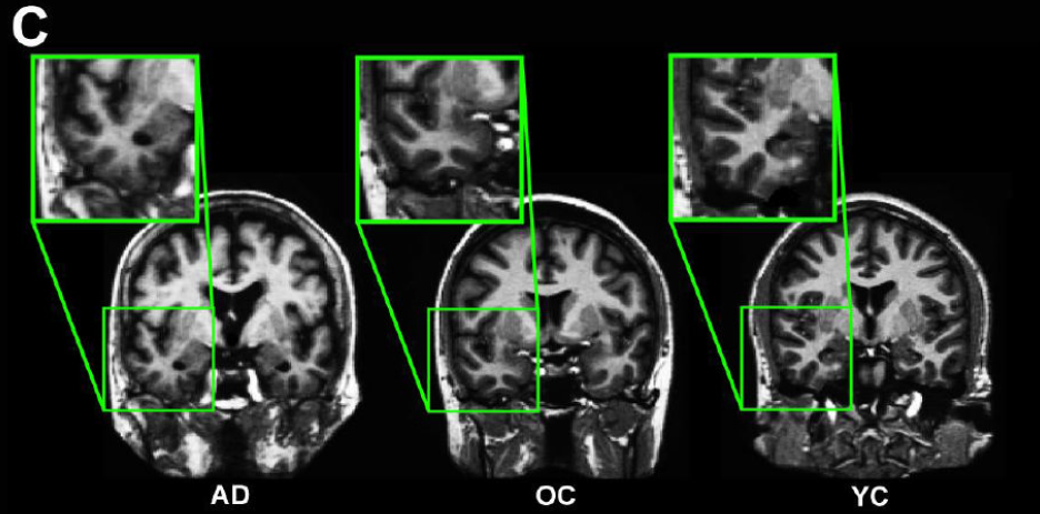
[cid:image002.png@01D9266F.C4FF47B0] Figure 1 in Salat et al., 2011 C. Demonstration of the differential regional gray matter/white matter tissue contrast in an individual with Alzheimer's disease (AD), an older adult (OA), and a younger adult (YA).
* In Jefferson et al., 2015, https://secure-web.cisco.com/1eI-SrjB_diK0qXUR9zdack9EnyRGND_v7pEjEKmRrcuoV7... "Gray to white matter signal intensity ratio (GWR) declines across the lifespan (Salat et al., 2009b). Furthermore, AD patients have more compromised GWR than cognitively normal elders (Westlye et al., 2009), and the spatial distribution of tissue contrast changes reflects regions associated with AD pathology, including the parahippocampal cortex (Salat et al., 2011)." "The GWR measurement was numerically calculated as 100*(white matter intensity-gray matter intensity)/(white matter intensity+gray matter intensity)."
* In Putcha et al., 2022, https://secure-web.cisco.com/1huiAvfl50zthEgxWkQ_JZwqolF1rRJoyryCp4ZW7AdxaTG... "The gray matter to white matter signal intensity ratio (GWR), which quantifies the signal contrast between these tissue compartments, has emerged as a promising marker of AD-related neurodegeneration that can be measured using conventional MRI scans. Reports indicate that a decrease in the contrast between the tissue classes is observed with increasing disease severity." 100*(W-G) GWR = --------- 0.5*(W+G)
"GWR" in Jefferson et al., 2015 and Putcha et al. 2022 could be very close each other. So my question is that whether the definition of "GWR" in Jefferson et al., 2015 and Putcha et al. 2022 is different from "GWR" in Salat et al., 2011. When you see figure 1 in Salat et al., 2011 for method demonstration, the "GWR" in Salat et al., 2011 looks like "GM intensity /WM intensity" Could you please confirm?
Thanks and best regards,
Xiao
From: freesurfer-bounces@nmr.mgh.harvard.edu freesurfer-bounces@nmr.mgh.harvard.edu On Behalf Of Douglas N. Greve Sent: Thursday, January 12, 2023 9:32 AM To: freesurfer@nmr.mgh.harvard.edu Subject: Re: [Freesurfer] Gray to White Matter Signal Ratio (GWR)
On 1/10/2023 2:26 PM, Xiao Da wrote:
External Email - Use Caution Dear FreeSurfer team,
I am currently using Gray to White Matter Signal Ratio (GWR) to analyze our longitudinal AD/MCI MRI data. I feel a little confused about the interpretation after reading some recent papers like Putcha et al, 2022 or Jefferson et al, 2015 and some early papers like Salat et al, 2011 and I hope Freesurfer team could help me confirm if my understanding is correct.
1, Freesurfer "recon-all" generates the GWR map by default using "pctsurfcon" and the T1 image. The formula is 100*(W-G) GWR = --------- 0.5*(W+G)
The white matter is sampled 1mm below the white surface. The gray matter is sampled 30% the thickness into the cortex. This is correct. Is there a question?
2, Gray to White Matter Signal Ratio (GWR) generated by "recon-all" is the same as Gray and White Matter tissue contrast. So Gray to white matter signal intensity ratio (GWR) declines across the lifespan? AD patients have smaller GWR than cognitively normal elders? Decrease of GWR is observed with increasing AD disease severity? Aren't those questions addressed in the papers you mention above?
Thank you so much and best regards,
Xiao Da Senior Data Scientist Cognito Therapeutics, Inc. 1218 Massachusetts Ave, Cambridge, MA 02138 xda@cognitotx.commailto:xda@cognitotx.com [A picture containing text, sign Description automatically generated]
_______________________________________________
Freesurfer mailing list
Freesurfer@nmr.mgh.harvard.edumailto:Freesurfer@nmr.mgh.harvard.edu
https://secure-web.cisco.com/1BILr4MWYiHqWlOaf6w2l1Vo4GOE_2L33Fr-J8s1E0vymdb...https://secure-web.cisco.com/1YJfK3TPwkkLDhKjd5iabwJiPUBqIBQ43GwpW8uplwsuvSPDYUxB6IK6jgFZkJWWKExRNayXvrqf81opOKOMGEeDhgUfgnjE0nLYoVB-7IcevdCpMTExpfYXQFnOpiweI5cLDSpmeUfthnq34mmJ8zwFiI6cMfddr-z-owHfFvTHpZCQFFT8CpWKfy_W9m82Sy3ky18ca1DV4Gcn5fKyqKOCA13aWin2GZkc7CJtCeuQTLaOcNHouqSq1ZsAH7PklnkCLpqQ1L-ClT0VEfj7hw5Oc5uGTibmeaXEGHGRgJqd9KNTCMGELpvSa-Ss_QPJN/https%3A%2F%2Fnam10.safelinks.protection.outlook.com%2F%3Furl%3Dhttps%253A%252F%252Fmail.nmr.mgh.harvard.edu%252Fmailman%252Flistinfo%252Ffreesurfer%26data%3D05%257C01%257Cxda%2540cognitotx.com%257C0529306b49324ed3ae6c08daf4a9c820%257C2c7c4c5919194231896c4a0123e4c3ed%257C0%257C0%257C638091307300689508%257CUnknown%257CTWFpbGZsb3d8eyJWIjoiMC4wLjAwMDAiLCJQIjoiV2luMzIiLCJBTiI6Ik1haWwiLCJXVCI6Mn0%253D%257C3000%257C%257C%257C%26sdata%3DytikLxYkDnayo%252FKdHyACKRO1mhfS1cXhQ6%252BLUEkQrRw%253D%26reserved%3D0
freesurfer@nmr.mgh.harvard.edu
-
 Douglas N. Greve
 -
 Xiao Da