
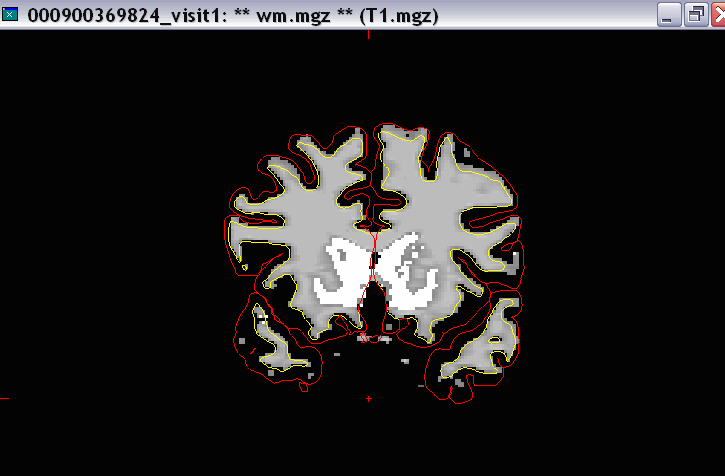
Hi there experts-When we load up wm.mgz with the lh.white and rh.white on one subject, the white matter mask overlaps into the the gray matter ribbon (see attached). When we look at the T1 underneath it, the red and green lines look just fine, but the white matter mask is a bit generous. Do I need to clean that up?
Thanks,
Jess
------------------------
Jessica Turner, Ph.D.
Project Manager, FBIRN (www.nbirn.net)
Department of Psychiatry and Human Behavior
University of California, Irvine
Phone: (949) 824-3331
Fax: (949) 824-3324

nope, that's why we do the surface deformation after the wm segmentation -to correct this type of inaccuracy. On Wed, 18 Apr 2007, Turner, Jessica wrote:
Hi there experts-When we load up wm.mgz with the lh.white and rh.white on one subject, the white matter mask overlaps into the the gray matter ribbon (see attached). When we look at the T1 underneath it, the red and green lines look just fine, but the white matter mask is a bit generous. Do I need to clean that up?
Thanks,
Jess
Jessica Turner, Ph.D.
Project Manager, FBIRN (www.nbirn.net)
Department of Psychiatry and Human Behavior
University of California, Irvine
Phone: (949) 824-3331
Fax: (949) 824-3324

Dear Freesurfers, I have a couple of questions ... 1. the yellow borders that outline the white matter surface look fine when I'm looking at the brainseg and wm volumes, but they don't correctly outline cortical white matter when I'm looking at the aseg segmentation as an overlay on the aforementioned volumes. Is this something to worry about? 2. I have some skull still remaining but is not included within the red pial borders, or areas that appear as white matter (on wm.mgz) and are not included within the yellow borders. Do I still need to make edits and remove these areas? I guess what I'm asking is that when stats like volumes and cortical thickness are calculated, are they calculated based on the red pial and yellow wm borders, in which case I wouldn't need to worry about removing those extra areas? appreciate your help,
Binyam
Binyam Nardos Cognitive Rehabilitation Research Group Washington University School of Medicine Box # 8505 4444 Forest Park Blvd St. Louis, MO 63108 Office: (314) 454-7795 Fax: (314) 286-1601
The contents of this e-mail message and any attachments are intended solely for the addressee(s) named in this message. This communication is intended to be and to remain confidential and may be subject to applicable physician/patient and/or work product privileges. If you are not the intended recipient of this message, or if this message has been addressed to you in error, please immediately alert the sender by reply e-mail and then delete this message and its attachments.
1. The ?h.white and ?h.pial are more accurate cortical representations than the aseg, so trust them if there is a mismatch in general.
2. Bits of skull can be around and not bother any of the processing as long as they aren't included in the pial surface, or mislabeled in the aseg as say cerebellum.
cheers, Bruce
On Wed, 18 Apr 2007, Binyam Nardos wrote:
Dear Freesurfers, I have a couple of questions ...
- the yellow borders that outline the white matter surface look fine when I'm looking at the brainseg and wm volumes, but they don't correctly outline cortical white matter when I'm looking at the aseg segmentation as an overlay on the aforementioned volumes. Is this something to worry about?
- I have some skull still remaining but is not included within the red pial borders, or areas that appear as white matter (on wm.mgz) and are not included within the yellow borders. Do I still need to make edits and remove these areas? I guess what I'm asking is that when stats like volumes and cortical thickness are calculated, are they calculated based on the red pial and yellow wm borders, in which case I wouldn't need to worry about removing those extra areas?
appreciate your help,
Binyam
Binyam Nardos Cognitive Rehabilitation Research Group Washington University School of Medicine Box # 8505 4444 Forest Park Blvd St. Louis, MO 63108 Office: (314) 454-7795 Fax: (314) 286-1601
The contents of this e-mail message and any attachments are intended solely for the addressee(s) named in this message. This communication is intended to be and to remain confidential and may be subject to applicable physician/patient and/or work product privileges. If you are not the intended recipient of this message, or if this message has been addressed to you in error, please immediately alert the sender by reply e-mail and then delete this message and its attachments.
freesurfer@nmr.mgh.harvard.edu
-
 Binyam Nardos
 -
 Bruce Fischl
 -
 Turner, Jessica