
Dear all,
In some of our subjects the border of ctx_lh_parahippocampal (green color) is inconsistent with the border of pial surface (red color) in the sense that some small region within the pial surface is segmented as cerebellum (orange) coming from the subcortical segmentation stream. This problem is present in all three planes. It is hard to decide whether the colored border of ctx_lh_parahippocampal or the pial surface is more realistic, but it would be nice to know which one is used for the parahippocampal statistics in lh.aparc.stats file. Do I need to correct for this inaccuracy? How should I correct this type of missegmentation?
Best Regards, Gabor petzinger.gabor@gmail.com

This is due to an inconsistency between the volume labeling and the surface placement. The volume labeling thinks those voxels are cerebellum. In the lh.aparc.stats, those voxels would be counted as parahippo. If the problem is with the volume labeling, then there is nothing you need to do as the aparc.stats is correct. If the problem is with the surface placement, I'm not sure what you can do. Maybe edit those voxels out of the brain.finalsurfs.mgz and re-run -autorecon2-wm and autorecon3
On 01/12/2017 02:37 AM, Gabor Perlaki wrote:
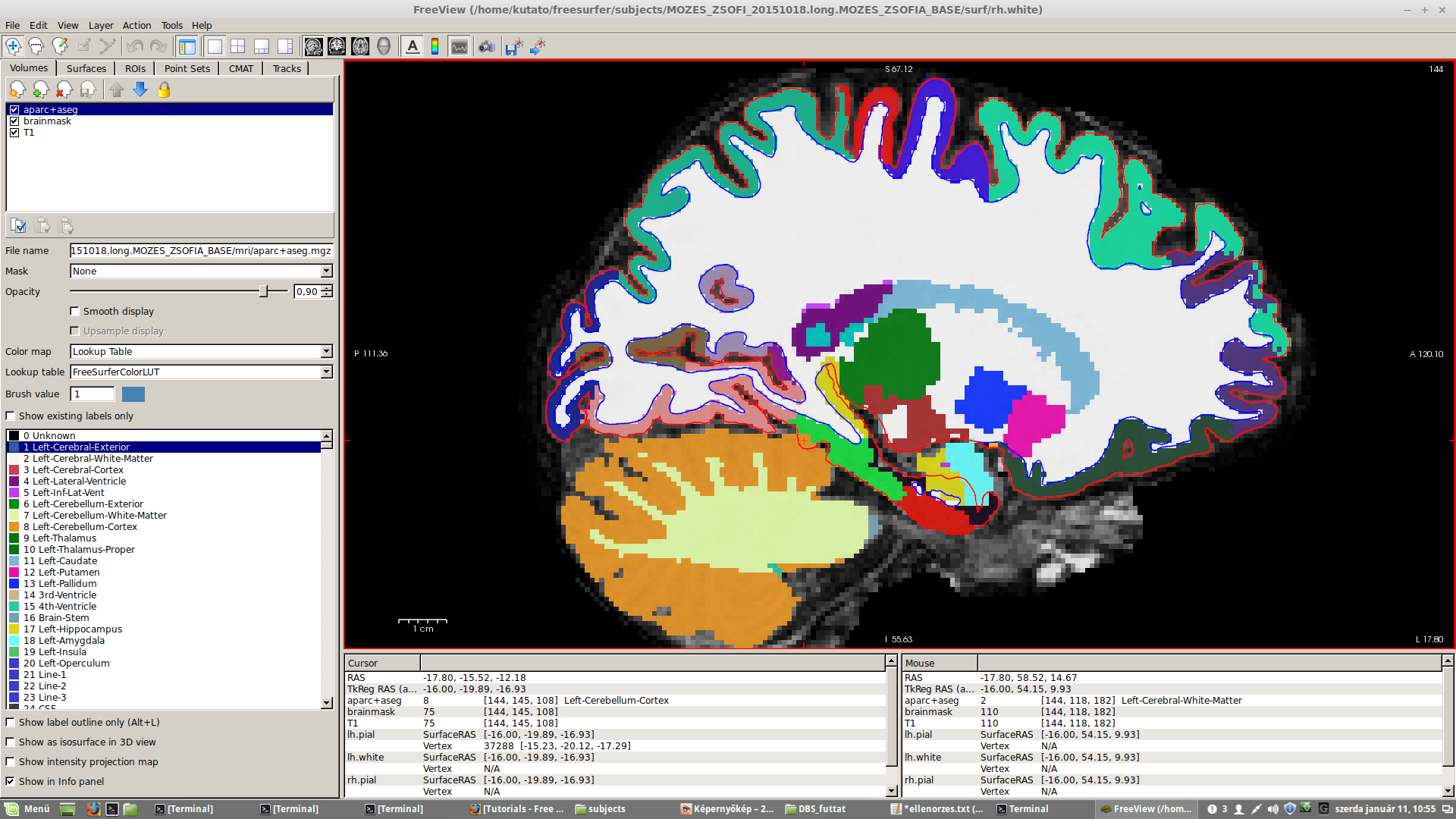
Dear all,
In some of our subjects the border of ctx_lh_parahippocampal (green color) is inconsistent with the border of pial surface (red color) in the sense that some small region within the pial surface is segmented as cerebellum (orange) coming from the subcortical segmentation stream. This problem is present in all three planes. It is hard to decide whether the colored border of ctx_lh_parahippocampal or the pial surface is more realistic, but it would be nice to know which one is used for the parahippocampal statistics in lh.aparc.stats file. Do I need to correct for this inaccuracy? How should I correct this type of missegmentation?
Best Regards, Gabor
Freesurfer mailing list Freesurfer@nmr.mgh.harvard.edu https://mail.nmr.mgh.harvard.edu/mailman/listinfo/freesurfer
freesurfer@nmr.mgh.harvard.edu
-
 Douglas N Greve
 -
 Gabor Perlaki