
Hi Bruce,
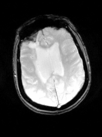
Don't worry about it! In addition, I was wondering about something else as well. I've been using MRIcron to analyze some of my Susceptibility Clinical Scans and even though they used to open fine, some of them (like the one I have attached in this email) appear distorted in a way. In the images I attached the "Original" one is how the Susceptibility clinical image should appear like, and the "Distorted" is how it appears for some of my subjects. Would you happen to know why is this happening? Thanks again for your time and help!
Best, Panos ________________________________________ From: Bruce Fischl [fischl@nmr.mgh.harvard.edu] Sent: Wednesday, July 17, 2013 11:54 AM To: Fotiadis, Panagiotis Cc: freesurfer@nmr.mgh.harvard.edu Subject: Re: [Freesurfer] SWI acquisition question
Hi Panos
sorry, I think this is pretty far outside our expertise, and it would be hard in any case to try to figure out what happened to give you these strange looking results. I certainly don't have any insight
sorry, Bruce On Wed, 17 Jul 2013, Fotiadis, Panagiotis wrote:
Hi FS Community,
I was wondering whether anyone had a chance to look into this. Thanks again for your time!
Best, Panos ________________________________________ From: freesurfer-bounces@nmr.mgh.harvard.edu [freesurfer-bounces@nmr.mgh.harvard.edu] on behalf of Fotiadis, Panagiotis Sent: Tuesday, July 16, 2013 2:00 PM To: freesurfer@nmr.mgh.harvard.edu Subject: [Freesurfer] SWI acquisition question
Hi FreeSurfer community,
I am analyzing some SWI scans of some old subjects. For most of my subjects when a SWI was acquired, the resulting files that I would get would be a Magnitude image, a Phase image, a mIP image, and the SWI image. However, I have a couple of subjects for whom I get only the mIP and the SWI images, and the SWI image looks more blurry than it's supposed to be. I was wondering:
- Why do I get only the mIP and the SWI images for those subjects and not the magnitude and phase images as well,
- Is it normal for the mIP to look like that? Usually it looks different, and
- Why is the final SWI image more blurry than usual? By looking at the image, I believe that maybe the head coil was not properly connected to the scanner at the time of the acquisition but I don't know whether that would be a sufficient explanation.
I have attached two attachments: One of the resulting mIP and one of the equivalent blurry SWI of the same subject.
Thank you in advance for your help and time!
Best, Panos
freesurfer@nmr.mgh.harvard.edu
-
 Fotiadis, Panagiotis