
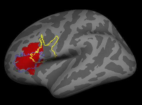
Hi freesurfers experts, this is (please check attached image) the best I got when trying to map a volume ROI (BA45 in MarsBar = MNI_Frontal_Inf_Tri_L_roi) to freesurfer using mri_vol2surf. I used /freesurfer/average/mni152.register.dat instead of spmregister since the ROI was already in mni152 2mm, the registration was perfect -checked with tkregister2.
- I obtained the red area of the image, which is outside of the ROI45.label. Considering that the source was not exactly the same, could I consider it acceptable for cortical thickness analysis? - Why are the 44 and 45 overlapping?
Thank you very much and sorry for the multiple questions, Gari

Hi Gari
sorry, I'm a bit confused. Are you using the individual subject estimates of 44 and 45 generated by FreeSurfer, or mapping them from MNI space? In either case you can get overlap unless you pick the most probably label at each point.
cheers Bruce
On Mon, 6 Aug 2012, Garikoitz Lerma Usabiaga wrote:
Hi freesurfers experts, this is (please check attached image) the best I got when trying to map a volume ROI (BA45 in MarsBar = MNI_Frontal_Inf_Tri_L_roi) to freesurfer using mri_vol2surf. I used /freesurfer/average/mni152.register.dat instead of spmregister since the ROI was already in mni152 2mm, the registration was perfect -checked with tkregister2.
- I obtained the red area of the image, which is outside of the ROI45.label. Considering that the source was not exactly the same, could I consider it acceptable for cortical thickness analysis?
- Why are the 44 and 45 overlapping?
Thank you very much and sorry for the multiple questions, Gari
Hi Bruce, thanks for your answer.
I am mapping a volume ROI (BA45) from SPM in MNI 152 space to fsaverage (the result is the red ROI). The problem is that: - when I load the BA45.label from FS in order to compare, they are not quite similar. My main problem is to know if the differences are acceptable or not and what method should I use to check the validity. - Secondary problem: when I load both the lh.BA45.label and lh.BA44.label, they overlap. I didn't create those labels, they are the ones that come in fsaverage/label. I don't understand how can they overlap (in the lh.aparc.annot or lh.aparc.a2009s.annot those areas don't overlap at all).
Thanks again for your help, Gari
On 2012-08-06, at 15:27, Bruce Fischl fischl@nmr.mgh.harvard.edu wrote:
Hi Gari
sorry, I'm a bit confused. Are you using the individual subject estimates of 44 and 45 generated by FreeSurfer, or mapping them from MNI space? In either case you can get overlap unless you pick the most probably label at each point.
cheers Bruce
On Mon, 6 Aug 2012, Garikoitz Lerma Usabiaga wrote:
Hi freesurfers experts, this is (please check attached image) the best I got when trying to map a volume ROI (BA45 in MarsBar = MNI_Frontal_Inf_Tri_L_roi) to freesurfer using mri_vol2surf. I used /freesurfer/average/mni152.register.dat instead of spmregister since the ROI was already in mni152 2mm, the registration was perfect -checked with tkregister2.
- I obtained the red area of the image, which is outside of the ROI45.label. Considering that the source was not exactly the same, could I consider it acceptable for cortical thickness analysis?
- Why are the 44 and 45 overlapping?
Thank you very much and sorry for the multiple questions, Gari
The information in this e-mail is intended only for the person to whom it is addressed. If you believe this e-mail was sent to you in error and the e-mail contains patient information, please contact the Partners Compliance HelpLine at http://www.partners.org/complianceline . If the e-mail was sent to you in error but does not contain patient information, please contact the sender and properly dispose of the e-mail.
Hi Gari
they overlap as the labels represent every location that could possibly be in either label. Unless we could predict them perfectly they have to overlap, since they share a border. With 5.2 we will include different default thresholds, but at the moment you have to do it yourself. You could do a max probability in matlab, or load them in tksurfer and threshold them manually to remove overlap.
cheers Bruce
On Mon, 6 Aug 2012, Garikoitz Lerma Usabiaga wrote:
Hi Bruce, thanks for your answer.
I am mapping a volume ROI (BA45) from SPM in MNI 152 space to fsaverage (the result is the red ROI). The problem is that:
- when I load the BA45.label from FS in order to compare, they are not quite similar. My main problem is to know if the differences are acceptable or not and what method should I use to check the validity.
- Secondary problem: when I load both the lh.BA45.label and lh.BA44.label, they overlap. I didn't create those labels, they are the ones that come in fsaverage/label. I don't understand how can they overlap (in the lh.aparc.annot or lh.aparc.a2009s.annot those areas don't overlap at all).
Thanks again for your help, Gari
On 2012-08-06, at 15:27, Bruce Fischl fischl@nmr.mgh.harvard.edu wrote:
Hi Gari
sorry, I'm a bit confused. Are you using the individual subject estimates of 44 and 45 generated by FreeSurfer, or mapping them from MNI space? In either case you can get overlap unless you pick the most probably label at each point.
cheers Bruce
On Mon, 6 Aug 2012, Garikoitz Lerma Usabiaga wrote:
Hi freesurfers experts, this is (please check attached image) the best I got when trying to map a volume ROI (BA45 in MarsBar = MNI_Frontal_Inf_Tri_L_roi) to freesurfer using mri_vol2surf. I used /freesurfer/average/mni152.register.dat instead of spmregister since the ROI was already in mni152 2mm, the registration was perfect -checked with tkregister2.
- I obtained the red area of the image, which is outside of the ROI45.label. Considering that the source was not exactly the same, could I consider it acceptable for cortical thickness analysis?
- Why are the 44 and 45 overlapping?
Thank you very much and sorry for the multiple questions, Gari
The information in this e-mail is intended only for the person to whom it is addressed. If you believe this e-mail was sent to you in error and the e-mail contains patient information, please contact the Partners Compliance HelpLine at http://www.partners.org/complianceline . If the e-mail was sent to you in error but does not contain patient information, please contact the sender and properly dispose of the e-mail.

Gari, can you check whether you have ?h.BA.annot in your subject/labels folder? this is an annotation of all the BA labels. They are non-overlapping; overlaps are resolved by the one with the most probability. I can't remember whether this made it into 5.1 or not. doug
On 08/06/2012 01:18 PM, Bruce Fischl wrote:
Hi Gari
they overlap as the labels represent every location that could possibly be in either label. Unless we could predict them perfectly they have to overlap, since they share a border. With 5.2 we will include different default thresholds, but at the moment you have to do it yourself. You could do a max probability in matlab, or load them in tksurfer and threshold them manually to remove overlap.
cheers Bruce
On Mon, 6 Aug 2012, Garikoitz Lerma Usabiaga wrote:
Hi Bruce, thanks for your answer.
I am mapping a volume ROI (BA45) from SPM in MNI 152 space to fsaverage (the result is the red ROI). The problem is that:
- when I load the BA45.label from FS in order to compare, they are not quite similar. My main problem is to know if the differences are acceptable or not and what method should I use to check the validity.
- Secondary problem: when I load both the lh.BA45.label and lh.BA44.label, they overlap. I didn't create those labels, they are the ones that come in fsaverage/label. I don't understand how can they overlap (in the lh.aparc.annot or lh.aparc.a2009s.annot those areas don't overlap at all).
Thanks again for your help, Gari
On 2012-08-06, at 15:27, Bruce Fischlfischl@nmr.mgh.harvard.edu wrote:
Hi Gari
sorry, I'm a bit confused. Are you using the individual subject estimates of 44 and 45 generated by FreeSurfer, or mapping them from MNI space? In either case you can get overlap unless you pick the most probably label at each point.
cheers Bruce
On Mon, 6 Aug 2012, Garikoitz Lerma Usabiaga wrote:
Hi freesurfers experts, this is (please check attached image) the best I got when trying to map a volume ROI (BA45 in MarsBar = MNI_Frontal_Inf_Tri_L_roi) to freesurfer using mri_vol2surf. I used /freesurfer/average/mni152.register.dat instead of spmregister since the ROI was already in mni152 2mm, the registration was perfect -checked with tkregister2.
- I obtained the red area of the image, which is outside of the ROI45.label. Considering that the source was not exactly the same, could I consider it acceptable for cortical thickness analysis?
- Why are the 44 and 45 overlapping?
Thank you very much and sorry for the multiple questions, Gari
The information in this e-mail is intended only for the person to whom it is addressed. If you believe this e-mail was sent to you in error and the e-mail contains patient information, please contact the Partners Compliance HelpLine at http://www.partners.org/complianceline . If the e-mail was sent to you in error but does not contain patient information, please contact the sender and properly dispose of the e-mail.
Freesurfer mailing list Freesurfer@nmr.mgh.harvard.edu https://mail.nmr.mgh.harvard.edu/mailman/listinfo/freesurfer
Thanks Bruce and Doug!
I can't find the lh.BA.annot in fsaverage/label folder (version 5.1):
lh.BA6.label lh.aparc.a2005s.annot rh.BA44.label rh.PALS_B12_OrbitoFrontal.annot lh.MT.label lh.aparc.a2009s.annot rh.BA45.label rh.PALS_B12_Visuotopic.annot lh.BA1.label lh.Medial_wall.label lh.aparc.annot rh.BA4a.label rh.V1.label lh.BA2.label lh.PALS_B12.labels.gii lh.aparc.label rh.BA4p.label rh.V2.label lh.BA3a.label lh.PALS_B12_Brodmann.annot lh.cortex.label rh.BA6.label rh.aparc.a2005s.annot lh.BA3b.label lh.PALS_B12_Lobes.annot lh.entorhinal.label rh.MT.label rh.aparc.a2009s.annot lh.BA44.label lh.PALS_B12_OrbitoFrontal.annot rh.BA1.label rh.Medial_wall.label rh.aparc.annot lh.BA45.label lh.PALS_B12_Visuotopic.annot rh.BA2.label rh.PALS_B12.labels.gii rh.aparc.label lh.BA4a.label lh.V1.label rh.BA3a.label rh.PALS_B12_Brodmann.annot rh.cortex.label lh.BA4p.label lh.V2.label rh.BA3b.label rh.PALS_B12_Lobes.annot rh.entorhinal.label
So, how can I be sure that the label I got from converting a volume ROI to a surface label with mri_vol2surf is accurate? Is there a way other than visually checking (in pial surface instead of in inflated in order to compare to a SPM surface render) that it is in the same location?
many thanks again for your help, Gari
On 2012-08-06, at 20:00, Douglas N Greve greve@nmr.mgh.harvard.edu wrote:
Gari, can you check whether you have ?h.BA.annot in your subject/labels folder? this is an annotation of all the BA labels. They are non-overlapping; overlaps are resolved by the one with the most probability. I can't remember whether this made it into 5.1 or not. doug
On 08/06/2012 01:18 PM, Bruce Fischl wrote:
Hi Gari
they overlap as the labels represent every location that could possibly be in either label. Unless we could predict them perfectly they have to overlap, since they share a border. With 5.2 we will include different default thresholds, but at the moment you have to do it yourself. You could do a max probability in matlab, or load them in tksurfer and threshold them manually to remove overlap.
cheers Bruce
On Mon, 6 Aug 2012, Garikoitz Lerma Usabiaga wrote:
Hi Bruce, thanks for your answer.
I am mapping a volume ROI (BA45) from SPM in MNI 152 space to fsaverage (the result is the red ROI). The problem is that:
- when I load the BA45.label from FS in order to compare, they are not quite similar. My main problem is to know if the differences are acceptable or not and what method should I use to check the validity.
- Secondary problem: when I load both the lh.BA45.label and lh.BA44.label, they overlap. I didn't create those labels, they are the ones that come in fsaverage/label. I don't understand how can they overlap (in the lh.aparc.annot or lh.aparc.a2009s.annot those areas don't overlap at all).
Thanks again for your help, Gari
On 2012-08-06, at 15:27, Bruce Fischlfischl@nmr.mgh.harvard.edu wrote:
Hi Gari
sorry, I'm a bit confused. Are you using the individual subject estimates of 44 and 45 generated by FreeSurfer, or mapping them from MNI space? In either case you can get overlap unless you pick the most probably label at each point.
cheers Bruce
On Mon, 6 Aug 2012, Garikoitz Lerma Usabiaga wrote:
Hi freesurfers experts, this is (please check attached image) the best I got when trying to map a volume ROI (BA45 in MarsBar = MNI_Frontal_Inf_Tri_L_roi) to freesurfer using mri_vol2surf. I used /freesurfer/average/mni152.register.dat instead of spmregister since the ROI was already in mni152 2mm, the registration was perfect -checked with tkregister2.
- I obtained the red area of the image, which is outside of the ROI45.label. Considering that the source was not exactly the same, could I consider it acceptable for cortical thickness analysis?
- Why are the 44 and 45 overlapping?
Thank you very much and sorry for the multiple questions, Gari
The information in this e-mail is intended only for the person to whom it is addressed. If you believe this e-mail was sent to you in error and the e-mail contains patient information, please contact the Partners Compliance HelpLine at http://www.partners.org/complianceline . If the e-mail was sent to you in error but does not contain patient information, please contact the sender and properly dispose of the e-mail.
Freesurfer mailing list Freesurfer@nmr.mgh.harvard.edu https://mail.nmr.mgh.harvard.edu/mailman/listinfo/freesurfer
-- Douglas N. Greve, Ph.D. MGH-NMR Center greve@nmr.mgh.harvard.edu Phone Number: 617-724-2358 Fax: 617-726-7422
Bugs: surfer.nmr.mgh.harvard.edu/fswiki/BugReporting FileDrop: www.nmr.mgh.harvard.edu/facility/filedrop/index.html
Freesurfer mailing list Freesurfer@nmr.mgh.harvard.edu https://mail.nmr.mgh.harvard.edu/mailman/listinfo/freesurfer
freesurfer@nmr.mgh.harvard.edu
-
 Bruce Fischl
 -
 Douglas N Greve
 -
 Garikoitz Lerma Usabiaga