
Hi all,
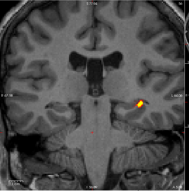
I just ran the hippo-subfield segmentation on a batch of subjects. Everything went perfect for most of my subjects, however I got an outlier in the left CA1 for one subject who had a very large volume. After checking the results in freeview for this subject, I saw that the problem was that CA1 was including a big portion of white matter in its volume (see attached png). To try to solve the problem, I just reran the recon-all -hippo-subfield for the left hemisphere of this subject (which by the way did not solve the white matter inclusion). The problem I have now is that kvlQuantifyPosteriorProbabilityImages does not seem to "see" anymore the resulting volume on the left side and does not output any volume for the left side (see error below), although the output was properly created in the mri folder (i.e. all the posterior_left_* images are there and I can load them in freeview without any problem).
Any guess?
Many thanks for the help,
Marie
[maries-iMac:LONGITUDINAL_Stats/5272/mri] marie% kvlQuantifyPosteriorProbabilityImages $FREESURFER_HOME/data/GEMS/compressionLookupTable.txt posterior_left_* volumeInVoxels: [maries-iMac:LONGITUDINAL_Stats/5272/mri] marie% kvlQuantifyPosteriorProbabilityImages $FREESURFER_HOME/data/GEMS/compressionLookupTable.txt posterior_right_* volumeInVoxels: right_CA1: 3050.22 right_CA2-3: 8579.62 right_CA4-DG: 4997.83 right_fimbria: 354.868 right_hippocampal_fissure: 302.39 right_presubiculum: 3784.79 right_subiculum: 5384.07 [maries-iMac:LONGITUDINAL_Stats/5272/mri] marie%