
12 Sep
2011
12 Sep
'11
5:04 p.m.
Dear Freesurfer List,
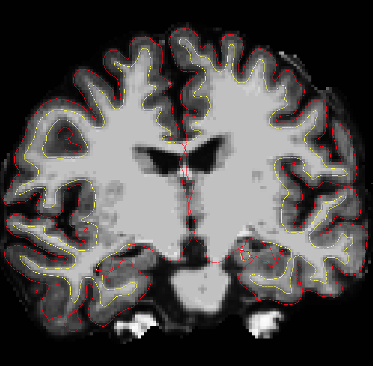
I am using recon-all to measure cortical thickness in a population of Alzheimer's patients and I have noticed that in many patients the pial surface does not include the whole brain. This seems to be because there are hippocampal lesions that are dark spots in the image which throw off the segmentation. I have tried adding control points, but that is only for the white matter boundary, and so I was wondering if you could provide any insight as to how to correct the pial surface so that the entire hippocampus is included. I have attached an example image from tkmedit showing the pial and white boundaries.
Thank you, Sheeva Azma