
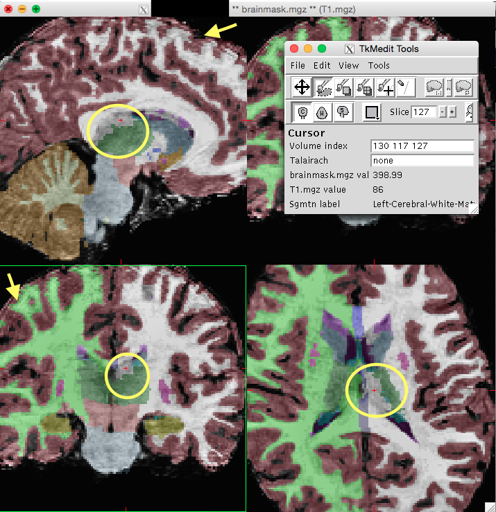
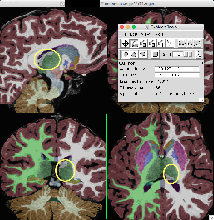
Hello, I’m an undergraduate at Duke University working on a study of pediatric structural imaging. We’ve recently come across some segmentation issues with the left thalamus (and very rarely the right thalamus) on many of our scans. Although our study isn’t looking at the thalamus as a region of interest, we’re concerned that it could be indicative of more global segmentation issues.
Freesurfer appears to be incorrectly labeling segments of the left thalamus as “left cerebral white matter." We initially thought that perhaps the errors were due to motion artifacts, but we have observed this phenomenon in some scans with striping and some scans without striping (as demonstrated in the two screenshots).
My questions are: What could be the possible reasons for this incorrect segmentation? Would scans with this problem need to be excluded? What steps could we take to fix it? Why might it only be occurring in the left thalamus?
Thanks so much for your help.
Best, Grant O'Brien Duke University Class of 2017 704-467-5449
[cid:BCEC92F0-59BE-4FBC-8E1B-B4DA90099CFB@dhe.duke.edu][cid:5CA3D954-63A9-4C96-B716-817AFCCB25D0@dhe.duke.edu]