
Dear Freesurfer Users, I'm a Ph.D. Student and I'm working on tractography using Tracula, with the aim of conducting statistical analysis on the FA values.
I have a cohort of 18 Amyotrophic Lateral Sclerosis Patients, and i'm doing all the process phases, starting from recon-all, ending to trac-all path.
I've attached a text file with info about DTI DICOM extracted by ImageJ.
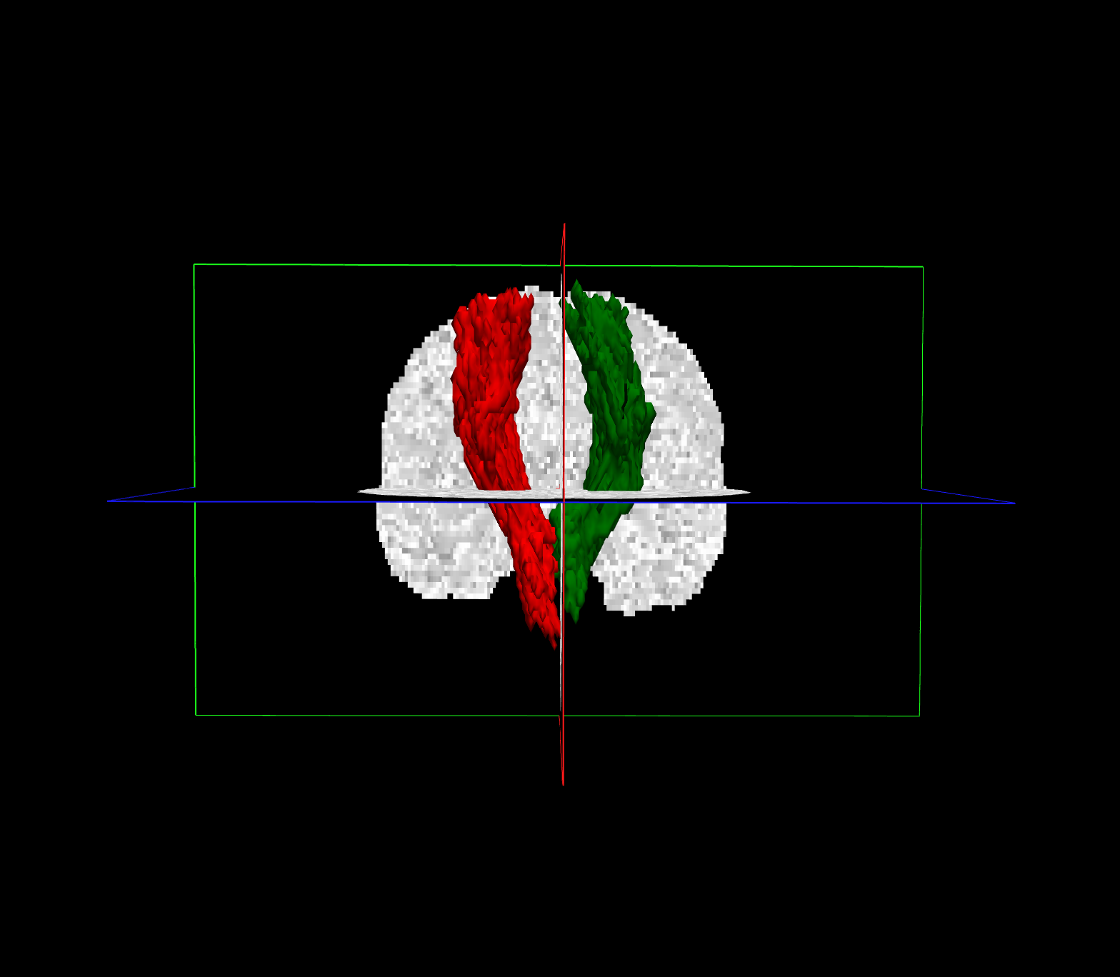
Now, my problem is about the dti mask, as you can see on the attached image, a pixelated one is obtained, even if the left and right cst tracts seem good. This problem only occurs in few patients, not in all of them, even if the script is the same for all.
This is the script i'm using:
#Patient_name
setenv SUBJECTS_DIR $TUTORIAL_DATA/Patient_recons set dtroot = $TUTORIAL_DATA/Patient_tracula set subjlist = (Patient_name) set runlist = (1) set dcmroot = $TUTORIAL_DATA/Patient_tracula set dcmlist = (Patient_name/orig/MR000000) set bvalfile = $TUTORIAL_DATA/Patient_tracula/Patient_name/bfiles/ Patient_name.bval set bvecfile = $TUTORIAL_DATA/Patient_tracula/Patient_name/bfiles/ Patient_name.bvec set nb0 = 1 set dob0 = 0 set doeddy = 1 set dorotbvecs = 1 set usemaskanat = 0 set thrbet = 0.3 set doregflt = 1 set doregbbr = 0 set doregmni = 1 set mnitemp = $FSLDIR/data/standard/MNI152_T1_1mm_brain.nii.gz set pathlist = (lh.cst_AS rh.cst_AS \ lh.ilf_AS rh.ilf_AS \ lh.unc_AS rh.unc_AS \ fmajor_PP fminor_PP \ lh.atr_PP rh.atr_PP \ lh.cab_PP rh.cab_PP \ lh.ccg_PP rh.ccg_PP \ lh.slfp_PP rh.slfp_PP \ lh.slft_PP rh.slft_PP)
set ncpts = 5 set nburnin = 200 set usetrunc = 1 set nkeep = 5 set nsample = 5000
I want to highlight that bval and bvec files are obtained from the tool dicom2nift, because Tracula is not able to extract them from DICOM header. I will really appreciate your help.
Best regards, Alessia.